Introduction
Sex steroid production is essential for follicle growth and ovulation. In amphibians, numerous published works have suggested that progesterone (P4) synthesized by ovarian follicles in response to pituitary gonadotropins acts directly on the oocyte to induce maturation in vitro (Smith et al., Reference Smith, Ecker and Subtelny1968; Maller & Krebs, Reference Maller and Krebs1980; Zelarayán et al., Reference Zelarayán, Oterino and Bühler1995). During maturation the oocyte undergoes significant morphological changes associated with meiosis progression including rupture of its large nucleus or germinal vesicle (GVBD).
However, there is controversy about the role of steroids in oocyte maturation and no direct evidence exists of P4 being the natural steroid involved in this process. In X. laevis oocytes both P4 and androgens are able to induce maturation (Lutz et al., Reference Lutz, Cole, Grupta, Kwist, Auchus and Hammes2001; Deng et al., Reference Deng, Carbajal, Evaul, Rasar, Jamnongjit and Hammes2009; Haccard et al., Reference Haccard, Dupré, Liere, Pianos, Eychenne, Jessus and Ozon2012). Besides, maturation and ovulation do not occur if the synthesis of ovarian androgens is inhibited (White et al., Reference White, Jamnongjit, Gill, Lutz and Hammes2005). In fish oocytes, the hydroxylated metabolites of P4 have been associated with maturation (Ponthier et al., Reference Ponthier, Shackleton and Trant1998; Canario et al., Reference Canario, Scott and Flint1989) while in higher vertebrates, progestins and androgens are involved in this process with various species-dependent effects (Deng et al., Reference Deng, Carbajal, Evaul, Rasar, Jamnongjit and Hammes2009).
Classically, steroids exert their effect by binding to receptors that modulate genetic expression in target cells. However, in amphibians, follicle cells respond to the LH surge by releasing P4, which acts on the oocyte surface to initiate non-transcriptional signalling mechanisms that induce oocyte maturation (Maller & Krebs, Reference Maller and Krebs1980; Deng et al., Reference Deng, Carbajal, Evaul, Rasar, Jamnongjit and Hammes2009; Zelarayán et al., Reference Zelarayán, Ajmat, Bonilla and Buhler2012). Numerous studies have focused on the interaction between P4 and the oocyte surface and on the cascade of signalling events (Sheng et al., Reference Sheng, Tiberi, Booth, Ma and Liu2001; Zelarayán et al., Reference Zelarayán, Ajmat, Bonilla and Buhler2012) that lead to activation of the key regulators of the G2–M phase transition (Maller & Krebs, Reference Maller and Krebs1980; Masui, Reference Masui1982; Liu et al., Reference Liu, Ma, Hamam and Liu2005): cytoplasmic maturation promoting factor (MPF), and a complex of cyclin-dependent kinase p34cdc2 and cyclin B that induces GVBD (Sánchez Toranzo et al., Reference Sánchez Toranzo, Bonilla, Zelarayán, Oterino and Bühler2006). Immature oocytes (Prophase I) contain the inactive form of MPF, pre-MPF, whose activation depends on the signalling events triggered by P4 action (Haccard & Jessus, Reference Haccard and Jessus2006).
Testosterone (T) seems to be the main androgen released by gonadotrophic stimulus in X. laevis follicles (El-Zein et al., Reference El-Zein, Boujard, Garnier and Joly1988; Deng et al., Reference Deng, Carbajal, Evaul, Rasar, Jamnongjit and Hammes2009). In this species, androgens have been considered the physiological maturation inducers as their actions are more potent than those of P4 (Lutz et al., Reference Lutz, Cole, Grupta, Kwist, Auchus and Hammes2001). Coincidentally, plasma concentrations of T and androstenedione (AD) are higher in animals stimulated with hCG, while plasma P4 levels remain low during ovulation (Lutz et al., Reference Lutz, Cole, Grupta, Kwist, Auchus and Hammes2001). In agreement with this finding, Medina et al. (Reference Medina, Ramos, Crespo, Gonzalez-Calvar and Fernandez2004) showed that plasma P4 levels decreased in R. arenarum (ex Bufo arenarum) during the ovulatory period. In this species, as in other amphibians, ovarian response in vitro is strongly associated with seasonal changes and is accompanied by changes in hormone serum levels (P4, estradiol and T). In R. arenarum oocytes, maturation can be induced in vitro by P4 throughout the year. However, our studies demonstrated that ovarian response to P4 is greater during the reproductive period in recently captured animals. The signalling mechanisms triggered in the oocyte in response to the steroid also seem to be related to seasonal variations (Ortiz et al., Reference Ortiz, Buhler and Zelarayán2013).
Considering that the role of steroids in meiotic resumption in amphibian oocytes has not been sufficiently clarified to date, we analyzed in this work the seasonal effect of different steroids on in vitro maturation of R. arenarum oocytes and the participation of MPF in this process.
Materials and methods
Animals
Female Rhinella arenarum specimens were collected in Tucumán, northwestern Argentina. They were kept in captivity at room temperature under natural light–dark cycles for a few hours until use. The animals were collected in March to August (non-reproductive period) and in September to February (reproductive period) for 3 consecutive years.
Reagents
All reagents were purchased from Sigma or Merck. The stock solutions of steroids were prepared in ethanol and added directly to the culture medium to give a final concentration of 1–10,000 nM. Amphibian Ringer solution (AR) (6.60 g NaCl/l, 0.15 g CaCl2/l, and 0.15 g KCl/l) with penicillin G-sodium (30 mg/l) and streptomycin sulphate (50 mg/l), pH 7.4, was used as a culture medium in all routine incubations.
In vitro cultures
Oocytes and follicles were selected and maturation assays performed on each preparation to determine its sensitivity to steroids.
Fully grown follicles (1.6–1.8 mm in diameter) were isolated from ovarian tissues using watchmaker's forceps. Denuded oocytes were obtained by manually pulling off the follicle epithelium and the theca layer with fine forceps. Follicle cells were removed by incubation of defolliculated oocytes in AR for 5 min with gentle shaking (100 oscillations/min) (Zelarayán et al., Reference Zelarayán, Oterino and Bühler1995). The incubations were performed at 26°C in multiwell culture dishes (Costar 3524, Cambridge, MA, USA) with randomized samples of 20 oocytes or follicles distributed into separate wells containing 2 ml of AR. Duplicates in two wells were run in each experimental group.
Dose–response curves of steroids were performed by incubating denuded oocytes or follicles for 22–24 h in the presence of different doses of the steroids (1–10,000 nM).
Time–response curves of steroids were conducted with oocytes and follicles using the dose that induced the best response. Samples were incubated for 2–24 h.
Maturation criteria
The criterion for maturation was GVBD judged by the appearance of a white spot surrounded by a pigmented area around the animal pole during the first few hours after hormone treatment and by GVBD as detected by dissecting the samples under a stereo-microscope, after fixation in Ancel & Vintemberger's solution (10% formol, 0.5% acetic acid and 0.5% NaCl) overnight at room temperature. Maturation rate was expressed as percentage of GVBD.
Mature cytoplasm transfers
Cytoplasm from mature oocytes was obtained using a modified Hedeimann's method (Bühler & Petrino, Reference Bühler and Petrino1983). Briefly, oocytes matured with different steroids (1000 nM) or with P4 (1000 nM) (control) were suspended at the interface of 60% Ficoll (v/v) in AR and then centrifuged (10,000 g, 20 min) at 4°C in an Eppendorf Centrifuge 5417C. After this procedure, oocytes displayed four distinct phases: yolk platelets, pigment granules, clear cytoplasm and a lipid layer. Microinjection was performed using ICSI micropipettes (Humagen™ Fertility Diagnostics) attached to a Leitz Wetzlar Micromanipulator. Denuded fully grown immature oocytes were microinjected with 40 nl of cytoplasm (Ortiz et al., Reference Ortiz, Arias-Torres and Zelarayán2014) obtained from the clear cytoplasm layer containing MPF (see Results). Then the oocytes were incubated in AR for 18–20 h and then fixed for GVBD scoring. A batch of immature oocytes microinjected with the same volume of cytoplasm obtained from immature oocytes (immature cytoplasm) was used as negative control while another batch microinjected with mature cytoplasm obtained from P4-treated oocytes (1000 nM) was used as positive control.
Statistical analysis
Results are expressed as mean ± standard deviation (SD) from experiments carried out in duplicate using different animals in each experiment. A Probit model was used to calculate the 50th percentile of oocyte and follicle maturation. Comparisons among different treatments in the microinjection experiments were carried out using Student's t-test. A value of P < 0.05 was considered to be statistically significant.
Results and Discussion
Assays were conducted to assess the ability of different steroids to induce oocyte and follicle maturation in vitro during the reproductive and non-reproductive period. At a first step the EC50 (EC50, ‘effective concentration’ that induces 50% maturation) of the steroids used was determined.
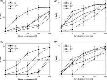
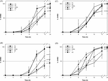
The results of Figs 1 and 2 indicated that all steroids assayed induced oocyte and follicle maturation in a dose- and time-dependent manner. In oocytes from animals captured during the reproductive period (Fig. 1 C), P4 and pregnenolone (P5) showed a similar response (P > 0.05) as maturation inducers (EC50 ≅ 6 and 8 nM respectively). However, androgens (DHEA, T and AD) were less effective (EC50 ≅ 50, 120 and 600 nM respectively).
Seasonal response of oocytes and follicles to steroids. Oocytes (A) and follicles (B) obtained during the non-reproductive period. Oocytes (C) and follicles (D) obtained during the reproductive period. The samples were incubated in the presence of increasing doses of P5, P4, DHEA, AD and T (1–10,000 nM). GVBD was assessed after 18–24 h of incubation. Values represent the mean ± standard deviation (SD) (n = 12) of experiments performed in duplicate on different animals. EC50 with different letters are significantly different (P < 0.05).

Time course of the effects of steroids on oocyte and follicle maturation. Oocytes (A) and follicles (B) obtained during non-reproductive period. Oocytes (C) and follicles (D) obtained during the reproductive period. Maturation was induced with P5, P4, DHEA, AD or T (1000 nM) in oocytes and follicles. Samples were fixed at 2, 4, 6, 8, 10, 12 or 24 h of incubation and GVBD was assessed. Data are the mean ± standard deviation (SD) (n = 12) of experiments performed in duplicate on different animals. EC50 with different letters are significantly different (P < 0.05).

In oocytes from the non-reproductive period (Fig. 1 A), the response to all steroids assayed was lower as higher steroid concentrations were required to induce GVBD. During this season, the oocyte response to androgens was significantly lower (P < 0.05) than in the reproductive period, as 10-fold greater doses of T and AD were required to induce 50% maturation (Fig. 1 A). In the same way, in the non-reproductive period, P4 (EC50 P4 ≅ 30 nM) was the most efficient steroid in inducing maturation.
In our experiments the sensitivity of R. arenarum follicles to GVBD varied between frogs in a similar way to the results obtained with X. laevis oocytes (Pickford & Morris, Reference Pickford and Morris1999; Haccard et al., Reference Haccard, Dupré, Liere, Pianos, Eychenne, Jessus and Ozon2012; Cao et al., Reference Cao, Xu, Zhanng, Zhao, Wei and Qin2014). We found that in oocytes and follicles the response to androgens varied among adult R. arenarum ovaries in the experiments carried out for 3 years. Therefore, we used a large number of recently captured specimens. Nevertheless, we observed individual variability in the sensitivity to the steroids tested, similar to those observed for X. laevis oocytes (Cao et al., Reference Cao, Xu, Zhanng, Zhao, Wei and Qin2014). This variability may result from different levels of endogenous gonadotropins that impact on ovarian physiology and on subsequent oocyte sensitivity to steroid-induced GVBD, as observed by LaMarca et al. (Reference LaMarca, Westphal and Rein1985) in X. laevis oocytes.
Follicle response to steroids was similar to that of oocytes for both seasonal periods. The comparison shown in Fig. 1 D between responses to the different steroids of follicles from the reproductive period revealed that follicle response to the androgens tested was variable, but in no case higher than the response induced by P4 or P5 (EC50 ≅ 30 nM). Follicle response to these steroids were not significantly different (P > 0.05). It seems likely that during this period the follicles of recently captured animals are still under the influence of environmental factors that stimulate gonadotropin release, so that P4 or P5 could metabolize to other maturation-inducing steroids.
With respect to androgens, T was the most efficient (EC50 ≅ 40 nM) in inducing follicle maturation while DHEA and AD were less efficient (P < 0.05) (Fig. 1 D). In contrast, Haccard et al. (Reference Haccard, Dupré, Liere, Pianos, Eychenne, Jessus and Ozon2012) demonstrated that DHEA is capable of inducing GVBD and is the main steroid produced by X. laevis follicles.
In brief, P4 proved to be a better oocyte and follicle in vitro maturation inducer than androgens, independently of the season. Our results are similar to the ones obtained for X. laevis oocytes and follicles (Thibier-Fouchet et al., Reference Thibier-Fouchet, Mulner and Ozon1976), in which continuous exposures to P4 and P5 induced similar GVBD percentages. The sensitivity of oocytes to steroids proved to be greater than that of follicles.
It seems likely that the differences found are based on the inhibitory role of follicle cells on oocyte maturation. It has been suggested that, as in mammals, gap junctions between oocyte and follicle cells would allow the passage of a maturation-inhibiting substance whose nature varies between species, like cAMP and purines in R. arenarum oocyte (Zelarayán et al., Reference Zelarayán, Oterino, Sánchez Toranzo and Bühler2000). In this sense we demonstrated that during the reproductive period R. arenarum oocytes are capable of meiosis resumption when deprived of the surrounding follicle cells (Zelarayán et al., Reference Zelarayán, Oterino and Bühler1995, Reference Zelarayán, Oterino, Sánchez Toranzo and Bühler2000). In X. laevis, steroid sulfotransferase has been suggested as the enzyme that regulates the accumulation of sulfated steroids in the prophase-arrested oocyte, preventing active steroid accumulation and therefore oocyte maturation (Haccard et al., Reference Haccard, Dupré, Liere, Pianos, Eychenne, Jessus and Ozon2012).
In X. laevis, Lutz et al. (Reference Lutz, Cole, Grupta, Kwist, Auchus and Hammes2001) showed that when ovaries are stimulated with β-HCG, androgens T and AD are stronger maturation inducers than P4. We found that in R. arenarum EC50 for P4 was even lower than the results obtained by those authors in X. laevis. As the effect of androgens on X. laevis oocytes maturation is correlated with the plasma levels reached after hormone stimulation, Lutz et al. (Reference Lutz, Cole, Grupta, Kwist, Auchus and Hammes2001) proposed them as physiological maturation inducers. However, our experiments, conducted in basal conditions in recently captured animals, showed that in R. arenarum P4 was a more potent inducer than either T or AD throughout the year, especially during the reproductive season. In agreement with that finding, P4 and T serum levels increased during the preovulatory period, when R. arenarum oocytes reached their maximum development in the ovary, and maturation and ovulation were about to occur (Medina et al., Reference Medina, Ramos, Crespo, Gonzalez-Calvar and Fernandez2004). During the postovulatory period (reproductive period) the serum levels of both steroids remained low in correlation with growth resumption of ovarian follicles in vivo. It is likely that T is used as a source of estrogens, which promote vitellogenesis and oocyte growth in a similar way to that observed in Rana esculenta (Gobbetti et al., Reference Gobbetti, Polzonetti-Magni, Zerani, Carnevali and Botte1985) and some fish (Sen et al., Reference Sen, Mukherjee, Bhattacharyya and Mukherjee2002).
In our in vitro experiments, the sensitivity of both oocytes and follicles in response to P4 (Fig. 1) could be related to the serum levels found by Medina et al. (Reference Medina, Ramos, Crespo, Gonzalez-Calvar and Fernandez2004), which in this species begin to increase again in September.
The results in Fig. 2 B, D indicate that follicles show a good temporal response to P4, P5 and T, independent of the period during which the females were captured. In fact, after incubation for 8–10 h, more than 50% of the follicles treated with these steroids resumed meiosis. Follicles treated with DHEA and AD matured more slowly, reaching 50% GVBD after 10–12 h. All steroids induced maximum response around 20 h of incubation.
Oocytes showed a response similar to that of follicles during the reproductive period (Fig. 2 C), while during the non-reproductive period their response to steroids was slower (Fig. 2 A). In both periods, P4 and P5 induced maturation faster than did androgens.
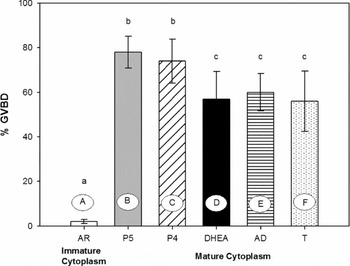
The release from meiotic arrest is regulated by the activation of pre-MPF to active MPF in response to hormone signalling. In our laboratory we earlier demonstrated that microinjection of mature cytoplasm from P4-treated oocytes is sufficient to activate pre-MPF, leading to GVBD (Sánchez Toranzo et al., Reference Sánchez Toranzo, Bonilla, Zelarayán, Oterino and Bühler2006). The fact that androgens are also capable of inducing maturation in R. arenarum follicles and oocytes induced us to question their competence to activate MPF. In order to address this issue we conducted experiments of microinjection of cytoplasm from oocytes matured with P5, DHEA, AD and T into immature oocytes compared with P4-treated oocytes. We demonstrated that small amounts of mature cytoplasm from androgen-treated oocytes are capable of inducing meiosis resumption in immature oocytes (Fig. 3). Cytoplasm transfer from androgen-matured oocytes produced lower results (i.e. T, 56 ± 13%) than those obtained with microinjection of cytoplasm from P4-treated oocytes (74 ± 9%). The oocytes microinjected with immature cytoplasm did not resume meiosis in either assay. Donor oocytes (those from which cytoplasm was obtained) reached similar GVBD values to those shown in Fig. 1 A. These results demonstrate that androgens such as DHEA, AD and T are just as competent as P4 to activate MPF in R. arenarum. To verify that phosphatase from gene cdc25 participates in MPF activation when immature oocytes are microinjected with mature cytoplasm, we studied the effect of cdc25 activity inhibition of MPF amplification by preincubating oocytes in NAVO3 before injection of mature cytoplasm (data not shown). The oocytes thus treated failed to resume meiosis.
MPF amplification in denuded oocytes. Immature oocytes were microinjected with 40 nl of (A) immature cytoplasm; (B) cytoplasm matured with P5 (1000 nM); (C) P4 (1000 nM); (D) cytoplasm matured with DHEA (1000 nM); (E) cytoplasm matured with AD (1000 nM); and (F) cytoplasm matured with T (1000 nM). After microinjection, oocytes were incubated in AR for 20 h and GVBD was scored. Each bar represents the mean ± standard deviation (SD) (n = 4) of experiments performed in duplicate on different animals. a,b,c means with different letters are significantly different (P < 0.05).

In this work we showed the relationship between seasonal periods and the response of R. arenarum follicles and oocytes to different steroids. Using in vitro GVBD assays, we demonstrated that P4 is the main steroid capable of inducing maturation in R. arenarum oocytes and follicles. In the second part of this work we demonstrated by cytoplasm microinjection experiments that androgens can activate pre-MPF as P4 does.
Acknowledgements
This work was supported by a grant from the Science Council of the National University of Tucuman (CIUNT), Argentina. The authors are grateful to Dr Federico Bonilla for his kind assistance in performing the microinjection experiments and Lic. Elena Bru for her assistance in statistical analyses.