Introduction
A pig (Betthauser et al., Reference Betthauser, Forsberg, Augenstein, Childs, Eilertsen, Enos, Forsythe, Golueke, Jurgella, Koppang, Lesmeister, Mallon, Mell, Misica, Pace, Pfister-Genskow, Strelchenko, Voelker, Watt, Thompson and Bishop2000; Onishi et al., Reference Onishi, Iwamoto, Akita, Mikawa, Takeda, Awata, Hanada and Perry2000; Polejaeva et al., Reference Polejaeva, Chen, Vaught, Page, Mullins, Ball, Dai, Boone, Walker, Ayares, Colman and Campbell2000) was successfully cloned 3 years after the birth of Dolly the sheep (Wilmut et al., Reference Wilmut, Schnieke, McWhir, Kind and Campbell1997), the first mammal cloned from a cultured cell. At present there are a limited number of countries and laboratories that have achieved viable piglets from fibroblast cells or undefined genital cells and cumulus cells of transfected or non-transfected cell lines (Bondioli et al., Reference Bondioli, Ramsoondar, Williams, Costa and Fodor2001; Boquest et al., Reference Boquest, Grupen, Harrison, McIlfatrick, Ashman, d'Apice and Nottle2002; Dai et al., Reference Dai, Vaught, Boone, Chen, Phelps, Ball, Monahan, Jobst, McCreath, Lamborn, Cowell-Lucero, Wells, Colman, Polejaeva and Ayares2002; De Sousa et al., Reference De Sousa, Dobrinsky, Zhu, Archibald, Ainslie, Bosma, Bowering, Bracken, Ferrier, Fletcher, Gasparrini, Harkness, Johnston, Ritchie, Ritchie, Travers, Albertini, Dinnyes, King and Wilmut2002; Hyun et al., Reference Hyun, Lee, Kim, Kim, Lee, Nam, Jeong, Kim, Yeom, Kang, Han, Lee and Hwang2003; Lai et al., Reference Lai, Park, Cheong, Kuhholzer, Samuel, Bonk, Im, Rieke, Day, Murphy, Carter and Prather2002a; Lee et al., Reference Lee, Wu, Tian, Barber, Hoagland, Riesen, Lee, Tu, Cheng and Yang2003; Park et al., Reference Park, Cheong, Lai, Im, Kuhholzer, Bonk, Samuel, Rieke, Day, Murphy, Carter and Prather2001; Ramsoondar et al., Reference Ramsoondar, Machaty, Costa, Williams, Fodor and Bondioli2003; Walker et al., Reference Walker, Shin, Zaunbrecher, Romano, Johnson, Bazer and Piedrahita2002; Watanabe et al., Reference Watanabe, Iwamoto, Suzuki, Fuchimoto, Honma, Nagai, Hashimoto, Yazaki, Sato and Onishi2005; Yin et al., Reference Yin, Tani, Yonemura, Kawakami, Miyamoto, Hasegawa, Kato and Tsunoda2002).
Somatic nuclear transfer (NT) in pig provides many potential applications for biomedicine, agriculture and research into basic biological mechanisms (Prather et al., Reference Prather, Hawley, Carter, Lai and Greenstein2003). In agriculture, NT in pigs combined with genetic modification could take us a step closer to our goals of (1) altering the carcass composition so that it is a healthier product, (2) producing pork faster or more efficiently, (3) creating animals that are resistant to specific diseases, (4) reducing the major losses normally observed during the first month of swine embryogenesis, and (5) creating animals that are more environmentally friendly. In biomedicine, making specific genetic modifications in the pig provides the possibility of producing recombinant products in animals for biomedical or nutraceutical uses and the possibility of producing models of human genetic disease for research and drug development (Lai & Prather, Reference Lai and Prather2003).
However, as with other species, cloning efficiency is still very low in pig NT (always less than 2% based on the percentage of pregnancies to term from oocytes used). It has generally been believed that many factors contribute to the problem, such as variations in technique between laboratories, oocyte source and quality, cell type of nuclear donors, treatment of donor cells prior to NT, methods of embryo culture, possible loss of somatic imprinting in the nuclei of the reconstructed embryo, failure to reprogram the transplanted nucleus adequately, and the failure of artificial methods of activation to emulate reproducibly those crucial membrane-mediated events that accompany fertilization. In the pig, there is an additional difficulty that at least four good embryos are required to induce and maintain a pregnancy. Therefore, to increase the chance of producing offspring, efforts must be made to minimize the inefficiencies at each step of the NT procedure (Lai & Prather, Reference Lai and Prather2003). Furthermore, among the factors mentioned above, many aspects related to donor cells are crucial and easily overlooked, such as cell cycle synchronization, cell shape and preparation methods. To date there have been no systematic studies combining all these factors.
The GFP (green fluorescent protein) gene is useful as a marker for identification of transgenic NT porcine embryos prior to embryo transfer. Cloned embryos produced by transfected somatic cells could in theory produce 100% transgenic animals. Use of the GFP reporter gene might significantly enhance the efficiency of transgenic animal production because expression of this gene allows us to select positive embryos before transfer to the surrogate mother (Uhm et al., Reference Uhm, Kim, Kim, Chung, Chung, Lee and Chung2000), and the descendants of a transgenic pig expressing GFP will probably provide a variety of genetically marked tissues, which would be very useful for basic research where such marked cells are required (Lai et al., Reference Lai, Park, Cheong, Kuhholzer, Samuel, Bonk, Im, Rieke, Day, Murphy, Carter and Prather2002b).
The current study was designed to evaluate the feasibility of producing transgenic blastocysts expressing enhanced GFP and to examine the effects of shape and preparation methods of GFP-transfected or normal donor cells on the in vitro developmental ability of pig nuclear transferred embryos (NTEs).
Materials and methods
Unless otherwise noted, all chemicals were purchased from Sigma-Aldrich (St Louis, MO).
Preparation of pig fibroblast cells
A Chinese miniature swine at 40 days (day 0 = natural mating) of gestation was killed and the uterus taken to the laboratory. Fetuses were collected aseptically. After removal of the heads and internal organs using iris scissors and forceps, the remains were rinsed twice in Ca2+- and Mg2+-free phosphate-buffered saline (DPBS; GIBCO, Life Technologies, Rockville, MD), and cut into 1 mm3 pieces. The pieces were seeded into a T-75 cell culture flask and cultured in an incubator at 39 °C, under 5% CO2 in air, and 100% humidity, for 4–6 h until tissues explants were attached. Then fresh culture medium, consisting of Dulbecco's Modified Essential Medium (DMEM; GIBCO), 20% fetal bovine serum (FBS; GIBCO) and 1% non-essential amino acids and penicillin and streptomycin, was added. After the cells had grown to 70–80% confluence, the old culture medium was discarded and DPBS was used to wash the cells three times followed by the use of 0.25% trypsin and 0.02% EDTA to harvest the cells. After centrifugation at 700 g for 5 min, the supernatants were removed and one group of pellets was resuspended in fresh cell culture medium, and seeded into new T-75 flasks. A second group of pellets was resuspended in cell cryopreservation medium consisting of DMEM, 10% dimethylsulfoxide (DMSO) and 20% FBS and aliquoted into cryovials. After being placed at −70 °C overnight, they were then stored at −196 °C in liquid nitrogen. For cell refrigeration, a proportion of the cell pellets were resuspended with serum-free DMEM, and aliquoted into 1.5 ml Eppendorf centrifuge tubes before being refrigerated at 4 °C.
Transfection of the EGFP gene into fetal fibroblasts
The plasmid pEGFP-N1 was purchased from Clontech Laboratories (Palo Alto, CA). Transfections were performed using Lipofectamine 2000 (Invitrogene, Life Technologies) as instructed by the manufacturer. Briefly, the day before transfection, confluent fetal fibroblasts (at passage 2–4) were trypsinized, counted, and seeded in growth medium without antibiotics in a 6-well culture plate to reach 90–95% confluence at the time of transfection. Four micrograms of pEGFP-N1 and 10 μl of Lipofectamine 2000 were diluted in 500 μl of serum-free DMEM. After 20 min of incubation at room temperature, 500 μl of the complexes containing cells and medium was added to each well, and the solution mixed gently by rocking the plate back and forth. After 6–8 h of culture, the medium was changed. Then a group of cells was selected by adding 0.5 mg/ml Geneticin (G418; GIBCO) to the cell culture medium for 10 days; after withdrawal of G418, cells were subcultured, trypsinized, and either cryopreserved or chilled at 4 °C as mentioned above. To prepare nuclear donors for nuclear transfer, subcultured cells at passage 5–9 were either subjected to serum starvation or maintained in cycling growth. For the serum starvation group, cells were cultured in normal culture medium for 2–3 days and then the old medium was replaced with DMEM plus 0.5% FBS and antibiotics and culture continued for another 2–5 days. For the cycling growth group, cells at 70% confluence were trypsinized and prepared as nuclear donors.
In vitro maturation of prepubertal gilt oocytes
Ovaries of prepubertal gilts were collected from a local slaughterhouse, stored in 0.9% NaCl supplemented with antibiotics at 30–35 °C and transported back to the laboratory within 1–2 h of slaughter. Cumulus–oocyte complexes (COCs) were aspirated from follicles 3–6 mm in diameter using Tyrode albumin lactate pyruvate medium (TL-HEPES) supplemented with 0.1% polyvinylalcohol (PVA; w/v) with a 20-gauge needle connected to a 20 ml disposable syringe. After being washed in the maturation medium three times, COCs with at least three layers of compacted cumulus and even cytoplasm were chosen. The in vitro maturation (IVM) medium for the first phase of culture consisted of NCSU-23 (BSA-free) plus 10% porcine follicular fluid (PFF; w/v), 0.6 mM L-cysteine, 10 ng/ml epidermal growth factor (EGF), 10 IU/ml human chorionic gonadotropin (hCG) and 10 IU/ml equine chorionic gonadotropin (eCG). The second-phase culture medium was the same as that for the first phase except that it was hormone-free. All maturation media were prepared and buffered in an incubator for at least 3–4 h before the start of IVM. Four 100 μl culture drops were placed in a 35 mm Petri dish, covered with mineral light oil, and 20–25 COCs allocated into each drop. The COCs were matured in the first-phase culture medium for 20 ± 2 h before being cultured in the second-phase IVM medium for a further 20 ± 2 h. PFF was made by the following protocol: Fluid was aspirated from follicles 5–7 mm in diameter then centrifuged at 1600 g for 30 min, after which the supernatant fluid was collected and filtered through 0.45 μm syringe filters before being stored at −20 °C until use.
After the end of maturation, COCs were transferred to 1 mg/ml hyaluronidase in DPBS and pipetted repeatedly for 2–3 min to denude cumulus cells. Oocytes free of cumulus cells and with an intact cytoplasmic membrane and clear perivitelline space were chosen as recipient cytoplasts.
Micromanipulation for nuclear transfer
Micromanipulation was conducted with TE2000U and TE300 inverted microscopes equipped with manipulation systems (Narishige, Japan).
The micromanipulation drop consisted of HEPES-buffered NCSU-23 supplemented with 7.5 μg/ml cytochalasin B (CB) and 0.4% bovine serum albumin (BSA) covered with mineral oil. Groups of 20–30 oocytes and nuclear donor cells were placed in the same drop. After incubation for 15–30 min, the oocyte was secured with a holding pipette (inner diameter 25–35 μm, outer diameter 80–100 μm). After being placed at the 12–1 o'clock position, the first polar body and 10% of the adjacent cytoplasm presumptively containing the metaphase plate were aspirated with a bevelled injection pipette (inner diameter 20 μm). A nuclear donor cell was then injected into the perivitelline space through the same slit. After NT, reconstructed couplets were transferred into drops of medium covered with mineral oil for recovery for 1–1.5 h until fusion and activation were conducted. The medium was NCSU-23 supplemented with 4 mg/ml BSA and 7.5 μg/ml CB.
Fusion and activation
Reconstructed couplets were washed three times after being balanced in the fusion solution for 4 min. Then groups of five couplets were placed in the fusion chamber (BTX, San Diego, CA; electrode width is 0.5 mm), which was filled with fusion solution. Couplets were aligned manually using a fine needle to make the contact plane parallel to the electrodes. A single, 30 μs, direct current pulse of 2.0 kV/cm was then applied using a BTX ECM2001 (BTX, San Diego, CA) to induce couplet fusion. The fusion medium was 0.25 M mannitol, 0.1 mM CaCl2, 0.1 mM MgCl2, 0.5 mM HEPES and 0.01% PVA. After culture in drops of NCSU-23 plus 4 mg/ml BSA and 7.5 μg/ml CB for 30–60 min, fusion results were examined under a stereomicroscope (SMZ1000, Nikon, Japan). Fused couplets were continued to the second activation 1.5 h after fusion, and then transferred to embryo culture medium to evaluate in vitro developmental ability.
Embryo culture
Embryo culture was conducted at 39 °C under 5% CO2 in air and 100% humidity. After 48 h and 168 h of in vitro culture, the cleavage rate and blastocyst rate were estimated under a stereomicroscope.
Experiments
Experiment 1
The effect of GFP transfection on the development of pig NTEs was evaluated. In order to reconstruct transgenic embryos, G418-selected cells that displayed green under a fluorescein isothiocyanate filter set were chosen and injected into perivitelline space of the oocyte to contact the oocyte membrane. NTEs from transfected cells were compared with those from non-transfected cells.
Experiment 2
The effect of different nuclear donor preparation methods on in vitro development of NTEs was investigated. GFP-transfected nuclear donors were prepared in three ways: (A) detaching cultured cells freshly using trypsin followed by reconstruction of cloned embryos; (B) freezing cells stored at 4 °C for 1–5 days in a refrigerator, followed by injection into the perivitelline space of the recipient oocytes; (C) thawing cells which had been cryopreserved for at least 1 day in liquid nitrogen before being used as donors.
Experiment 3
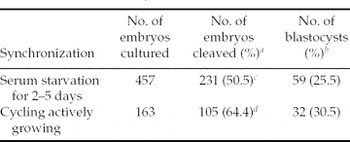
The effect of cell cycle synchronization methods on in vitro development potential of pig NTEs was examined. NTEs derived from cells serum-starved for 2–5 days and GFP-transfected were compared with NTEs from cycling growing subconfluent cells.
Experiment 4
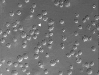
The effect of different shapes of cultured GFP-transfected fibroblast cells on cleavage and blastocyst formation of reconstructed embryos was evaluated. Cells with a smooth surface or a rough surface were compared as donor nuclei for nuclear transfer. The former had an intact smooth plasma membrane and uniform, clear cytoplasm, while the latter had a disrupted rough plasma membrane and a granular cytoplasm (Fig. 1).
Morphology and surface of pig fetal fibroblast nuclear donor cells. The arrowhead indicates cells with a rough surface. Round smooth cell lies to the left of R. ×300.

Statistical analysis
The rates of cleavage and blastocyst formation were analysed using a chi-square test. A value of χ2 corresponding to p < 0.05 was considered significant. At least three replicates were done in each experiment.
Results
Experiment 1: effect of GFP transfection
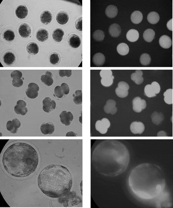
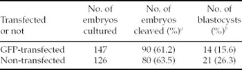
Transgenic pig embryos developed to blastocyst that expressed GFP were obtained 168 h after culture (Fig. 2C). As shown in Table 1, in vitro developmental ability of NTEs using transfected cells as donors was not compromised significantly when compared with NTEs using non-transfected cells as donors.
In vitro development of pig transgenic embryos. (A)–(C) Embryos under normal light; (A')–(C') Embryos under fluorescein isothiocyanate filters to identify EGFP (A)–(A') One-cell stage, ×200; (B)–(B'): 2- to 4-cell stage, ×200; (C)–(C') blastocyst stage, ×300.

Effects of GFP transfection on in vitro developmental ability of pig preimplantation embryos

aPercentage of cleaved embryos from embryos cultured.
bPercentage of blastocysts from embryos cleaved.
Experiment 2: effect of donor cell preparation
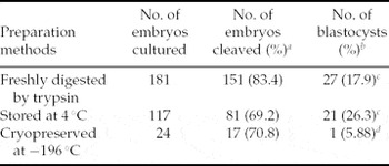
As shown in Table 2 there were no statistical significances among the three preparation methods as regards cleavage rate. The blastocyst rates of donor cells that had been treated at 4 °C and freshly digested cells were similar to each other (26.3% vs 17.9%). However, lower blastocyst rates were obtained when cells that had been cryopreserved at −196 °C were used as NT donors.
Effects of preparation methods of GFP-transfected donors on preimplantation developmental competence of nuclear transferred embryos

aPercentage of cleaved embryos from embryos cultured.
bPercentage of blastocysts from embryos cleaved.
c,dValues with different superscripts in the same column are significantly different (p < 0.05).
Experiment 3: effect of cell cycle synchronization
As shown in Table 3, the cleavage rate increased significantly when cycling cells were used as donors (50.5% vs 64.4%). However, no significant difference in blastocyst rate was observed between the two groups.
Effects of cell cycle synchronization treatment of GFP-transfected cells on in vitro development ability of pig nuclear transferred embryos

aPercentage of cleaved embryos from embryos cultured.
bPercentage of blastocysts from embryos cleaved.
c,dValues with different superscripts in the same column are significantly different (p < 0.05).
Experiment 4: effect of donor cell shape
As shown in Table 4, the fusion rate of rough cells was lower than that of round smooth cells (47.8% vs 76.8%). However, there was no significant difference in cleavage rate and blastocyst rate between the two types of cell.
Effects of shapes of GFP-transfected donor cell on in vitro developmental ability of preimplantation nuclear transferred embryos in pig

aPercentage of cleaved embryos from embryos cultured.
bPercentage of blastocysts from embryos cleaved.
c,dValues with different superscripts in the same column are significantly different (p < 0.05).
Discussion
Production of genetically modified pig embryos
NT provides a more powerful tool than any traditional method such as pronuclear injection, for which efficiency is still under 1%, for the production of transgenic livestock. Currently embryonic stem cell lines have been successfully established only in mouse (Evans & Kaufman, Reference Evans and Kaufman1981), human (Thomson et al., Reference Thomson, Itskovitz-Eldor, Shapiro, Waknitz, Swiergiel, Marshall and Jones1998) and non-human primates (Cibelli et al., Reference Cibelli, Grant, Chapman, Cunniff, Worst, Green, Walker, Gutin, Vilner, Tabar, Dominko, Kane, Wettstein, Lanza, Studer, Vrana and West2002). Thus NT is the only practical method for producing gene-targeted farm animals, including pig.
The present study demonstrated that GFP-transfected and G418-selected cells did not compromise in vitro developmental competence of reconstituted embryos. This suggests that GFP could serve as a useful, safety selective marker for screening positive cells just prior to NT, which would surely reduce the time and cost involved in the production of transgenic animals using a traditional method such as pronuclear transfer. Although some reports (Hyun et al., Reference Hyun, Lee, Kim, Kim, Lee, Nam, Jeong, Kim, Yeom, Kang, Han, Lee and Hwang2003; Uhm et al., Reference Uhm, Kim, Kim, Chung, Chung, Lee and Chung2000) have implied that no harmful influences were observed in bovine and pig cloning, there was nevertheless one report which indicated the existence of a possible deleterious effect of EGFP on embryo development, and this should be considered in future gene targeting studies (Arat et al., Reference Arat, Gibbons, Rzucidlo, Respess, Tumlin and Stice2002). To determine whether such a genetic modification would exert a negative effect on postimplantation development of cloned embryos derived using the current protocol would require further study.
Preparation methods of somatic nuclear donors for NT
Currently, there are several ways for preparing somatic cells for NT, including fresh isolation of cells from tissues (Wakayama et al., Reference Wakayama, Perry, Zuccotti, Johnson and Yanagimachi1998), fresh detachment of cells from cultured cell lines (Cibelli et al., Reference Cibelli, Stice, Golueke, Kane, Jerry, Blackwell, Ponce de Leon and Robl1998), thawing of cryopreserved cells (Lai et al., Reference Lai, Kolber-Simonds, Park, Cheong, Greenstein, Im, Samuel, Bonk, Rieke, Day, Murphy, Carter, Hawley and Prather2002a), and freezing of cells stored at 4 °C (Guo et al., Reference Guo, An, Li, Li, Li, Guo and Zhang2002; Liu et al., Reference Liu, Wang, Sun, Zhang, Jiang and Chen2001).
It was reported that refrigerated cells can support the development of nuclear transferred bovine embryos to blastocysts at a comparable rate to controls (Liu et al., Reference Liu, Wang, Sun, Zhang, Jiang and Chen2001). In goat, Guo and his co-workers successfully obtained a viable NT offspring from refrigerated donor cells, although these cells supported the development of cloned embryos to blastocyst embryos at a poorer rate than controls (Guo et al., Reference Guo, An, Li, Li, Li, Guo and Zhang2002). The present study shows that refrigerated cells have no negative effects on in vitro development of reconstructed embryos. Thus, cell refrigeration, when combined with other preparation methods, could save more somatic cells and reduce the cost of cell culture, especially for species that are rare and difficult to access.
To our knowledge, this is the first study employing pig refrigerated transgenic somatic cells as donors for NT that has successfully obtained cloned embryos developed to blastocyst stage expressing GFP.
Synchronization of the cell cycle
The cell cycle stage of donor cells for NT is a subject of debate; mainly because there is currently no system that provides a 100% synchronization of cells in a certain stage of the cell cycle (Boquest et al., Reference Boquest, Day and Prather1999). For example, the cloners of Dolly the sheep strongly believed that their attempts would not succeed without first inducing donor cells into the quiescent stage (Wilmut et al., Reference Wilmut, Schnieke, McWhir, Kind and Campbell1997), while many have stated that serum starvation is not necessary for the success of NT (Cibelli et al., Reference Cibelli, Stice, Golueke, Kane, Jerry, Blackwell, Ponce de Leon and Robl1998; Lai et al., 2002; Zhou et al., Reference Zhou, Jouneau, Brochard, Adenot and Renard2001). However, it is very interesting that many researchers preferred to use this protocol to synchronize donor cells when they first attempted to clone a new species (Baguisi et al., Reference Baguisi, Behboodi, Melican, Pollock, Destrempes, Cammuso, Williams, Nims, Porter, Midura, Palacios, Ayres, Denniston, Hayes, Ziomek, Meade, Godke, Gavin, Overstrom and Echelard1999; Chesne et al., Reference Chesne, Adenot, Viglietta, Baratte, Boulanger and Renard2002).
In the pig, it was proved that serum starvation for 48 h could efficiently make 80% of cells arrest at G0/G1 stage. Extending serum deprivation periods beyond 48 h does not further increase the proportion of cells at G0/G1 stage (p < 0.05), but can have deleterious effects on cell survival and lead to massive DNA fragmentation, a hallmark of apoptosis (Kues et al., Reference Kues, Anger, Carnwath, Paul, Motlik and Niemann2000).
Consistent with the results of Kühholzer et al. (Reference Kühholzer, Hawley, Lai, Kolber-Simonds and Prather2001), Hyun et al. (Reference Hyun, Lee, Kim, Kim, Lee, Nam, Jeong, Kim, Yeom, Kang, Han, Lee and Hwang2003) and Koo et al. (Reference Koo, Kang, Choi, Park, Han, Park, Kim, Lee, Son, Chang and Han2000), our study also demonstrated that for the transfected cells serum starvation does not have beneficial effects on in vitro development of reconstructed embryos while the use of cycling, actively growing cells could improve the cleavage rate, although there was no significant difference between the two groups. This implies that serum starvation of transfected cells is not required, at least for in vitro development.
Details of changing genetic and epigenetic patterns in response to various cell cycle synchronization protocols are needed and would be helpful to predict the fate of a certain embryo. Pioneer work related to the epigenetics of cloned embryos such as DNA methylation and histone acetylation has already been done in cattle (Enright et al., Reference Enright, Jeong, Yang and Tian2003a, Reference Enright, Kubota, Yang and Tianb), and similar work in other species including pig would be of great value.
Cell shapes
Wang et al. (Reference Wang, Zhou and Liu1999) reported that cells with a rough shape would affect the fusion rate of NT couplets. Tao et al. (Reference Tao, Boquest, Machaty, Peterson, Day and Prather2000) also suggested that rough cells have negative effects on in vitro development in terms of pronuclear formation. Our results were consistent with those of Wang et al. in that rough cells always led to poorer fusion results. However, our results did not support Tao et al.'s observation in that we found no significant decrease in cleavage and blastocyst formation rate between embryos derived from rough cells or smooth round cells. This inconsistency may result from the fact that different activation/fusion protocols were employed, since different concentration of Ca2+ and/or different electric parameters may have different effects on cloned embryos.
In conclusion, the present study indicated that: (i) refrigerated pig GFP-transfected cells could be used as donors in NT and that these NTEs could be effectively developed to blastocyst; (ii) serum starvation of GFP-transfected cells is not required for preimplantation development of pig NTEs; and (iii) a rough surface of GFP-transfected donor cells affects fusion rate negatively but has no influence on cleavage and blastocyst formation rates of pig NTEs.
Acknowledgements
We thank Ms Jing Fei for technical assistance, and we are grateful to Dr Yan-Xin Li and Mr Gang Wang for collection of pig ovaries. This work was supported by the National High-Tech Research and Development program and National Scientific Foundation of Beijing.