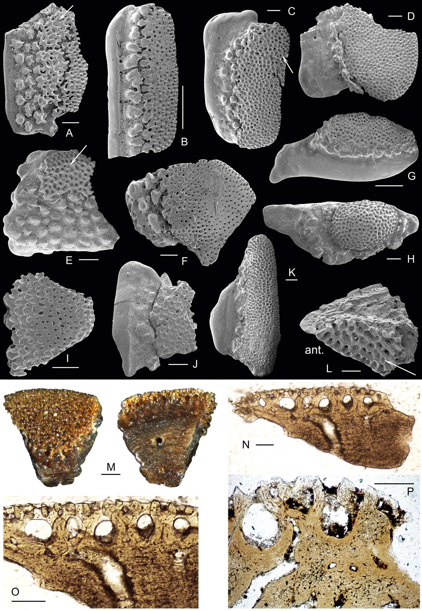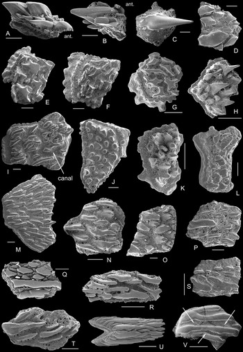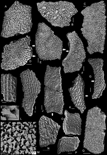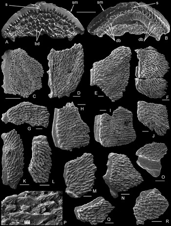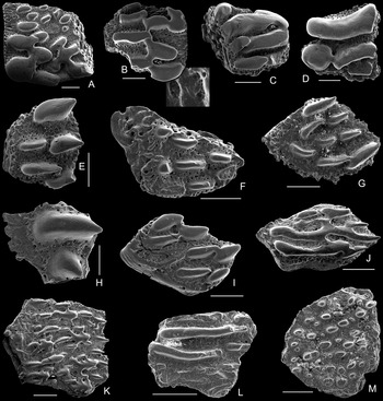


Since the first description of Thyestes verrucosus Eichwald in Reference Eichwald1854, the rocks of Saaremaa Island, Estonia have revealed some of the most diverse and well-preserved osteostracan fossils in the world, permitting study of the articulated head shields and occasional tail squamations. Osteostracan taxonomy has been based mainly on the shape, size and features of the head shield, while isolated elements, such as scales, platelets and fragments of head shields, have generally been described in open nomenclature (e.g. Gross Reference Gross1968a), if mentioned at all. The recovery of early vertebrates by acetic acid preparation has further promoted studies of microscopic remains of osteostracans (Gross Reference Gross1961, Reference Gross1968a). Lack of detailed taxonomic work on Estonian osteostracan material has resulted in poor documentation of their biostratigraphic and geographic distribution. This has, in turn, hindered their utility in palaeogeographic reconstructions and phylogenetic studies.
Disarticulated osteostracan scales and plate fragments are fairly abundant in the Silurian marine sediments of the Baltic Sea region, including outcrops of Saaremaa, Estonia, and the drill cores of Estonia and Latvia, suggesting their potential to improve the resolution of osteostracan biostratigraphy. Identification of the disarticulated material has proven difficult. Scales and fragments can only be distinguished based upon a limited number of preserved characters, which relate to overall shape, sculpture and microstructure. These characters have been insufficiently studied in taxa previously established from articulated specimens.
The present study builds on established knowledge on the morphology of articulated head shields of osteostracans from Saaremaa and maps the previously poorly known variations in the sculpture of their dermal skeleton. Additional material has been obtained from many Silurian outcrops and continuous drill core sections of Estonia and western Latvia (Fig. 1; Appendix 1). This material contributes significantly to the resolution of the stratigraphical and geographical distribution, as well as to interregional correlation in the Baltica (southern Sweden and Gotland Island; Ringerike District; the Central Urals, Timan-Pechora Region) and Kara continents (Severnaya Zemlya Archipelago). With this monographic treatment, we seek to identify taxonomic criteria for the scales and fragments, to evaluate microstructural changes during ontogeny and to contribute to early vertebrate diversity and evolution studies.
Geographical distribution of East Baltic (Estonia and Latvia) osteostracans: (1) Viita; (2) Vesiku; (3) Elda; (4) Karala; (5) Silma; (6) Himmiste; (7) Riksu; (8) Lahetaguse; (9) Sõmera; (10) Paadla; (11) Suurlahe; (12) Kaarmise; (13) Irase; (14) Pähkla; (15) Kuressaare; (16) Uduvere; (17) Laadjala; (18) Tahula; (19) Reo; (20) Pihtla; (21) Kuusiku; (22) Sutu; (23) Nässumaa; (24) Sakla; (25) Kõiguste; (26) Laimjala; (27) Kailuka; (28) Tehumardi; (29) Kaavi; (30) Ohesaare; (31) Loode; (32) Varbla; (33) Seliste; (34) Kihnu; (35) Ruhnu; (36) Kolka; (37) Ventspils; (38) Pavilosta. Locations: circles=drill cores; triangles=outcrops.

1. Historical review
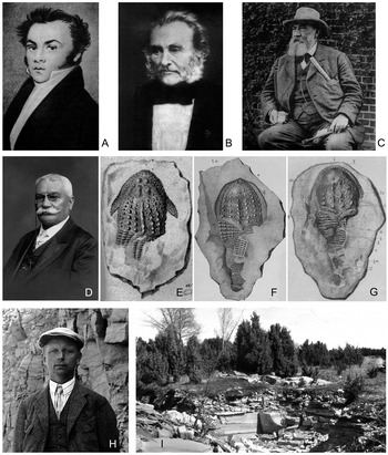
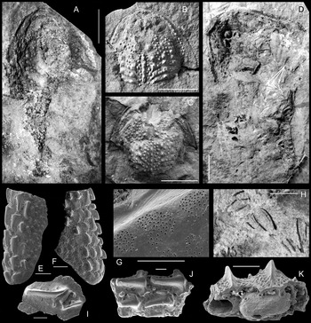
The first record of Silurian osteostracans from Saaremaa was published more than 150 years ago by K. E. Eichwald (Reference Eichwald1854), (Fig. 2A) who described and illustrated a specimen of Thyestes verrucosus with the head and anterior portion of the trunk preserved (Eichwald Reference Eichwald1854, pl. 2, fig. 1) (Fig. 2E). Eichwald obtained some of his research material from A. G. von Schrenk, the lecturer on mineralogy at Dorpat (Tartu) University, who discovered well preserved osteostracan shields and eurypterids in the Viita Quarry when investigating Silurian bedrocks on Saaremaa (Rõõmusoks Reference Rõõmusoks1983, p. 152). An examination of the same specimen of T. verrucosus was published a few years later by C. H. Pander (Fig. 2B) in his study (Pander Reference Pander1856, pl. 4, fig. 1) (Fig. 2F) devoted to the Silurian fossil fish of Saaremaa. Pander (Reference Pander1856) also established several new taxa in his monograph, which were later recognised as Osteostraci (Lankester 1868; Rohon Reference Rohon1892 for the Baltic material). Pander's taxa were, however, ignored by succeeding researchers, who believed his microscopic material was insufficient for comparison with more complete specimens. Researchers have used replacement names for over 50 years, so all of Pander's names (except for W. schrenkii (Pander)) must now be discarded in accordance with Article 23.9.1 of the International Code of Zoological Nomenclature (see remarks under ‘Systematic Palaeontology’ for the individual taxa).
(A–D) Early geologists who have contributed to Estonian osteostracan study: (A) Karl Eduard Eichwald (1795–1876) was one of the very first lecturers of palaeontology in Russia, who gave the first description of Thyestes verrucosus from Estonia; (B) Christian Heinrich Pander (1794–1865) was the founder of micropalaeontology, and the first to describe conodonts, but also the Silurian and Devonian fishes from East Baltic; (C) Carl Friedrich Schmidt (1832–1908) subdivided the Palaeozoic (Cambrian to Silurian) strata of Estonia and adjacent regions, created the stratigraphic scheme, which basically is also exploited today, and studied some Silurian fish material; (D) Josef Victor Rohon (1845–1923) was a neuroanatomist and a palaeontologist, who published monographic description of Silurian fishes of Estonia. (E–G) Drawings of holotype PSM SPU 145/1 Thyestes verrucosus as given by three different scientists: (E) Eichwald (Reference Eichwald1854, pl. 2, fig. 1; (F) Pander (Reference Pander1856, pl. 4, fig. 1a); (G) Rohon (Reference Rohon1892, pl. 2, fig. 1). (H) Artur Luha (1892–1953). He was the discoverer of the world famous agnathan containing layer in Himmiste Quarry (I).

C. F. Schmidt (Reference Schmidt1866) (Fig. 2C) collected and described many additional specimens of Thyestes verrucosus and established a new genus, Tremataspis Schmidt. J. V. Rohon (Fig. 2D) continued to study the Schmidt collection, which is now housed in the Borissiak Paleontological Institute of the Russian Academy of Sciences, and also the Eichwald (Reference Eichwald1854) collection. Together with the re-description of species of Thyestes Eichwald and Tremataspis Schmidt, Rohon (Reference Rohon1892) established several new taxa, some of which are valid today (Tremataspis schmidti and Saaremaaspis mickwitzi). He also figured the same specimen of Thyestes verrucosus (Rohon Reference Rohon1892, pl. 2, fig. 1) (Fig. 2G) as did Eichwald and Pander. Rohon devoted several subsequent publications to early fishes of Saaremaa (Rohon Reference Rohon1893, Reference Rohon1894, Reference Rohon1895, Reference Rohon1896).
During our study, with the help of museum workers in St. Petersburg and Tartu, we located both the Eichwald and part of the Rohon (Reference Rohon1892) collections in the Palaeontological-Stratigraphical Museum (PSM SP) at St. Petersburg State University. Thanks to the special care of numerous people, the collections have surprisingly survived revolutions, wars, strife and even the Leningrad blockade. Now with the type specimen in hand, it is interesting to discover how much the drawings of T. verrucosus in early publications differ from each other. The main differences occur in the shield margin features, a number of exposed scale rows and configuration of underlying rock sample, mostly following later preparation (compare Eichwald Reference Eichwald1854, pl. 2, fig. 1; Pander Reference Pander1856, pl. 4, fig. 1a; and Rohon Reference Rohon1892, pl. 2, fig.1; and Fig. 2E, F, G).
The main achievement of W. Patten was to assemble a good collection of agnathan specimens, building on the first specimens that he had obtained from A. Simonson, the son of the head of the Oesel Museum (from local newsletter ‘Meie Maa’, 1928, No. 101), during a visit to the island (Patten Reference Patten1903a, p. 226), and published a paper on the material of that trip (Patten Reference Patten1903b). Later, between 1928 and 1932, Patten made four expeditions to Saaremaa; the first two times he excavated in Viita (Rootsiküll) Quarry, and during the two last expeditions he spent the majority of his time in the Himmiste (Himmiste–Kuigu) Quarry, in which a rich layer containing agnathans was discovered by A. Luha (Fig. 2H) in 1929. Although many geologists and fossil collectors from different countries have sampled at Himmiste (Fig. 2I), the quarry became particularly famous after Patten's last two visits in 1930 and 1932 when he, with the help of up to 27 local workers, collected over 3500 agnathan specimens (Patten Reference Patten1931a, Reference Pattenb). The agnathan exoskeletons were generally observed on slabs dominated either by the thelodont Phlebolepis elegans Pander, Reference Pander1856, or by various osteostracans; however, 2–3 larger specimens of anaspids were also found and later described (Robertson Reference Robertson1937, Reference Robertson1941; Ritchie Reference Ritchie1980). After being shipped to the US, the material collected by Patten was first deposited in the Dartmouth College Museum and later transferred to the American Museum of Natural History. Patten's expeditions exhausted the main argillaceous dolomite lens with early vertebrates in Himmiste. Shortly after his expedition in 1932 to Saaremaa, W. Patten died. He established the species Tremataspis milleri Patten, Reference Patten1931a, Tr. mammillata Patten, Reference Patten1931a, Oeselaspis pustulata (Patten, Reference Patten1931a) and Dartmuthia gemmifera Patten, Reference Patten1931a, all of which are valid today.
Patten's osteostracan collection at Dartmouth College was repeatedly investigated by G. M. Robertson, who published 17 papers on that group from Saaremaa (Robertson Reference Robertson1935a, Reference Robertsonb, Reference Robertsonc, Reference Robertson1938a, Reference Robertsonb, Reference Robertson1939a, Reference Robertsonb, Reference Robertson1940a, Reference Robertsonb, Reference Robertson1945, Reference Robertson1947, Reference Robertson1949, Reference Robertson1950, Reference Robertson1954, Reference Robertson1955, Reference Robertson1957, Reference Robertson1970) and R. Denison (Reference Denison1947, Reference Denison1951a, Reference Denisonb, Reference Denison1952, Reference Denison1956, Reference Denison1963). Robertson re-described the species previously addressed by Patten and Rohon, among others, and established several new taxa, including the genera Oeselaspis Robertson, Reference Robertson1935a; Saaremaaspis Robertson, Reference Robertson1938b; Witaaspis Robertson, Reference Robertson1939b; and a species later called Procephalaspis oeselensis (Robertson, Reference Robertson1939a). Denison (Reference Denison1947, Reference Denison1951a, Reference Denisonb) described in detail the histology of the exoskeletons of osteostracans and classified and discussed the evolution of the group.
As mentioned above, the famous Himmiste Quarry was discovered by A. Luha (Fig. 2H), Tartu University, during geological mapping of Saaremaa in 1929, when he found there a rich bed with upper Silurian articulated agnathans. Luha's collection is now housed at Tartu University and includes the world's largest number of articulated thelodont specimens (Phlebolepis elegans Pander), but not as many osteostracans. Luha (Reference Luha1940) argued that this collection represented the best collection of Silurian osteostracans in the world.
In Estonia, osteostracan head shields, internal structure, taxonomy and systematics have been studied more thoroughly by E. Bölau (Reference Bölau1949, Reference Bölau1951, Reference Bölau1957), who discussed the distribution of osteostracans in the Kaarma Stage and studied the sensory canal system in Tremataspis. Bölau collected over 1000 specimens of agnathans, mainly the osteostracan Tremataspis mammillata, from the Himmiste Quarry, Saaremaa; however, his material is not as well preserved as earlier material. Little is known about E. Bölau. He graduated cum laude from Tartu University in 1940 and later, in 1942, was at the Geologisch-Paläontologisches Institut und Museum der Universität Berlin as a scholar, working with H. Stille, W. Quenstedt and W. Gross. From 1942 to 1944, he spent every summer on Saaremaa collecting osteostracans for his doctoral studies, and in the spring of 1944 he defended his dissertation entitled “Über Ostrakodermen aus der Karmel-Stufe Oesels (Obersilur, Estland) und das Kosmin-Problem”. In 1944, E. Bölau left Estonia for Stockholm to continue his studies on the tremataspid sensory system. Schultze (Reference Schultze2009, p. 32) wrote about E. Bölau, “He had reached the same conclusion as Denison (Reference Denison1947), that the tremataspids form an intermediate stage between an early diffuse sensory system and a defined linear sensory line (Bölau Reference Bölau1951) while Stensiö argued that the defined linear sensory system was primary.” Because of his scientific disagreements with E. Stensiö, he had to leave research and continue as a geologist at a coal and ceramic clay company in southern Sweden (Schultze Reference Schultze2009).
The osteostracans from Saaremaa Island have been an important source for many researchers around the world, and these collections have been distributed to many research institutes and museums, promoting work by Wängsjö (Reference Wängsjö1944, study based on his own collection in 1938), Obruchev (Reference Obruchev and Orlov1964), Janvier (Reference Janvier1974, Reference Janvier1975, Reference Janvier1978, Reference Janvier1985a, Reference Janvierb), Janvier & Lelièvre (Reference Janvier and Lelièvre1994), Afanassieva (Reference Afanassieva1985, Reference Afanassieva1986, Reference Afanassieva1991, Reference Afanassieva1996, Reference Afanassieva, Novitskaya and Afanassieva2004) and many others. These papers mainly address the shields, whereas the microstructure of the exoskeletons has been more thoroughly studied by Denison (Reference Denison1947, Reference Denison1951a, Reference Denisonb), Gross (Reference Gross1956, Reference Gross1961, Reference Gross1968b), Afanassieva (Reference Afanassieva1985, Reference Afanassieva1986, Reference Afanassieva1991, Reference Afanassieva1995, Reference Afanassieva, Novitskaya and Afanassieva2004, Reference Afanassieva2014), and Afanassieva & Märss (Reference Afanassieva and Märss1997, Reference Afanassieva and Märss2014). In two monographs on the osteostracans of Russia and adjacent countries, Afanassieva (Reference Afanassieva1991, Reference Afanassieva, Novitskaya and Afanassieva2004) devoted considerable attention to the exoskeleton of Estonian forms, and used features of the external morphology and internal structure at various levels of generalisation for diagnoses of the various rank taxa. The comparative study of the microstructure of the exoskeleton of osteostracans from Saaremaa allowed Afanassieva (Reference Afanassieva2000), for the first time within this group, to establish a new taxon based on a small, isolated fragment when describing the new genus Septaspis, with the type species S. pectinata, from the upper Silurian deposits of the Severnaya Zemlya Archipelago. In this work, Afanassieva also suggested a scheme for description of the microremains of osteostracans (including histocharacteristics).
The scale-covered trunk and tail behind the solid head shield of Estonian osteostracans have been less investigated because they are only known from a few genera: Thyestes (Eichwald Reference Eichwald1854; Pander Reference Pander1856; Rohon Reference Rohon1892); Tremataspis (Patten Reference Patten1903a, Reference Patten1931b; Rohon Reference Rohon1894; Robertson Reference Robertson1938a; Märss Reference Märss1982, Reference Märss1986 and herein; Janvier Reference Janvier1985a) and Dartmuthia (Janvier Reference Janvier1985a). The tail of a taxon identified as Saaremaaspis by Robertson (Reference Robertson1938a, p. 288; Reference Robertson1938b, pl. 60, fig. 3) and Janvier (Reference Janvier1985a) is here re-identified as Thyestes (see the description of the tail below). Despite these reconstructions of the entire body of Silurian osteostracans, no complete specimen with head, trunk and tail has been found in Estonia.
The osteostracan material at Pander's disposal came from just one locality, the Rootsiküla (Rootsikülle), which is the name of the village in which the farm and Viita Quarry were situated. The Viita Quarry, levelled during redevelopment in the 1970s, is the type locality for Thyestes verrucosus Eichwald, Witaaspis schrenkii (Pander), Tremataspis schmidti Rohon and Saaremaaspis mickwitzi (Rohon). The adjacent Viita trench has provided the only head shield of Aestiaspis viitaensis (In Janvier & Lelièvre Reference Janvier and Lelièvre1994) and tail of Tremataspis schmidti (In Märss Reference Märss1986). Rohon (Reference Rohon1892) obtained his research material from Viita (Wita-Steinbruch bei Rotziküll), Vesiku (Wesiko) and Hoheneichen. The name Hoheneichen comes from the manor, which until 1778 belonged to the Antarctic explorer Bellingshausen's family and is well known today. There are two quarries close to one another, Pilguse and Himmiste; however, Pilguse Quarry, which is closer to the manor, has not produced fish fossils. The fish locality name was tied to the better-known manor, even though it was situated 1.7 km away, rather than to a farmstead in the vicinity. The Himmiste Quarry is the source of the richest and most diverse osteostracan assemblage including Tremataspis mammillata Patten, T. milleri Patten, Tremataspis rohoni Robertson, Dartmuthia gemmifera Patten, Oeselaspis pustulata (Patten) and Procephalaspis oeselensis (Robertson). Historically there has been confusion regarding the origin of the type material, especially that of Tr. milleri and O. pustulata. Patten (Reference Patten1931a, pp 2–3) wrote, “After exhausting the old site near Kiehelkond, we moved to Atla, in which there is a peasant quarry recently explored by Professor A. Luha, an Esthonian geologist from the University of Dorpat. Here we found six new species of ostracoderms”, whereby he described Tr. milleri, Tr. mammillata, Didymaspis (now Oeselaspis) pustulata and Dartmuthia gemmifera in that work; two ‘ostracoderms’, Phlebolepis elegans and an anaspid, remained undescribed. The site mentioned as Atla is actually the same Himmiste Quarry fish locality; in Atla Quarry, which produced biohermal limestones for lime burning, no fish have ever been found. So, these six taxa were first described from the Himmiste Quarry and not from Atla, and therefore the former must be considered as the type locality for all these taxa.
In 1974 a new fossil fish locality, Elda Cliff, was discovered by T. Märss and R. Einasto, Tallinn, on the western coast of Saaremaa in the Kuusnõmme Beds of the Rootsiküla Stage. Here the preservation of the material is rather poor, and the head shields of Tremataspis (Tr. schmidti, Tr. milleri and Tr. rohoni), Saaremaaspis mickwitzi, Oeselaspis pustulata and Thyestes verrucosus are compressed and full of cracks. This material is still suitable for studying the surface morphology and some new taxa are described here.
Since the late 1960s, the collection of osteostracans from the western islands of Estonia, and later from Latvia, expanded substantially to also include thousands of osteostracan scales and fragments. This results from the research by Märss, which began with a ‘term paper’ on Silurian osteostracan bibliography, continued as an undergraduate project examining the distribution of early vertebrate microremains in the Kingissepa–GI drill core and transferred to a diploma thesis including samples of three more drill cores (Kaugatuma-GI, Ohesaare-GI and Sakla-GI). Subsequent work resulted in a PhD Degree on samples with different fish remains from 26 drill core sections and 57 outcrops (Märss Reference Märss1983), most of which was published (Märss Reference Märss1986). The samples from drill cores were mainly taken for biostratigraphical purposes from the whole Silurian sequence. The availability of drill core data goes back to the 1960s, when the geological mapping of the East Baltic area was initiated and continued until the 1990s, when projects stopped because of lack of funding for drilling. Sadly, many drill cores were lost following a fire at a storehouse. Consequently, the vertebrate micromaterial previously obtained from these cores is unique and extremely important. The scales and shield fragments of osteostracans found in these samples form the basis of this study.
2. Material and methods
2.1. Location and age of material
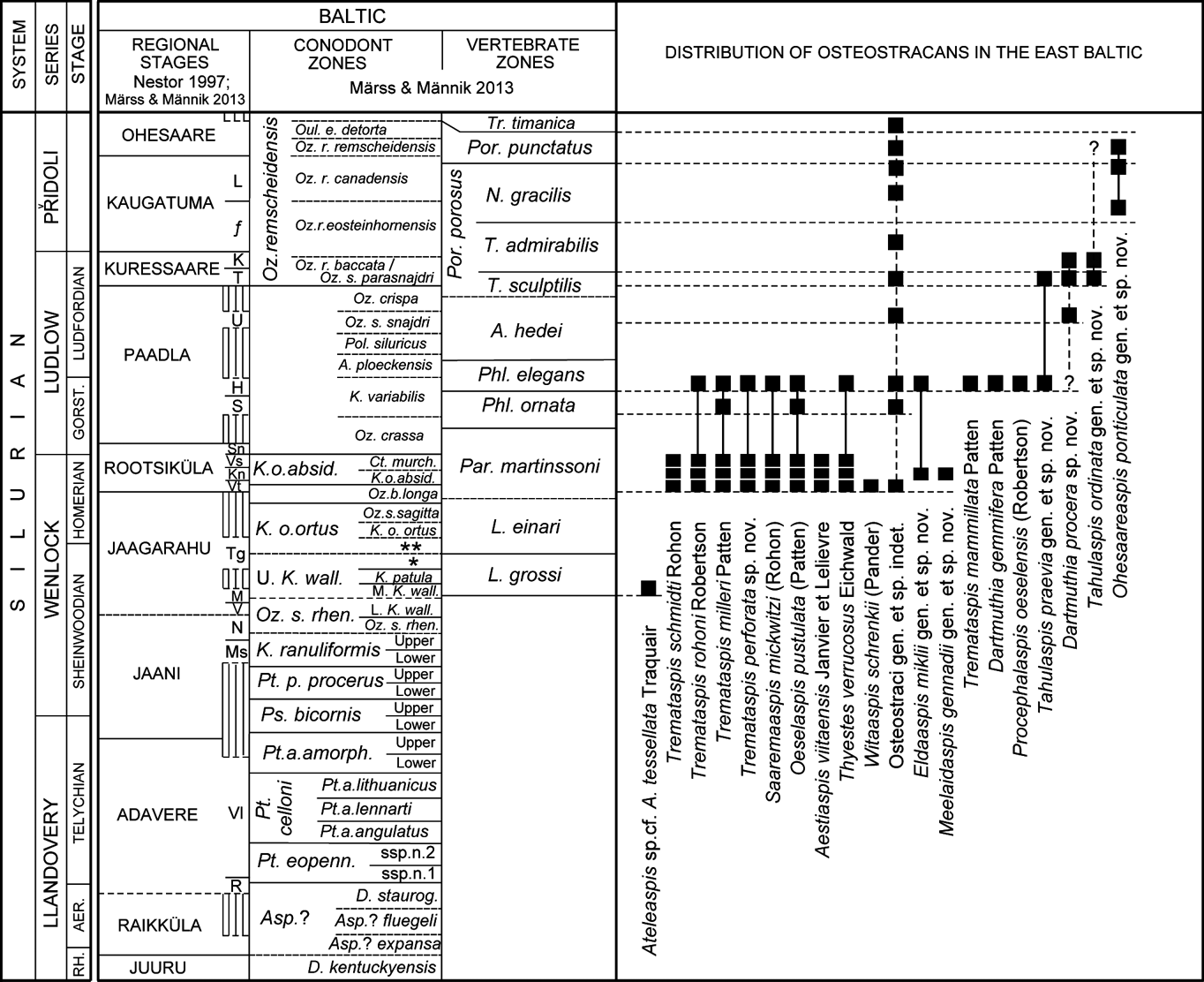
The material studied herein comes from 12 outcrops and 32 drill cores of a lower Silurian (Wenlock) to upper Silurian (upper Přidoli) interval of Estonia and Latvia, East Baltic (Fig. 1; see Appendix 1 for the distribution of osteostracans in drill cores and outcrops). Osteostracan microremains have been found from the interval starting with the Maasi Beds of the Jaagarahu Stage, lower Wenlock, the Loganellia grossi Vertebrate Zone (VZ) running up to Trimerolepis timanica VZ, uppermost Přidoli (Fig. 3; see also Märss Reference Märss1986, table on pp 79–80). The articulated head shields of osteostracans come from five localities: Viita Quarry and Viita trench, Elda Cliff; Rootsiküla Stage; Wenlock; and Himmiste Quarry and Silma Cliff, Saaremaa, Paadla Stage, Ludlow.
Distribution of osteostracans in the East Baltic (Estonia and Latvia) Silurian sequence. Abbreviations: Aer.=Aeronian; Gorst.=Gorstian; H=Himmiste Beds; K=Kudjape Beds; Kn=Kuusnõmme Beds; L=Lõo Beds; M=Maasi Beds; Ms=Mustjala Beds; N=Ninase Beds; R=Rumba Formation; Rh.=Rhuddanian; S=Sauvere Beds; Sn=Soeginina Beds; T=Tahula Beds; Tg=Tagavere Beds; U=Uduvere Beds; V=Vilsandi Beds; Vl=Velise Formation; Vs=Vesiku Beds; Vt=Viita Beds; Ä=Äigu Beds. Dashed line marks the lower boundaries of zones in Estonia; a star (*) means the uppermost walliseri range, and two stars (**) the post-K. walliseri interregnum in conodont zonal scheme. Biostratigraphy is taken from Märss & Männik (Reference Märss and Männik2013).

2.2. Head shield studies
In order to explore the full taxonomic diversity of osteostracans in the East Baltic by also including disarticulated material, it was necessary to understand the sculpture varieties of articulated head shields and the trunk squamation, as well as the histology of exoskeletons. It was thus necessary to study the head shields of the type collection of Estonian osteostracans housed in different institutions (see section 2.6, “Repositories”) and identify diagnostic characters of the sculpture of the external surface. Unfortunately, this study was hindered by missing type specimens, the lack of SEM facilities needed for detailed studies of exoskeletons, and because, understandably, the AMNH does not lend type material. More importantly, not all types (holo-, lecto- or neotypes) have the external sculpture preserved, and some show the visceral surface of the head shields only. These limitations were remedied by detailed studies of other type material from type localities and the selection of specimens that could unambiguously be assigned to the species of interest and preserved even a little external sculpture.
Head shields of most taxa were studied at Uppsala University using a Zeiss Supra 35-VP field emission SEM, which is ideal to avoid the non-acceptable coating of the material. SEM pictures were taken at both low and high vacuum in order to obtain the best results, and avoid charging and other problems related to SEM work. The holotype of Aestiaspis was too fragile for the SEM, so its sculpture was illustrated using a standard digital imaging system attached to a stereomicroscope, whereby the specimen was coated with ammonium chloride. Photographs of macroscopic specimens, both head shields and tail squamations, were taken in reflective light.
2.3. Micropalaeontological analysis and SEM studies
The disarticulated scales, platelets and head shield fragments have been extracted from different limestones, dolostones or terrigenous rocks with carbonate cement by dissolving the samples with 10–15% acetic acid, the residue washed with water and dried. To facilitate the work, the residue was strained through sieves with apertures of 0.1 mm, 1.0 mm and 2.0 mm, before the vertebrate remains were extracted. Bromoform was used to separate fish bones from the residue; however, this was discontinued because the holes and cavities holding air kept the bits and pieces in the light fraction, which at the first stage remained unstudied. A few specimens were treated in buffered 10% formic acid to remove some matrix that was still between the sculpture. Many of these samples produced a large number of scales, shield fragments, spines and teeth of different early vertebrates, including thousands of osteostracan scales and fragments. The number of vertebrate remains per sample, which differ in weight (about 20 g–1 kg from drill cores and up to 3 kg from outcrops), varies from one to thousands and many samples are barren. The total number of samples with agnathan and fish microremains in the collection of the Institute of Geology at Tallinn University of Technology is over 2000, but only about 100 samples contain osteostracan fragments.
The best microscopic specimens were chosen for study and illustration with the scanning electron microscopes JEOL JSM-840A and Zeiss EVO MA15 at 10 kV. Some of the specimens, especially those with highly porous surfaces, were imaged using back-scattered electrons, which generated fine, high-contrast pictures. This laboratory work was conducted at the Institute of Geology and Laboratory of Material Research at TUT, Tallinn.
2.4. Histological study
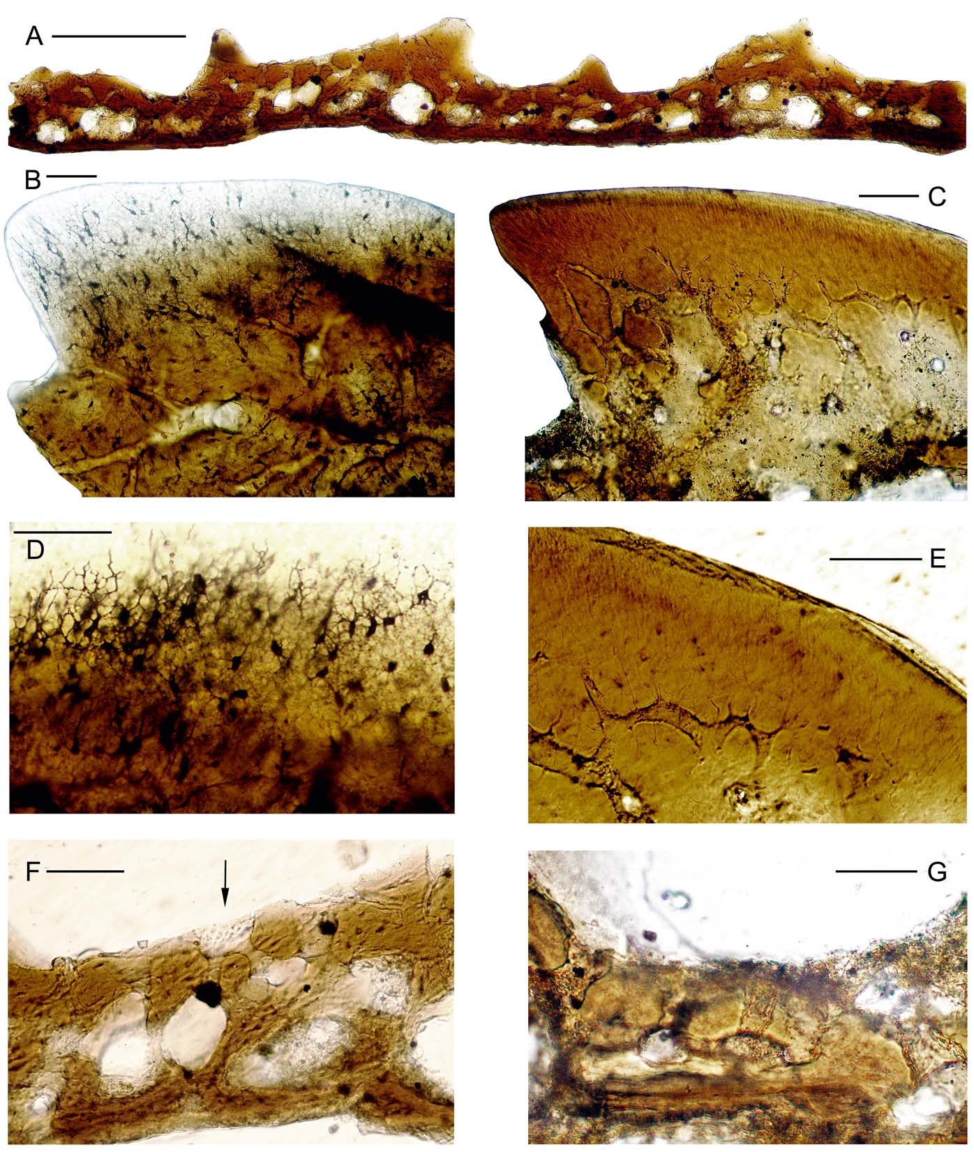
The histology of Saaremaa osteostracans has been described and illustrated by several authors (Gross Reference Gross1935; Denison Reference Denison1947, Reference Denison1951b; Afanassieva Reference Afanassieva1991, Reference Afanassieva, Novitskaya and Afanassieva2004). Histological descriptions of the rich new material complement and complete the monographic descriptions presented here. New taxa received the most attention. The East Baltic Silurian fossil vertebrate material is generally suitable for histological studies, as it is typically well preserved. Thin sections were prepared following the procedure used in fossil fish studies (e.g., Gross Reference Gross1947). A specimen was glued to a glass slide in a desired orientation with Canada balsam, polished, then heated until the balsam liquefied, allowing the scale to be turned upside down and oriented, and then the other side was polished until the element became transparent but preserved fine canaliculi. At the beginning of polishing we used 7 μm, then 3 μm aluminium oxide powder, and at the end just water, with no powder, to achieve a clean surface. Finally, the thin section was covered with warm liquid balsam and glass. This laboratory work was mostly done at the Institute of Geology at TUT, Tallinn, and partly at the Borissiak Paleontological Institute of the Russian Academy of Sciences, Moscow. The pictures of the best specimens were taken with a Nikon ECLIPSE 50i microscope with a Nikon DS-Fi1 camera, in Tallinn.
2.5. Taxonomic procedures
Synonymy lists of most taxa are presented by Afanassieva (Reference Afanassieva1991, Reference Afanassieva, Novitskaya and Afanassieva2004) and are not repeated here, unless additions or changes were necessary. Synonyms for new taxa are included.
As one of the main objectives of this study was to establish diagnostic characteristics for the sculpture and microstructure of the dermal skeleton of East Baltic osteostracans, the articulated shields were not measured (except length and width), nor were ratios calculated by us. For such data we refer to previous publications (Denison Reference Denison1951a; Afanassieva Reference Afanassieva1991, Reference Afanassieva, Novitskaya and Afanassieva2004) where they have been given for each species having a complete shield. Here we mainly stress the most characteristic features of sculpture and histology in diagnoses.
2.6. Repositories
The osteostracans described here are housed in the Institute of Geology at Tallinn University of Technology (collection GIT 502; both micro- and macroremains); in the Museum of Geology, University of Tartu (TUG, macroremains); in the Borissiak Paleontological Institute of the Russian Academy of Sciences, Moscow (PIN, macroremains); the Palaeontological-Stratigraphical Museum at St. Petersburg State University (PSM SPU, some type material) and in the American Museum of Natural History, New York, USA (AMNH, macroremains including some type material). Some type specimens and important reference material in the AMNH have not been available for study, since part of the Patten collection was borrowed for PhD work in the UK and never returned, and at present cannot be located (see below at species treatments).
2.7. Authors' responsibilities
The text of this monograph was prepared as follows: the introductory chapters by T. Märss and H. Blom; the descriptions of sculpture on scattered scales and shield fragments by T. Märss; descriptions of the sculpture on articulated shields and trunk squamations by H. Blom; and the histology by O. Afanassieva. The final text was read and accepted by all of us.
3. Distribution
The Silurian sequence of the northern East Baltic has been studied in discontinuous outcrop sections on Saaremaa Island, Estonia, and relatively continuous drill core sections of western Estonia and Latvia (see list in Appendix 1). The stratigraphy of tightly drilled core sections (see locations in Fig. 1) partly overlaps, giving us a good stratigraphical framework for the Silurian in the region.
In the East Baltic, the earliest record of osteostracans is in the Maasi Beds of the Jaagarahu Stage, upper Sheinwoodian, lower Wenlock (Fig. 3). Ateleaspis cf. A. tessellata Traquair was found in the Ohesaare-GI drill core, depth 174.10–174.50 m, at the same level as the first findings of zonal thelodont Loganellia grossi Fredholm. Ateleaspis cf. A. tessellata is also present at higher depths, 173.50–173.81 m, found at one-metre intervals.
Osteostracans in the Viita Beds of the Rootsiküla Stage, upper Homerian, upper Wenlock, the Paralogania martinssoni Vertebrate Zone (VZ) (Fig. 3), are best known from the Viita Quarry, which is also the type locality of Tremataspis schmidti, Saaremaaspis mickwitzi, Thyestes verrucosus and Witaaspis schrenkii, all known by their well preserved head shields. The quarry was levelled and the fish beds buried in the 1970s. Another locality, the Viita trench, lies 250 m to the west of the quarry location. A well-preserved trunk and tail of Tremataspis schmidti and head shield of Aestiaspis viitaensis originate from this trench. In addition to the taxa listed above, T. milleri, T. rohoni, T. perforata sp. nov. and Oeselaspis pustulata occur in the Viita Beds. The same taxa are also known in the Vesiku-507 drill core, depth 8.65–8.85 m; the core mouth is approximately 2.5 km to the southwest of the Viita trench. In the Ohesaare-GI drill core at a depth of 150.40–150.50 m, only Tremataspis sp. indet. has been found, and in the Kihnu-526 drill core, at a depth of 101.70–101.85 m, Osteostraci gen. et sp. indet. has been found in Viita Beds.
The osteostracan species higher up in the sequence, in the Kuusnõmme and Vesiku beds of the Rootsiküla Stage, upper Homerian, upper Wenlock, are similar to those in the Viita Beds, with two new taxa, Eldaaspis miklii gen. et sp. nov. and Meelaidaspis gennadii gen. et sp. nov., found in the Elda Cliff section (Kuusnõmme Beds) in addition to Tremataspis schmidti, T. milleri, T. rohoni, T. perforata sp. nov., Saaremaaspis mickwitzi, Oeselaspis pustulata, Aestiaspis viitaensis and Thyestes verrucosus. In addition to small fragmented material, pressed and cracked head shields of Tremataspis schmidti, T. milleri and T. rohoni, a few Saaremaaspis mickwitzi and Oeselaspis pustulata come from this locality.
The bonebed at Vesiku Brook (Vesiku Beds) has similar osteostracan fossils as the Kuusnõmme Beds, with the exceptions that Eldaaspis miklii gen. et sp. nov. and Meelaidaspis gennadii gen. et sp. nov. are absent in the Vesiku Beds.
As a whole, the Sauvere Beds of the Paadla Stage, Gorstian, lower Ludlow, the Phlebolepis ornata VZ (Fig. 3), are poor in vertebrate remains, including osteostracans. Only rare fragments of Tremataspis milleri, Oeselaspis pustulata and indefinite Osteostraci gen. et sp. indet. can be listed.
The Himmiste Beds (Paadla Stage, upper Gorstian, lower Ludlow, Phlebolepis elegans VZ (Fig. 3)) contain characteristic osteostracan taxa, which occur in the lower parts of the following localities: Himmiste Quarry (the stratotype of the beds, the section stratigraphically situated in the uppermost part of the Himmiste Beds, and type locality of a number of osteostracans); Silma Cliff; Silma Brook; Paadla Quarry; Pähkla Quarry; as well in several drill core sections (Kaarmise-GI, Paadla-GI, Himmiste-982, Uduvere-968, Irase-680, Kailuka-817, Kingissepa-GI, Kuressaare-804, Suurlahe-738, Varbla-502, Varbla-522, Nässumaa-825; for the depths see Appendix 1). The beds typically contain Tremataspis milleri, T. rohoni, Tremataspis perforata sp. nov., Saaremaaspis mickwitzi, Oeselaspis pustulata, Thyestes verrucosus and Eldaaspis miklii gen. et sp. nov. In addition, the Himmiste Beds mark the first appearance of taxa such as Tremataspis mammillata, Dartmuthia gemmifera, Procephalaspis oeselensis and Tahulaspis praevia gen. et sp. nov. The presence of Dartmuthia procera sp. nov. in these beds is questionable at present because so little material is available.
In the lower part of Pähkla Quarry, the Uduvere-968 drill core, depth 4.5 m, and the Varbla-502 drill core, depth 31.80 m, some fragments of Tremataspis have small pores and are similar to Tremataspis rohoni in the distance between the pores (as described by Robertson Reference Robertson1938a). Witaaspis tesserae-like elements in the Vesiku and Himmiste beds (Mark-Kurik & Noppel Reference Mark-Kurik and Noppel1970; Märss Reference Märss1986, table on p. 79–81) should be re-identified as belonging to Saaremaaspis mickwitzi.
The Uduvere Beds of the Paadla Stage, mid Ludfordian, upper Ludlow, Andreolepis hedei VZ, contain Dartmuthia procera sp. n. in the Tahula-709 drill core, depth 11.6 m. This sample occurs with A. hedei itself.
The basal layer of the Tahula Beds of the Kuressaare Stage, upper Ludfordian, upper Ludlow, Thelodus sculptilis VZ (Fig. 3), is characterised by accumulations of vertebrate remains in many drill cores. Osteostracan shield and scale fragments occur together with thelodonts, heterostracans, anaspids and acanthodians. The most common osteostracan taxa are Tahulaspis ordinata gen. et sp. nov. and Dartmuthia procera sp. nov., although Tahulaspis praevia gen. et sp. nov. is rare. In many cases, Tahulaspis ordinata gen. et sp. nov. and Dartmuthia procera sp. nov. occur together. Such is the case in the following drill cores: Kõiguste-833, depth 4.4 m; Kingissepa-GI, depth 10.40–18.25 m; Reo-927, depth 8.8–10.6 m; Kuressaare-804, depth 8.0–8.5 m; Varbla-502, depth 19.10–22.0 m; and Nässumaa-825, depth 41.3–41.9 m. In addition to listed occurrences, in many localities, either one or the other taxon is present alone: Tahulaspis ordinata gen. et sp. nov. (Laadjala Bridge outcrop, Ohesaare-GI, depth 94.45–94.48 m) or Dartmuthia procera sp. nov. (Sakla-GI, depth 10.52–10.58 m; Kuusiku-605, depth 23.0–23.7 m (before digitisation of data, the drill core was named as Sutu-605, see Märss Reference Märss1986, fig. 34); Sutu-606, depth 9.6 m; Tahula-709, depth 7.6–8.4 m; Kolka-54, depth 282.2–282.5 m). Tahulaspis ordinata gen. et sp. nov. and Tahulaspis praevia gen. et sp. nov. are distributed together in Pihtla-816, depth 15.7 m. Thus, Tahulaspis praevia gen. et sp. nov. has its last occurrence in the Tahula Beds, and Tahulaspis ordinata gen. et sp. nov. starts in these beds. Two taxa, Dartmuthia procera sp. nov. and Tahulaspis ordinata gen. et sp. nov., continue upwards into the Kudjape Beds of the Kuressaare Stage. Higher in the section, the osteostracan fauna is depauperate if compared with the Kuressaare Stage, but especially with the Rootsiküla and Paadla stages.
The Kaugatuma Stage, lower Přidoli, which is rich in gnathostome acanthodian scales, is rather poor in agnathan remains. Osteostracans are represented by Ohesaareaspis ponticulata gen. et sp. nov. identified in the Ruhnu-500 drill core, depth 201.2 m in the upper Äigu Beds, Nostolepis gracilis VZ (Fig. 3), and at a depth 176.4 m in the same core, in the high Lõo Beds [see the stratigraphy of the Ruhnu drill core in Põldvere (Reference Põldvere2003, p. 47–76)].
In the Ohesaare Stage, upper Přidoli, the Poracanthodes punctatus VZ (Fig. 3), Ohesaareaspis ponticulata gen. et sp. nov. occurs in the Ohesaare Cliff section and in the Ruhnu-500 drill core, depth 165.2 m. The Ohesaare Cliff section lies stratigraphically at the base of the Ohesaare Stage. In the Kolka-54 drill core, depth 245.5 m, it occurs together with Paralogania tarranti and Poracanthodes cf. punctatus. In this stage, there are some indications of the presence of Tahulaspis ordinata gen. et sp. nov. fragments.
In addition to those presented above, the Kolka-54 drill core yields scales and shield fragments of Osteostraci gen. et sp. indet. on several other levels. Below is the list of the depths and the zonal taxon found with osteostracan(s) (given in the brackets), which also have been used to ease the stratigraphical placement of findings: Kolka-54 drill core, depth 244.3 m (Th. sculptilis VZ level); 243.4–243.7 m (Th. admirabilis VZ level); 205.6–205.9 m (?Por. punctatus VZ level); 180.2–181.0 m (Por. punctatus VZ level); 161.3–161.6 m and 160.2–160.4 m (Tr. timanica VZ level). Southwards, in the Pavilosta-51 drill core, depth 676.4–676.8 m, Pagegiai Formation, Osteostraci gen. et sp. indet. occurs in the T. sculptilis VZ.
4. Environment
The Silurian sequence of the northern East Baltic is represented lithologically by rather different sedimentary rocks, including limestones, dolostones, marlstones, domerites and siltstones (Nestor Reference Nestor, Kaljo and Nestor1990a, Reference Nestor, Kaljo and Nestorb, Reference Nestor, Raukas and Teedumäe1997), which attest to the complex basin history influenced by several transgressions and regressions (e.g., Nestor & Einasto Reference Nestor, Einasto and Kaljo1977, Reference Nestor, Einasto, Raukas and Teedumäe1997). Vertebrates inhabited all facies belts: the lagoonal, shoal, open shelf, slope and depression (Märss & Einasto Reference Märss and Einasto1978; Märss Reference Märss1991), whereas osteostracans were restricted only to the three shallow water environments (Märss Reference Märss1986, table on pp 79–80).
The poorly sorted argillaceous skeletal nodular limestones intercalate with marls in the Maasi Beds of the Jaagarahu Stage in the Ohesaare-GI drill core. The beds have been interpreted as deposits of the distal portion of shoal or open shelf facies belt (Nestor Reference Nestor, Kaljo and Nestor1990a, Reference Nestor, Kaljo and Nestorb; Nestor & Einasto Reference Nestor, Einasto and Kaljo1977, Reference Nestor, Einasto, Raukas and Teedumäe1997). In that part of the sea dwelt the earliest osteostracan Ateleaspis sp. cf A. tessellata Traquair in Estonia.
In the Rootsiküla Stage, articulated shields and tail squamations of osteostracans mainly derive from three outcrops: Viita Quarry and trench and Elda Cliff. In Viita Quarry (Viita Beds) the one metre-thick section had the argillaceous horizontally micro-laminated dolostone beds in the lower part, wavy micro-laminated Eurypterus-dolostone in the middle part, and horizontal flaggy dolostone with oolites, pebles, vertical and horizontal trace fossils in the upper part; discontinuity surfaces separated these three parts (TM pers. obs. 1969). The lectotype of Tremataspis schmidti (Fig. 7A) and Thyestes verrucosus tail squamations (Fig. 28A, holotype in PSM PSU; 28D; and 29I, the latter identified as Saaremaaspis mickwitzi from the Wenlock of Saaremaa by Janvier Reference Janvier1985a, fig. 17) are lying on the argillaceous, horizontally micro-laminated dolostone, all most likely collected from the Viita Quarry. A tail specimen of Tremataspis sp., which originally was referred to the Wenlock of Saaremaa (Janvier Reference Janvier1985a, fig. 29A, B), we identified as Tremataspis rohoni; this specimen may also originate from the Viita Quarry. Two more tail specimens of Tremataspis were available from the Viita locality for the reconstruction (Rohon Reference Rohon1894, pl.1, figs 14, 15, 16). The middle portion of the Viita trench section (Viita Beds), which lies stratigraphically a little lower than the quarry section, has yielded a Tremataspis schmidti tail squamation (Fig. 7B, C) and a head shield of Aestiaspis viitaensis (Janvier & Lelièvre Reference Janvier and Lelièvre1994, fig. 1A, B) (Fig. 20D). In both localities (in the Viita trench and Quarry), the sedimentary rock around the articulated shield and tail specimens is the argillaceous, horizontally micro-laminated dolostone.
In Elda Cliff (Kuusnõmme Beds), approximately 1.30 m from the top of the section, a bonebed with both osteostracan shields and smaller remains was discovered. The rippled surface in the argillaceous domerites is sporadically covered with their shields and scales; however, the preservation of the shields is quite poor: they are pyritised and full of cracks.
Agnathan microremains have been found in a few levels in the Viita Beds; however, the Viita trench section, bed 3 (Märss Reference Märss, Kaljo and Nestor1990a, fig. 47), composed of thin bioclastic pelletal limestone, is quite rich in agnathan remains and in osteostracans. In the Vesiku-507 drill core, such a rich agnathan fossil level is in the calcitic argillaceous unsorted skeletal dolomites of the Viita Beds (Märss Reference Märss1986, fig. 27). In the Vesiku Brook section (Vesiku Beds), the Eurypterus dolostones intercalate with thin layers of conglomerates with ooids and flat pebbles and are full of gastropod shells. Also in this layer, the accumulation of agnathan small remains forms a bone-bed.
The finds of osteostracans in the Rootsiküla Stage come from two main rock types, from the microlaminated Eurypterus-dolostones of quiet-water lagoonal origin and from the skeletal, oolitic pelletal grainstones of the agitated water shoal origin. Less material originates from the nodular unsorted skeletal pack- and wackestones of the open-shelf facies belt.
In the Paadla Stage, osteostracans are common fossils in the Himmiste Beds, upper Gorstian, lower Ludlow, but become much rarer in the Uduvere Beds of that stage, lower Ludfordian, upper Ludlow. A few thousand shield specimens, mainly Tremataspis mammillata, but fewer Dartmuthia gemmifera and others, come from the Himmiste Quarry, and have been housed in different museums; the scale-covered trunks and tails are rare (Janvier Reference Janvier1985a, fig. 28B1, B2; Fig. 7I; Janvier Reference Janvier1985a, figs 12, 13; Fig. 12A, B). Robertson (Reference Robertson1938a, p. 185) noted that ‘Slabs with numerous specimens jumbled together and many shield fragments are common’ and suggested that the rolling and sorting action of currents may have occurred. The study of sedimentation of the thelodont Phlebolepis elegans Pander in the Himmiste Quarry (Märss Reference Märss, Kaljo and Nestor1990b) indicated that the micro-laminated dolostones, which contained their squamations, are the product of sedimentation of grey carbonate mud in the Paadla lagoon connected with the open sea over the reef belt in the west (Märss et al. Reference Märss, Perens and Klaos2003). Only rare shields of Tremataspis mammillata and Dartmuthia gemmifera were found on large slabs together with that thelodont. The Uduvere-968 drill core, depth 4.7 m, revealed a shield of Dartmuthia gemmifera in the marlstone, with skeletal limestone nodules.
Vertebrate microremains do form accumulations in the argillaceous limestones in the Kingissepa-GI drill core and in Eurypterus-dolostones in the Kaarmise-GI drill core and Silma Cliff. Isolated findings of Tremataspis milleri fragments occur either alone or with Oeselaspis sp. in the argillaceous nodular dolostones in the Sõmera-I and Paadla-GI drill cores thought to be at the Sauvere Beds level, Gorstian, lower Ludlow. Calcareous domerites of undivided Sauvere and Himmiste Beds in the Varbla-502 drill core and Eurypterus-dolostones of Himmiste Beds in the Kingissepa-GI and Kaarmise-GI drill cores have elements of osteostracan taxa typical for this level. Nodular unsorted skeletal limestones of the open shelf facies belt in the Ventspils-D3 drill core and calcareous domerites, perhaps of lagoonal origin in Tahula-709 drill core, Uduvere Beds, in the interval of Andreolepis hedei VZ, contain Dartmuthia procera sp. nov. and unidentified osteostracan remains.
In the Paadla Stage, the agnathan findings are restricted to the lagoonal microlaminated dolostones or Eurypterus-dolostones (articulated shields and trunk squamations) or the shoal and proximal portions of open-shelf environments (scattered exoskeleton elements). Notable is that articulated skeletons or shields of osteostracans, except single Tremataspis and Dartmuthia, in the Himmiste Quarry are not found on the same bedding plane with the thelodont P. elegans, which may reflect different lifestyles and habitats influenced by the adjacency of the Paadla reef (Märss et al. Reference Märss, Perens and Klaos2003).
In the Kuressaare Stage, upper Ludfordian, upper Ludlow, up to the Ohesaare Stage, Přidoli, osteostracans occur only as broken shield fragments and scattered scales. The Tahula Beds of the Kuressaare Stage are characterised mainly by bioclastic calcareous and argillaceous marlstones and domerites, with thin interlayers of unsorted skeletal limestones (Einasto In Märss Reference Märss1986; Perens et al. Reference Perens, Perens and Lelgus1994). Remains of rich assemblages of different lower vertebrate, both agnathans and gnathostomes, form bonebeds in these marlstones, domerites and skeletal limestones. These bonebeds contain the osteostracans Tahulaspis praevia gen. et sp. nov., Tahulaspis ordinata gen. et sp. nov. and Dartmuthia procera sp. nov. Such is the case in the Laadjala Bridge outcrop and several drill cores, e.g.: Sakla-GI; Kuressaare-804; Kõiguste-833; Nässumaa-825; Ohesaare-GI, Reo-927; Kuusiku-605; and Sõrve-514 (for depths see Appendix 1). These domerites and marlstones are the sedimentation products of the lagoonal environments, perhaps of their distal parts with some wave influence, whereas the skeletal grainstones interlayered with marls come from the shoal belt (Nestor Reference Nestor, Kaljo and Nestor1990b).
In the Kaugatuma and Ohesaare stages, Přidoli, the scales and fragments of osteostracans have been found in small quantities in the calcareous and argillaceous domerites, and nodular argillaceous limestones with clayey intercalations in the Ohesaare and Loode Cliff outcrops and the Ruhnu-500 and Kolka-54 drill core sections. These rock types correspond to the three shallower water environments in the sea, the lagoonal, shoal and open shelf ones (Nestor Reference Nestor, Kaljo and Nestor1990b).
To summarise, the lagoonal environment and the ensuing quiet water post-mortem depositional conditions left us the complete head shields and occasional trunk squamations of osteostracans. The shoal and proximal portions of the open shelf environments with agitated waters caused the deposition of disintegrated and/or broken exoskeletal particles, the bone-beds (Märss & Einasto Reference Märss and Einasto1978).
5. Terminology
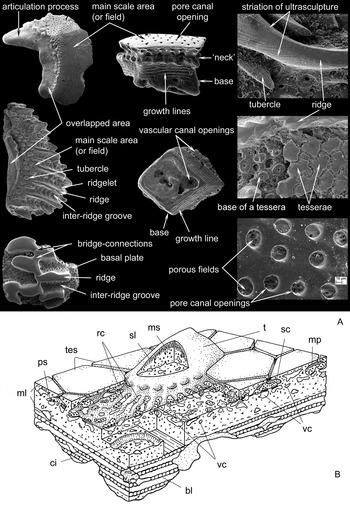
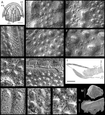
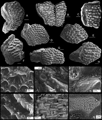
In osteostracan study, the terminology for description of the exoskeleton sculpture is insufficient. Over the long period that osteostracans have been studied, the most used term for their sculpture has been “granulated”, because microscopes with low magnification did not allow researchers to see the small details. Today we have described many new taxa, and we know that the surfaces of their exoskeletal elements, such as plates, tesserae and scales, have many variable patterns formed by tiny elements. These tiny elements must be named (Fig. 4). The surface can be smooth with pores, and these pores can be roundish, elongated and deep or funnel-shaped. The external surface may be covered with different shapes of higher sculpture elements, such as tubercles, ridges and ribs. The visceral surface is pierced by openings of vascular canals; growth lines are often recognisable.
Terminology for description of exoskeletal elements: (A) sculpture elements; (B) three-dimensional reconstruction of exoskeleton of Dartmuthia gemmifera (from Afanassieva Reference Afanassieva, Novitskaya and Afanassieva2004). Abbreviations: bl=basal layer; ci=circular canal under tubercle; ml=middle layer; mp=microapertures of perforated septum or porous field; ms=mesodentine; ps=perforated septum or porous field; rc=radiating canals; sc=sensory canal; sl=superficial layer; t=tubercle; tes=tesserae; vc=vascular canals.

Tubercles. Morphologically, the simplest sculpture element is a round tubercle, which may become slightly elongated to strongly elongated (two to three times longer than the width) or which have uneven margins that then are referred to as nodules.
A tubercle may have a pointed apex (spine) or be dome-like, or the tubercle may be of the same height or rise posteriorly; the margins of tubercles can be smooth, serrated or cren(ul)ated; they may have indentations forming shapes such as crosses, stars, oak leaves, water drops or triangles. The sculptural elements may be separated from one another or may partly overlap.
Ridges and ribs. If the elongated tubercle becomes stretched more than approximately four times its width, it is referred to as a ridge. The ridge may have a sharp crest or be flat; the latter is called a rib. The ridge may have side branches, each branch having a sharp crest or flat surface. The ridges may be straight or sinuous (or meandering); they may be tightly packed or widely spaced and separated by grooves that are called inter-ridge grooves.
Tesserae. Polygonal tesserae may be placed between the ridges, or occur as separate units. They may be flat or carry fine ridgelets.
For histology, the terminology of Denison (Reference Denison1947, Reference Denison1951a, Reference Denisonb) has been adopted.
6. Classification and phylogenetic framework
Osteostracan classifications have varied over the many years since Lankester (Reference Lankester1868–1870) first recognised the group and created the name Osteostraci. The following are reflections of the different hypotheses of osteostracan interrelations that have been suggested by numerous authors based on substantially different methodologies (see Sansom Reference Sansom2009 for review). The articulated materials from the East Baltic have been of particular interest because they form the most informative and diverse osteostracan fauna, which has been essential to our understanding of the ancestral state of osteostracans (Janvier Reference Janvier1981, Reference Janvier1985a, Reference Janvierb, Reference Janvier1996). This material has had a profound effect on the manner in which osteostracan evolution has been interpreted (Denison Reference Denison1951a; Obruchev Reference Obruchev and Orlov1964; Janvier Reference Janvier1985b; Afanassieva Reference Afanassieva1991, Reference Afanassieva, Novitskaya and Afanassieva2004; Sansom Reference Sansom2008, Reference Sansom2009) and therefore also on our understanding of early vertebrate evolution. By applying phylogenies to the stratigraphical record of a particular clade dominated by the East Baltic taxa, Sansom (Reference Sansom2008) demonstrated the early evolution and origin of the Osteostraci, supporting previous observations that the fossil record of osteostracans was strongly influenced by facies control (Märss & Einasto Reference Märss and Einasto1978; Märss Reference Märss1986). Sansom (Reference Sansom2009) subsequently presented an unranked classification for all osteostracans based on a phylogenetic hypothesis constructed from an expanded data set and the use of parsimony and out-group analysis which, in certain areas, differs rather substantially from previous classifications (Janvier Reference Janvier1981, Reference Janvier1985a, Reference Janvierb, Reference Janvier1996; Afanassieva Reference Afanassieva1991, Reference Afanassieva, Novitskaya and Afanassieva2004), including all of the articulated Estonian osteostracans in Thyestida Berg, Reference Berg1940 (see below regarding the priority of Thyestinae, Thyestidae, Thyestida). When also considering disarticulated material from the Silurian of the East Baltic, we observe a broader representation of taxa, which may be considered for future phylogenetic optimisation and discussions regarding osteostracan evolution. The ranked classification used in this study follows that of Afanassieva (Reference Afanassieva1991, Reference Afanassieva, Novitskaya and Afanassieva2004) with the exclusion of suborder and subfamily levels.
Subclass Osteostraci Lankester, Reference Lankester1868–1870
Order Ateleaspidiformes Tarlo, Reference Tarlo, Harland, Holland, House, Hughes, Reynolds, Rudwick, Satterthwaite, Tarlo and Willey1967
Family Ateleaspididae Traquair, Reference Traquair1899
Genus Ateleaspis Traquair, Reference Traquair1899
Ateleaspis sp. cf. Ateleaspis tessellata Traquair, Reference Traquair1899
Order Tremataspidiformes Berg, Reference Berg1937
Family Tremataspididae Woodward, Reference Woodward1891
Genus Tremataspis Schmidt, Reference Schmidt1866
T. schmidti Rohon, Reference Rohon1892
T. rohoni Robertson, Reference Robertson1938a
T. mammillata Patten, Reference Patten1931a
T. milleri Patten, Reference Patten1931a
T. perforata sp. nov.
Genus Dartmuthia Patten, Reference Patten1931a
D. gemmifera Patten, Reference Patten1931a
D. procera sp. nov.
Genus Saaremaaspis Robertson, Reference Robertson1938a
S. mickwitzi (Rohon, Reference Rohon1892)
Genus Oeselaspis Robertson, Reference Robertson1935a
O. pustulata (Patten, Reference Patten1931a)
Genus Aestiaspis Janvier et Lelièvre, Reference Janvier and Lelièvre1994
A.viitaensis Janvier et Lelièvre, Reference Janvier and Lelièvre1994
Family Thyestidae Rohon, Reference Rohon1892
Genus Thyestes Eichwald, Reference Eichwald1854
T. verrucosus Eichwald, Reference Eichwald1854
Family Procephalaspididae Stensiö, Reference Stensiö and Grassé1958
Genus Procephalaspis Denison, Reference Denison1951a
P. oeselensis (Robertson, Reference Robertson1939a)
Family Witaaspididae Afanassieva, Reference Afanassieva1991
Genus Witaaspis Robertson, Reference Robertson1939b
W. schrenkii (Pander, Reference Pander1856)
Family incertae sedis
Genus Tahulaspis gen. nov.
T. ordinata gen. et sp. nov.
T. praevia gen. et sp. nov.
Genus Eldaaspis gen. nov.
E. miklii gen. et sp. nov.
Order ?Cephalaspidiformes Berg, Reference Berg1937
Family incertae sedis
Genus Meelaidaspis gen. nov.
M. gennadii gen. et sp. nov.
Genus Ohesaareaspis gen. nov.
O. ponticulata gen. et sp. nov.
7. Systematic palaeontology
Subclass Osteostraci Lankester, Reference Lankester1868–1870
Order Ateleaspidiformes Tarlo, Reference Tarlo, Harland, Holland, House, Hughes, Reynolds, Rudwick, Satterthwaite, Tarlo and Willey1967
Family Ateleaspididae Traquair, Reference Traquair1899
Genus Ateleaspis Traquair, Reference Traquair1899
Content. Ateleaspis tessellata Traquair, Reference Traquair1899; Ateleaspis (Aceraspis) robusta (Kiaer, Reference Kiaer1911); Ateleaspis sp. Afanassieva & Karatajūtė-Talimaa, Reference Afanassieva and Karatajūtė-Talimaa1998.
Occurrence. Lower Wenlock of Scotland (Robertson Reference Robertson1989; Walton & Oliver Reference Walton, Oliver and Craig1991; Rolfe Reference Rolfe, Lawson and Weedon1992; Wellman & Richardson Reference Wellman and Richardson1993; Märss & Ritchie Reference Märss and Ritchie1998); Wenlock to lower Ludlow of the Oslo Region, Norway (Heintz Reference Heintz1969; Worsley et al. Reference Worsley, Aarhus, Bassett, Howe, Mørk and Olaussen1983; Davies et al. Reference Davies, Turner and Sansom2005); Lochkovian, Lower Devonian of Severnaya Zemlya Archipelago (Afanassieva & Karatajūtė-Talimaa Reference Afanassieva and Karatajūtė-Talimaa1998).
Ateleaspis sp. cf. A. tessellata Traquair, Reference Traquair1899
(Fig. 5)
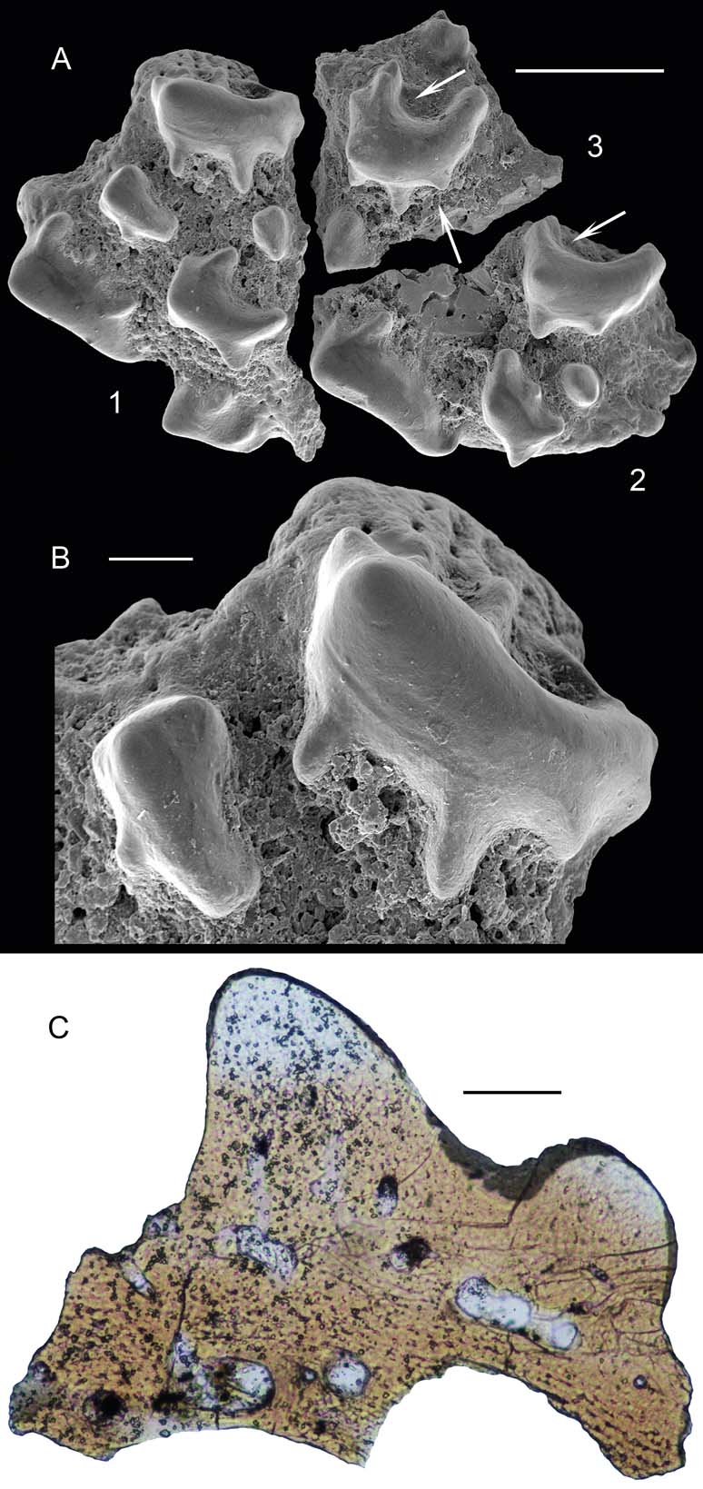
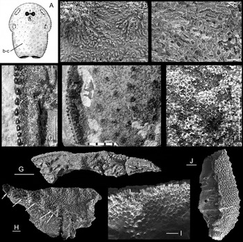
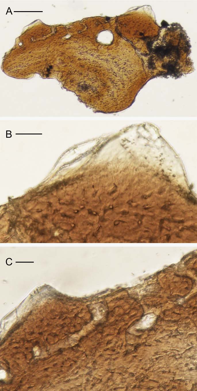
Ateleaspis sp. cf. A. tessellata Traquair: (A) SEM photographs of tessera broken into three pieces, all in external view, GIT 502-224-(1–3); (B) GIT 502-224-1, close-up of (A); (C) vertical section of fragment, GIT 502-224-3, from (A). Scale bars=500 µm (A); 100 µm (B, C). Location: Ohesaare-GI drill core, depth 174.10–174.50 m, Maasi Beds of Jaagarahu Stage, Sheinwoodian, lower Wenlock. Arrows in (A) point to porous fields shown in high magnification.

Ateleaspis tessellata occurs in the Slot Burn Formation, Waterhead Group in the Lesmahagow Inlier; in the Fish Bed Formation of Glenbuck Group in the Hagshaw Hills Inlier; and in the Lyne Water Fish Bed, Henshaw Formation, in the North Esk Inlier, Midland Valley, Scotland. The beds are thought to be lower Wenlock (Walton & Oliver Reference Walton, Oliver and Craig1991; Wellman & Richardson Reference Wellman and Richardson1993; Märss & Ritchie Reference Märss and Ritchie1998). In Ringerike Area, Norway, another species of the genus, Ateleaspis (Aceraspis) robusta (Kiaer Reference Kiaer1911), comes from the Wenlock to lower Ludlow level (Heintz Reference Heintz1969; Worsley et al. Reference Worsley, Aarhus, Bassett, Howe, Mørk and Olaussen1983; Davies et al. Reference Davies, Turner and Sansom2005). Ateleaspis sp. Afanassieva & Karatajūtė-Talimaa Reference Afanassieva and Karatajūtė-Talimaa1998 from October Revolution Island, Severnaya Zemlya Archipelago, was found in the Severnaya Zemlya Formation, Lochkovian, Lower Devonian. In the studied collection, three fragments of a tessera (Fig. 5A,1–3) come from the Ohesaare-GI drill core, depth 174.10–174.50 m; more come from a depth of 173.75–173.81 m, Maasi Beds of the Jaagarahu Stage, upper Sheinwoodian, lower Wenlock. This material falls within the known range of the genus.
Description. Sculpture. The fragments bear small crescent-shaped or linear elongated tubercles on the basal plate of the tessera; the tubercles have short but rather strong side branches; small oval or waterdrop-like tubercles lie in between the larger ones. The tubercles are rather high with one end slightly higher than the other. The surface of the tubercles is smooth (Fig. 5B). The upper surface of the basal plate is uneven, pierced by vascular canal openings. Porous fields are recognisable in a few spots in Figure 5A (indicated with arrows).
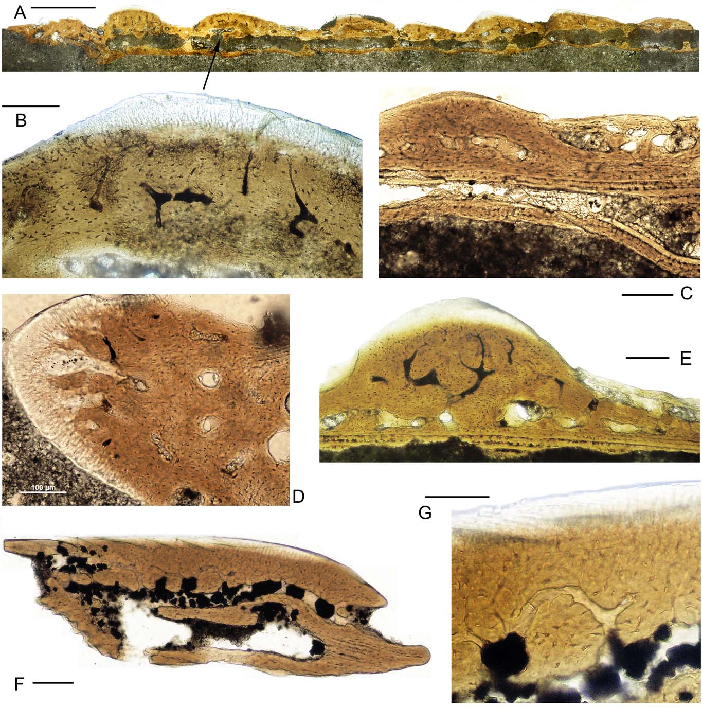
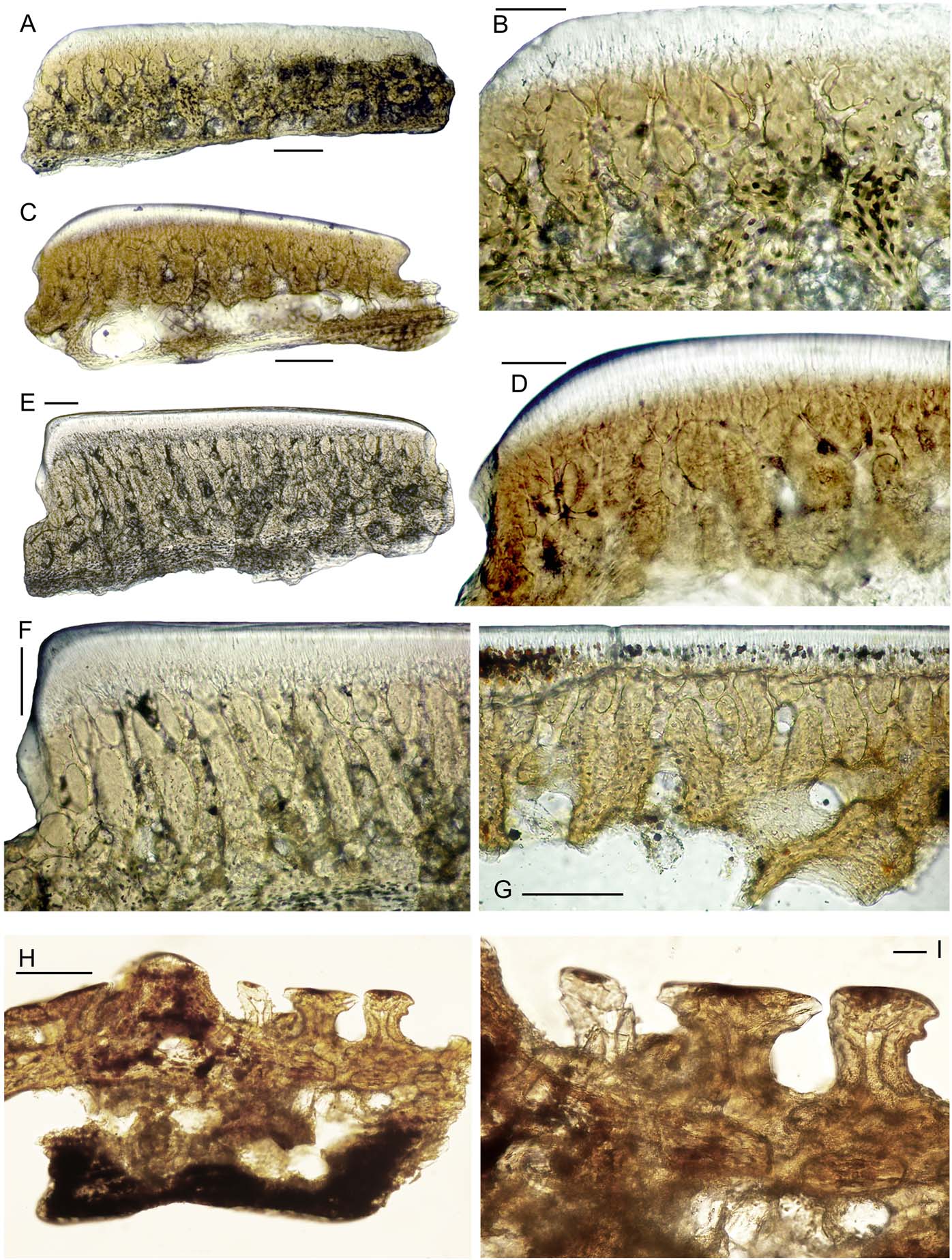
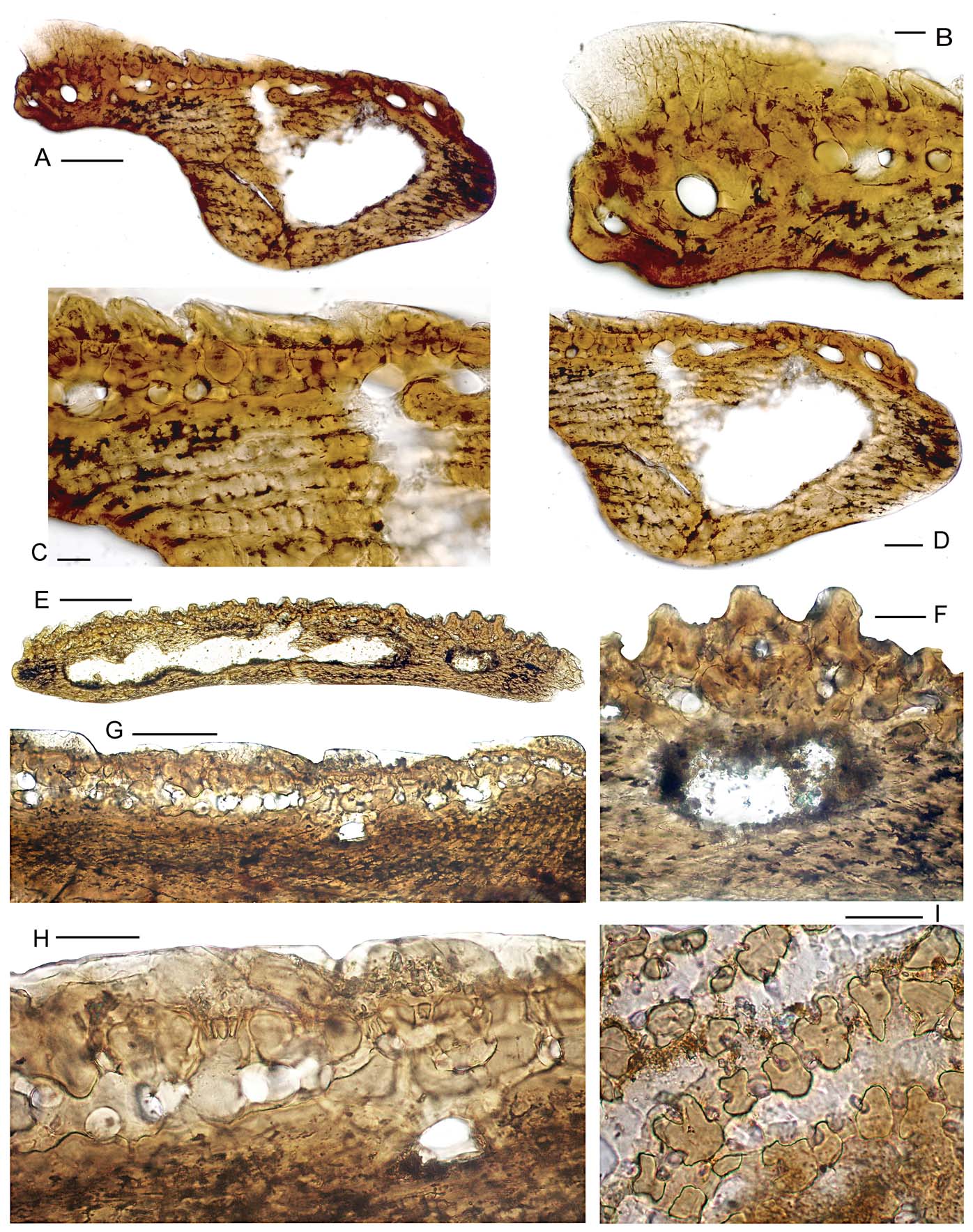
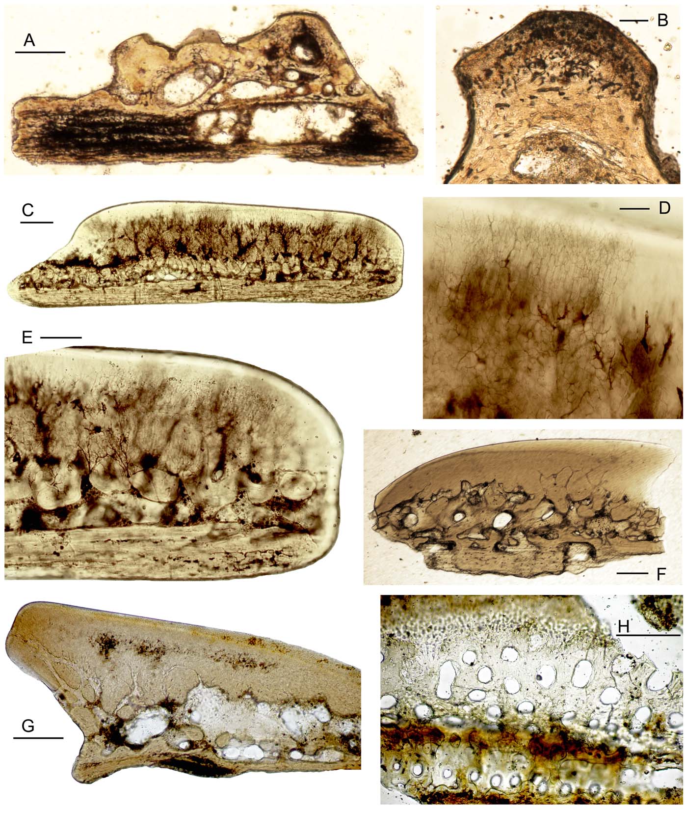
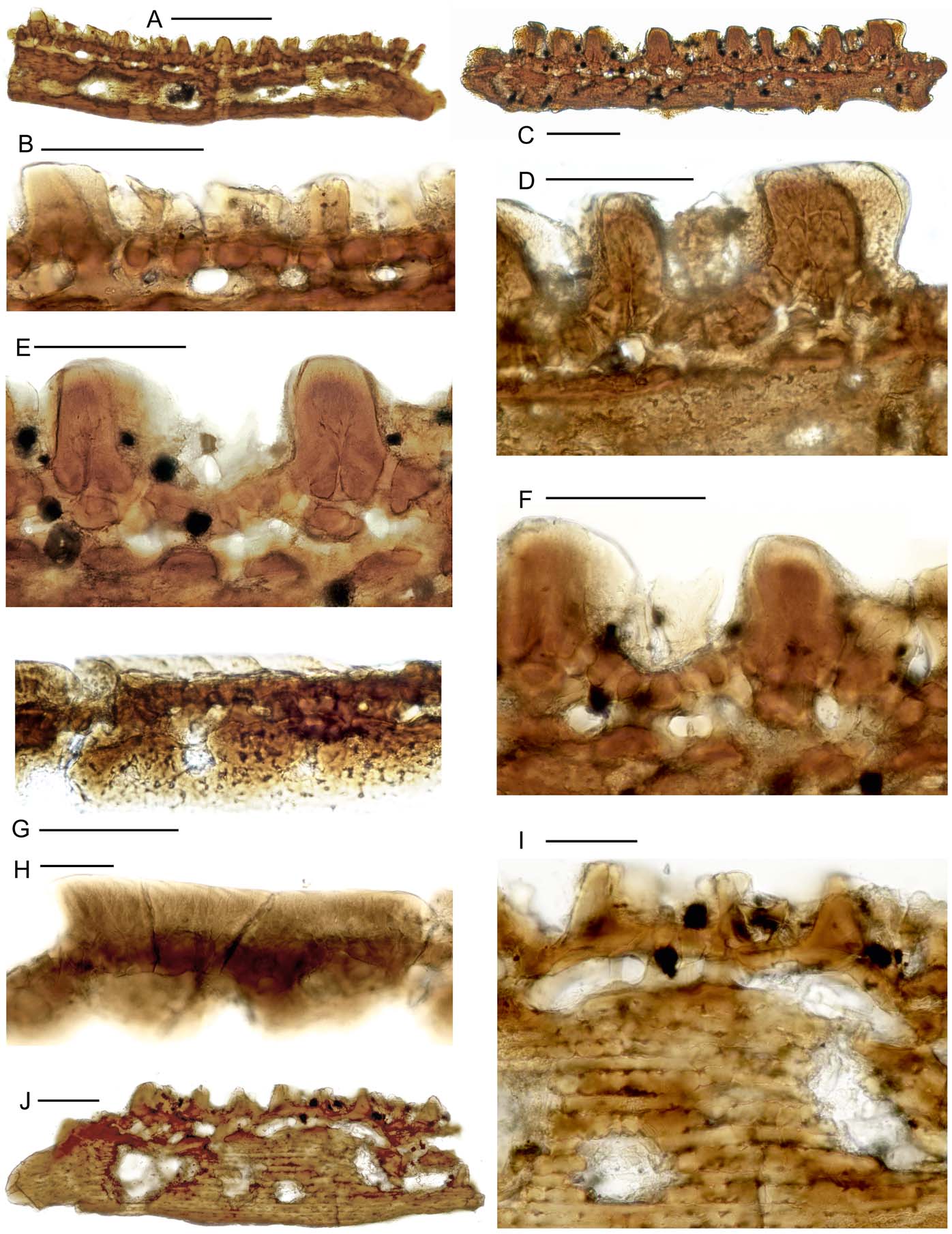
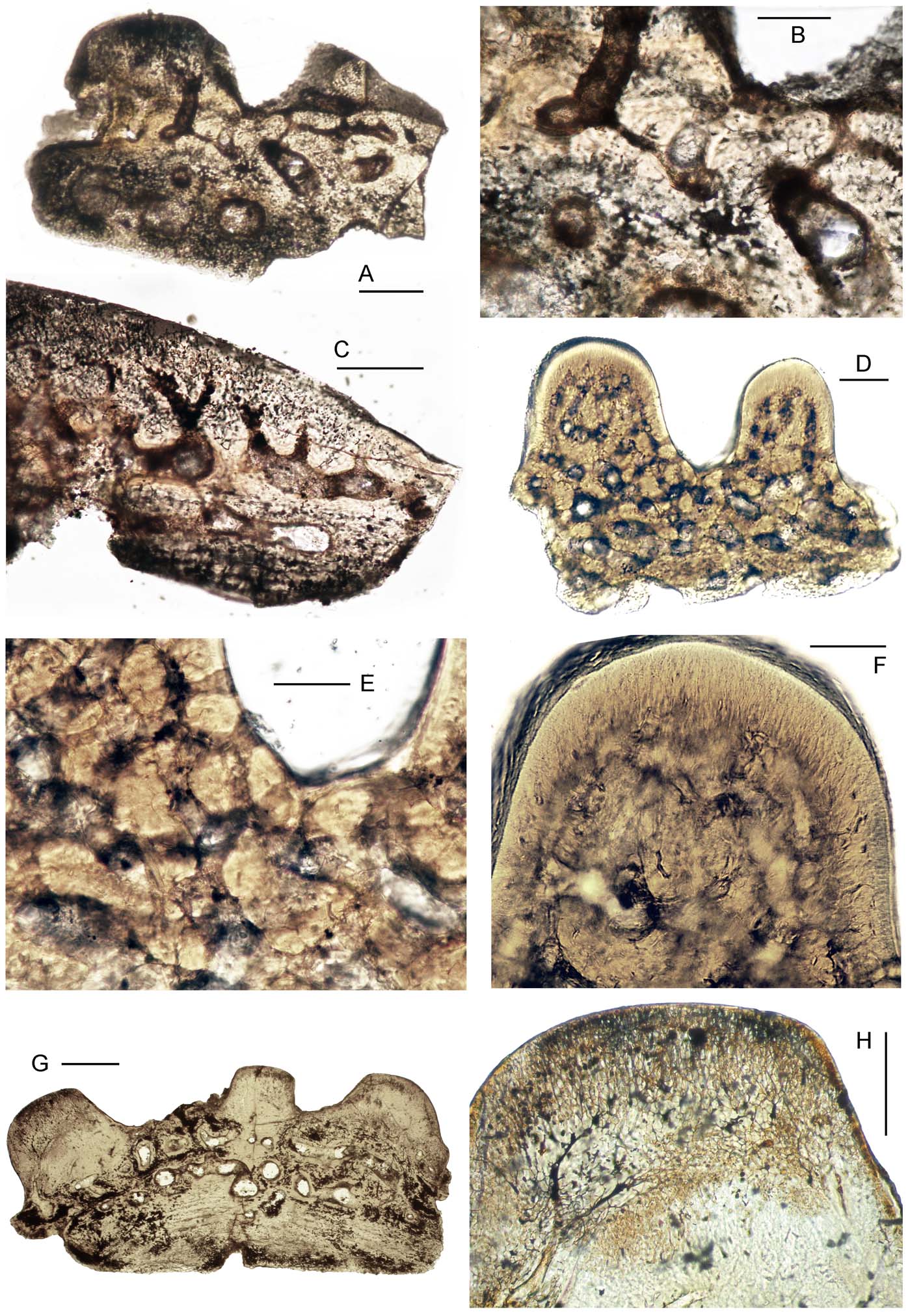
Histology. The material of Estonian Ateleaspis is very rare; nevertheless, we succeeded in creating the only thin section of a fragment of a specimen under investigation (GIT 502-224). The vertical section of a piece (GIT 502-224-3) crossed tessera through two parts of the large robust convex tubercle (see Fig. 5A) so that two tubercles (the large and the small ones) are visible in the section (Fig. 5C). The exoskeleton of Ateleaspis sp. cf. A. tessellata is well developed and is characterised by the presence of all three layers typical of osteostracans. The layers are comparatively equal in thickness. The superficial layer is present in the upper portion of the tubercles and is well developed (about 100 µm thick in large tubercles). The superficial layer contains a network of tubules and odontocyte cavities connected by branches, which is consistent with mesodentine sensu Ørvig (Reference Ørvig1951). The tissue of the superficial layer is transparent and comparatively dense, i.e., odontocyte cavities are not numerous. The length of dentine tubules branching off from odontocyte cavities depends on the size of the tubercles and the degree of the layer development. In the middle layer, the cavities of the osteocytes are clearly visible, and the matrix of bony tissue is comparatively dense. The vascular canal network is developed; the vascular canals reach the surface of the tessera and are opened by apertures of different sizes between the tubercles. It is difficult to distinguish the arrangement of radiating canals in a single section. The probable radiating canals are visible under the small tubercle; the diameter of radiating canals is approximately 30 µm. No perforated septa are visible in the thin section under study. The basal layer is strongly developed. There are several basal cavities of medium size in the basal layer. In the section under study, the walls of the vascular canals and different cavities are dense.
Comparison. The sculpture on our fragments is comparable to Ateleaspis tessellata sculpture, the species described from the Wenlock of Scotland (Ritchie Reference Ritchie1967, pl. 4, fig. 1, R.S.M. 1966.48.2). Its sculpture varies substantially from round tubercles on tesserae-like units of the head shield to elongated, subparallel ridges on the scales; the former area distinctly includes crescent-shaped tubercles arranged in rings laterally from the orbital area of the head. According to Ritchie (Reference Ritchie1967), the ornamentation of another Ateleaspis, A. robusta (Kiaer) from Rudstangen, Ringerike, S. Norway, is of rounded tubercles anterior to the orbits and of low spines behind this line. Despite rather good similarities in sculpture between our form and A. tessellata, we leave the species identified in open nomenclature because of scanty material found so far.
Order Tremataspidiformes Berg, Reference Berg1937
Family Tremataspididae Woodward, Reference Woodward1891
Genus Tremataspis Schmidt, Reference Schmidt1866
Type species. Tremataspis schmidti Rohon, Reference Rohon1892.
Content. T. schmidti Rohon, Reference Rohon1892; T. mammillata Patten, Reference Patten1931a; T. milleri Patten, Reference Patten1931a; T. rohoni Robertson, Reference Robertson1938a; T. obruchevi Afanassieva et Karatajūtė-Talimaa, Reference Afanassieva and Karatajūtė-Talimaa1998; T. perforata sp. nov.
Diagnosis. Small cephalic shields, 3.0–4.8 cm long and 2.0–3.8 cm wide; low tubercles on posterior half of dorsal head shield, which most posteriorly form a narrow median ridge; short median projection posterior to dorsal shield; two pairs of small, oval lateral fields and an unpaired small median field; pectoral sinuses, cornua and fins absent; well-separated openings of endolymphatic ducts posterior to median dorsal field; external surface smooth, pierced by pores of different diameters and densities; ultrasculpture of small irregular or honeycomb-like hollows; exoskeleton of three well-developed layers: superficial layer generally continuous, pierced by numerous regular pores; middle layer with the polygonal canals of the sensory system divided by perforated septa into upper and lower parts, sensory canal network simple; basal layer thick and strongly laminated. (Shield diagnosis adapted from Robertson Reference Robertson1938a; Denison Reference Denison1951a, Reference Denisonb; Janvier Reference Janvier1985a; Afanassieva Reference Afanassieva1991, Reference Afanassieva, Novitskaya and Afanassieva2004).
Occurrence. Upper Wenlock and Ludlow, Silurian of Estonia; Halla and Hemse beds of Gotland Island, Sweden (Fredholm Reference Fredholm1988, Reference Fredholm1990 as Tremataspididae sp.); Paralogania martinssoni VZ and Andreolepis hedei VZ, Kuba Beds of the Central Urals (Modzalevskaya & Märss Reference Modzalevskaya and Märss1991; Märss Reference Märss1992); Ust'-Spokoinaya Formation, Ludlow of Severnaya Zemlya Archipelago (Afanassieva & Karatajūtė-Talimaa Reference Afanassieva and Karatajūtė-Talimaa1998).
Remarks. (1) Schmidt (Reference Schmidt1866) established the genus Tremataspis based on the type species Cephalaspis schrenkii Pander, Reference Pander1856. Robertson (Reference Robertson1939b) recognised the significant difference between this taxon and Tremataspis species and established a new genus Witaaspis for Cephalaspis schrenkii. This resulted in problems involving the genotype of Tremataspis, which was addressed to the International Commission on Zoological Nomenclature (ICZN), who in 1948 agreed to designate Tremataspis schmidti Rohon, Reference Rohon1892 as the type species of Tremataspis Schmidt, Reference Schmidt1866 (Denison Reference Denison1951a, p. 180).
(2) Several Tremataspis species were listed and established by Robertson (Reference Robertson1938a) including T. patteni. The material of T. patteni was studied to test its validity/invalidity in terms of the details of its exoskeleton; this supports the view of Janvier (Reference Janvier1985a, p. 143) that T. patteni should be attributed to T. mammillata. Janvier (Reference Janvier1985a) also considered T. scalaris Robertson, Reference Robertson1938a and T. panderi Robertson, Reference Robertson1938a to belong to T. mammillata because their superficial layer had just been distorted by growth disturbances. T. simonsoni Rohon, Reference Rohon1892, assigned by Schmidt (Reference Schmidt1894) to Cephalaspis (Witaaspis) schrenkii, is based on an indeterminable specimen with ornament similar to Saaremaaspis, and Janvier (Reference Janvier1985a, p. 145) considered T. simonsoni as nomen dubium. Consequently, only T. schmidti, T. milleri, T. mammillata and T. rohoni have remained valid and will be included with Tremataspis perforata sp. nov. under the genus Tremataspis. The location of the holotype (by monotypy) and only articulated shield specimen of T. rohoni is not known at present and could not be used for this study. The Tremataspis type material originally housed in the AMNH, which was not available for this study, is in need of revision.
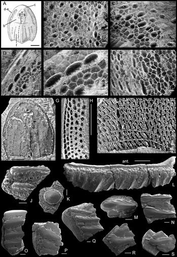
(3) Pores in the superficial layer of the exoskeleton, their diameter and the distance between them have been regarded as diagnostic features of Tremataspis species similar to the sculpture of ridges and tubercles of other taxa. Whilst the sculpture ridges on the exoskeleton of other osteostracans had mainly direct hydrodynamic functions, the function of these pores is unclear. They may be part of a sensory canal system, as suggested by Denison (Reference Denison1947), or as proposed by Stensiö (Reference Stensiö1927, p. 38–39), as ‘part of a mucous canal system’, in which the pores led mucous to the surface to minimise the friction between the fish body and the water. In the latter case the pores thereby had hydrodynamic function. Streamlined longitudinal relief on the scale surface of Tremataspis is expressed only on their most posterior margin with stretched-out pores in that area and spines overhanging the margin. Nevertheless, as the ridges and pores do not belong to exactly the same category of surface pattern, we have used term ‘sculpture’, in inverted commas if formed by pores.
Tremataspis schmidti Rohon, Reference Rohon1892
(Figs 6A–C, J, 7A–C, 8A–D, 9A, B)
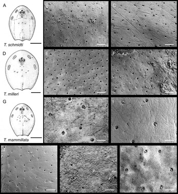
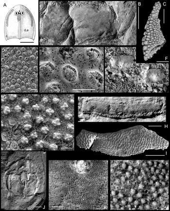
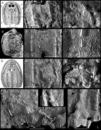
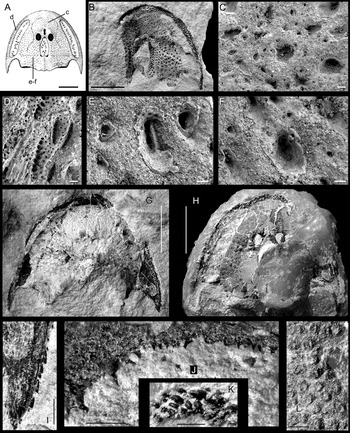
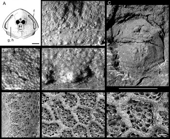
Head shield reconstructions and SEM photographs of head shield sculpture of various Tremataspis species in external view. (A–C) Tremataspis schmidti Rohon: (A) head shield reconstruction; (B) dorsal head shield surface, AMNH 20208; (C) ventral head shield surface; note also the scratches, AMNH 20209. (D–F) Tremataspis milleri Patten: (D) head shield reconstruction; (E) dorsal head shield surface, AMNH 20202; (F) ventral head shield surface, AMNH 20203. (G–I) Tremataspis mammillata Patten: (G) head shield reconstruction; (H) dorsal head shield surface, AMNH 13073; (I) ventral head shield surface, AMNH 13072. (J) Tremataspis schmidti Rohon, scale surface on tail, GIT 232-270-1. (K) Tremataspis rohoni Robertson, surface on scale of tail, AMNH 20211. (L) Tremataspis patteni Robertson, dorsal head shield surface, AMNH 20207, showing pore density similar to Tremataspis mammillata. Scale bars=10 mm (A, D, G); 0.1 mm (B, C, E, F, H–L). Reconstructions (A), (D) & (G) from Janvier (Reference Janvier1985a, fig. 33A–C), reproduced by permission of Philippe Janvier and Elsevier Masson, Paris.

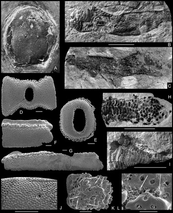
(A–C) Tremataspis schmidti Rohon: (A) lectotype PSM SPU 75/26 (Rohon Reference Rohon1892, pl. 2, fig. 7), head shield in dorsal view; (B–C) squamation of posterior part of trunk and tail, GIT 232-270-1 and GIT 232-270-2 (part and counterpart). (D–G) Tremataspis milleri Patten: (D) pineal plate, GIT 502-330; (E) sclerotic ring, GIT 502-331; (F) scale, GIT 502-324; (G) scale, GIT 502-418. ( H–I) squamation of posterior part of trunk and tail: (H) Tremataspis mammillata Patten, GIT 502-436; (I) Tremataspis rohoni Robertson, AMNH 20211. (J) Tremataspis mammillata Patten, ultrasculpture, GIT 502-236. (K–L) Tremataspis milleri Patten, fragment of shield with partly overgrown superficial layer (reparation of shield?), arrows point to small tubercles of dorsal shield, GIT 502-269. All images in external view. Scale bars=1 cm (A–C, H, I); 200 µm (D–G, K); 100 µm (J, L). Locations: (A) most possibly Viita Quarry; (B–G) bed 3, Viita trench; Viita Beds of Rootsiküla Stage, upper Homerian, upper Wenlock; (H) lower beds in Himmiste Quarry; Himmiste Beds of Paadla Stage, Gorstian, lower Ludlow; (I) most possibly Himmiste Quarry; (J) Varbla-502 drill core, depth 32.3 m, western Estonian mainland, undivided Sauvere and Himmiste Beds, Paadla Stage, Gorstian, lower Ludlow; (K, L) bed 5, Viita trench, Viita Beds of Rootsiküla Stage, upper Homerian, upper Wenlock. All specimens originate from Saaremaa Island, Estonia.

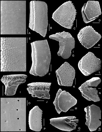
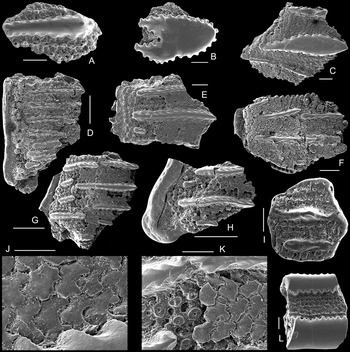
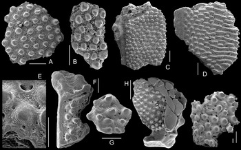
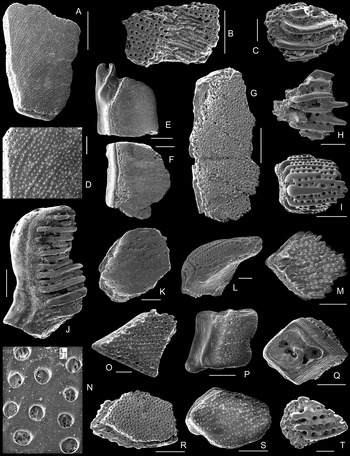
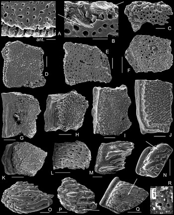
Scales of Tremataspis species. (A–D) Tremataspis schmidti Rohon: (A, B) GIT 502-197; (C) GIT 232-377; (D) GIT 502-270. (E–L) Tremataspis milleri Patten: (E, F) GIT 502-196; (G) GIT 502-415; (H) GIT 502-252; (I) GIT 502-327; (J) GIT 502-328; (K) GIT 502-271; (L) GIT 502-249. (M–Q) Tremataspis mammillata Patten: (M) GIT 502-276. (N) 502-239; (O, P) GIT 502-198; (Q) GIT 502-235. (R) Tremataspis rohoni Robertson, GIT 502-452. All scales in external view except (I) and (J), which are in side view. Scale bars=100 µm (A, E, O); 500 µm (B, C, F, I, P); 200 µm (D, G, H, J–N, Q, R). Locations: (A, B, E–G) Vesiku Brook, Vesiku Beds of Rootsiküla Stage; (C–D, I–K, R) bed 3 in Viita trench, Viita Beds of Rootsiküla Stage, upper Homerian, upper Wenlock; (H, L) lower beds in Pähkla Quarry; (M) Kingissepa-GI drill core, depth 30.82–30.90 m; (N–P) lower beds in Silma Cliff; Himmiste Beds of Paadla Stage, Gorstian, lower Ludlow. All specimens originate from Saaremaa.

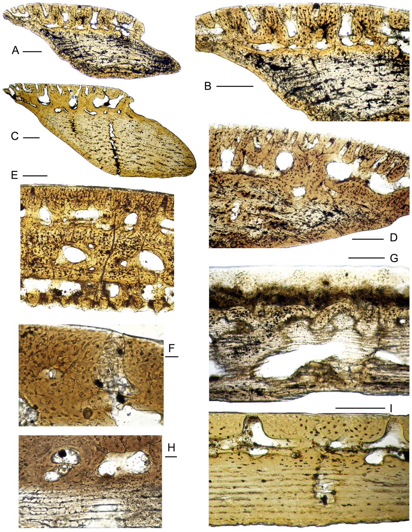
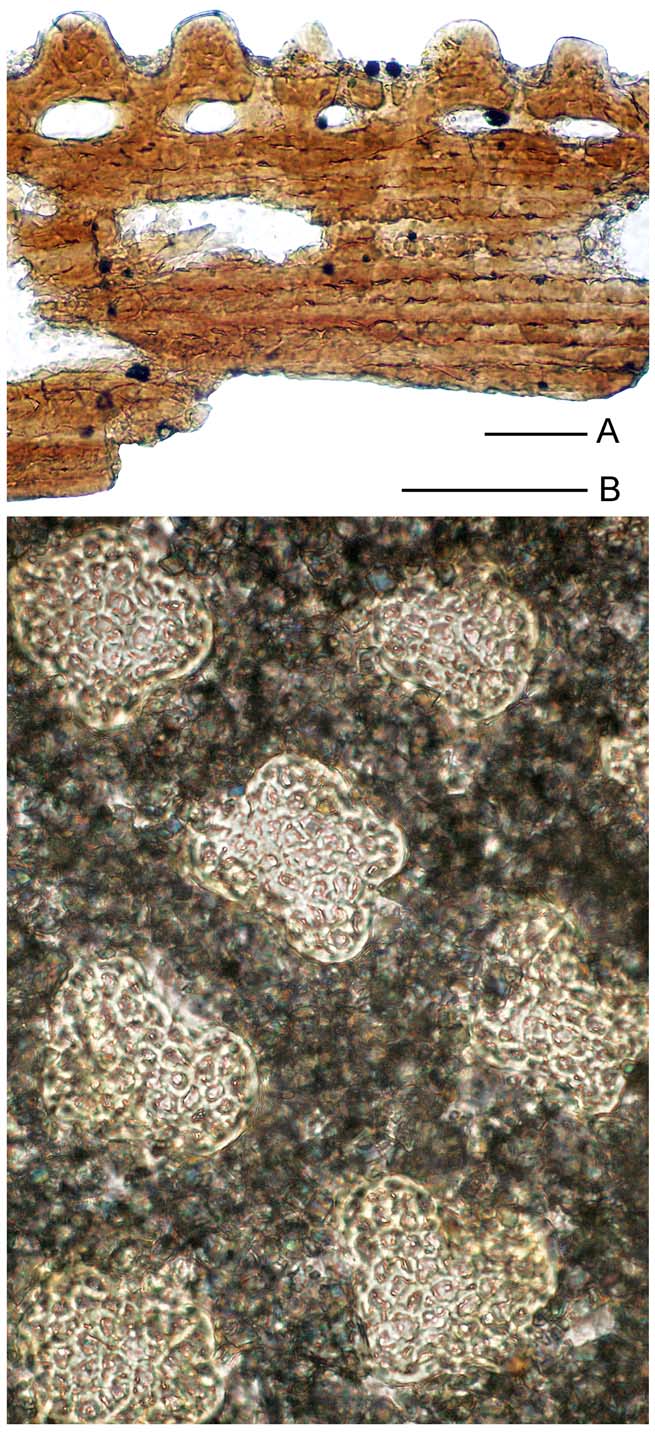
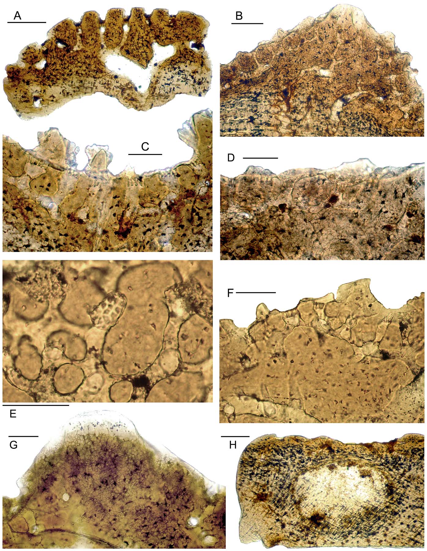
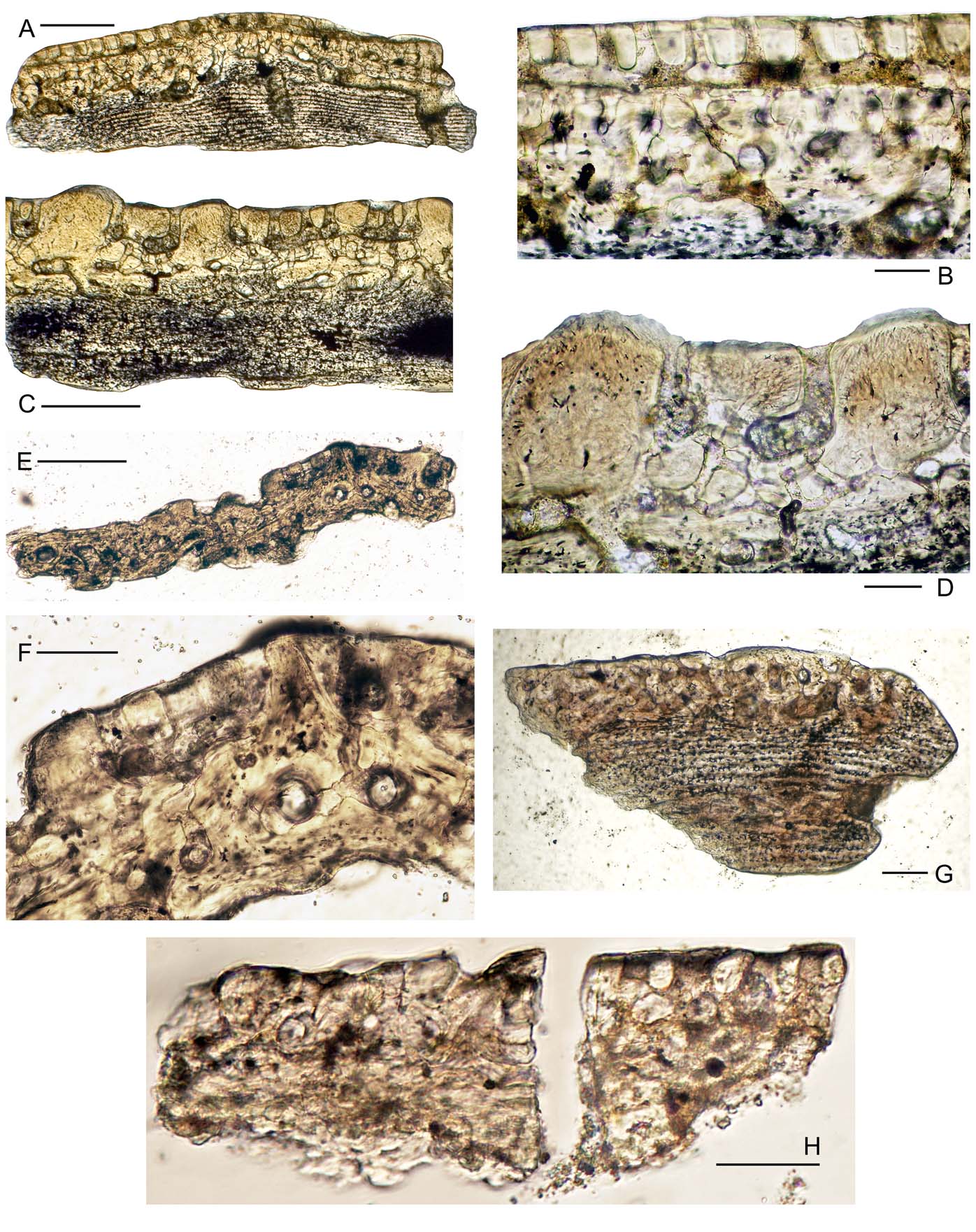
Vertical longitudinal sections of the scales of Tremataspis species. (A, B) Tremataspis schmidti Rohon, GIT 502-54. (C, D) Tremataspis milleri Patten: (C) GIT 502-59; (D) GIT 502-58. (E) Tremataspis sp., GIT 502-55. (F, H) Tremataspis rohoni Robertson, GIT 502-78. (G, I) Tremataspis mammillata Patten: (G) GIT 502-62; (I) GIT 502-63. Locations: (A–B) bed 3 in Viita trench, Viita Beds of Rootsiküla Stage; (C–E) Vesiku Brook, Vesiku Beds of Rootsiküla Stage; (F, H) Elda Cliff, Kuusnõmme Beds of Rootsiküla Stage; (G, I) Silma Cliff, Himmiste Beds of Paadla Stage. Scale bars=100 μm (A–E, G, I); 20 μm (F, H).

For synonymy, see Afanassieva (Reference Afanassieva, Novitskaya and Afanassieva2004, p. 227).
Holotype. Not designated by Rohon (Reference Rohon1892).
Lectotype. PSM SPU 75/26, Figure 7A herein (=Rohon Reference Rohon1892, taf. 2, fig. 7), housed in the Palaeontological-Stratigraphical Museum at St. Petersburg State University; most possibly Viita Quarry, Saaremaa Island, Estonia; Viita Beds of the Rootsiküla Stage, upper Homerian, upper Wenlock, lower Silurian.
Rohon (Reference Rohon1892, p. 39) had 12 specimens from Viita, Vesiku and Himmiste (Hoheneichen by him). The first he described is the specimen with four tubercles on the head shield (Rohon Reference Rohon1892, p. 42). The description was accompanied by four figures for the species: one reconstruction of T. schmidti (Rohon Reference Rohon1892, pl. 1, fig. 11); two more figures depicting fish from the dorsal and ventral sides jointly (Rohon Reference Rohon1892, figs 13, 15); and a representational specimen showing four tubercles on the dorsal side of the shield (Rohon (Reference Rohon1892, pl. 2, fig. 7). The holotype was not indicated and a lectotype must be chosen from the available Rohon syntypes. The specimen AMNH 38.71.9410 indicated as the lectotype (Janvier Reference Janvier1985a, p. 145, and subsequent authors), therefore loses its status according to the ICZN 1999, 74.2.
Diagnosis. Cephalic shield 3.0–3.7 cm long and 2.0–2.7 cm wide (lectotype: 3.7 cm long and 2.7 cm wide; specimen a little crushed); low and round nasohypophysial depression near the rostral margin; small orbits; four tubercles behind the dorsal field; postero-median dorsal ridge short and low with a few small tubercles; surface pore pattern with approximately 100 pores per mm2 and distance of approximately 0.1 mm; pore diameter 20–25 μm; polygons of the middle layer subdivided by narrow canals into small fields (shield diagnosis from Denison Reference Denison1947, Reference Denison1951b; Janvier Reference Janvier1985a; Afanassieva Reference Afanassieva1991, Reference Afanassieva, Novitskaya and Afanassieva2004).
Material. The exact number of T. schmidti specimens is not known. From approximately 2000 shields in AMNH, New York, only approximately 3% belong to this species (Denison Reference Denison1947, p. 344). A few specimens of T. schmidti are in the Museum of Geology, University of Tartu, and the Paleontological Institute in Moscow. Approximately 25 larger shield fragments come from Elda Cliff; hundreds of microscopic exoskeletal elements picked out of the acetic acid residue are in the Institute of Geology in Tallinn. In the residue, the large rectangular anterior trunk scales are generally broken whereas smaller rhomboidal, pentagonal and hexagonal scales from the posterior portion of the trunk and from the tail are intact. The material comes from the following outcrops: Viita Quarry, Viita trench, Elda Cliff, Vesiku Brook, and the Vesiku-507 drill core, depth 8.65–8.80 m.
Occurrence. The Viita, Kuusnõmme and Vesiku beds of the Rootsiküla Stage, upper Homerian, upper Wenlock of Estonia; the Halla Beds, Wenlock of Gotland, Sweden; the Kuba Beds in the Mikhailovsk Bond section, Central Urals.
Description. Pore pattern (‘sculpture’). The head shield of T. schmidti (Figs 6A, 7A), which has previously been described several times, has a shiny, smooth surface, with pores of the sensory canal system piercing the surface (Fig. 6B–C). The size and density of the openings are the main characteristic features of the surface (Robertson Reference Robertson1938a, p. 288; Gross Reference Gross1968a). The pore diameter barely changes along the dorsal side of the shield and is on average 20 μm. The density of the pores is close to 100 per mm2, which is equivalent to an average distance of approximately 0.1 mm. Nevertheless, along the margins where the shield turns down, the density is a little higher and the pores somewhat larger. Four dorsal tubercles from behind the median dorsal field are 0.2–0.5 mm in diameter; the tubercles forming a short and low postero-median longitudinal dorsal ridge are also tiny (approximately 0.5–1.0 mm long and 0.5 mm wide) and often grown together.
The scale-covered trunk and tail of T. schmidti is known from a few specimens; two were presented by Rohon (Reference Rohon1894, pl. 1, figs 14, 15) and one by Janvier (Reference Janvier1985a, fig. 29, specimen AMNH 38.71.9626), the latter specimen originating from the Wenlock of Saaremaa. If we are correct, this specimen must be from the Viita Quarry, the Viita Beds of the Rootsiküla Stage, upper Homerian, upper Wenlock. Another specimen, GIT 232-270, with part and counterpart (Fig. 7B, C), is quite complete, with the majority of the trunk and tail preserved. It was identified as T. schmidti based on the size and density of the pore canal openings (see closeup in Fig. 6J). The anterior trunk scales are large and rectangular (Fig. 7B) but become gradually smaller and rhomboidal, pentagonal and hexagonal more posteriorly (Fig. 7B, C). The pores in the scale surface are small, a bit smaller and situated a little closer than in the head shields of that species. The diameter of the openings often varies, even on the same scale, although the openings are separated by the same distance. Somewhat wider openings occur closer to the anterior margin of the scales (see also Märss Reference Märss1986, fig. 25, 5–7). The pore characteristics may change along the trunk and, like sculpture elements, become somewhat smaller backwards.
A few isolated scales have been illustrated in Figure 8A–D. High and short scales are rare in the residue, whereas low and short rhomboidal or pentagonal or hexagonal scales are more common. In addition, young, thin scales are less common than thick, mature scales. The large and flattened anterior trunk scales have a smooth overlapped anterior area (Fig. 8A, B). The overlapped area is followed by the main scale area, with the higher anterior margin slightly scalloped and having openings for vascular canals. The main scale surface is slightly domed. The transitional area between the overlapped area and the main surface may be knobby. Smaller rhomboidal scales with deep bases are identified as posterior trunk scales. The anterior margin of smaller rhomboidal to hexagonal scales is rather smooth (Fig. 8B, D) or serrated (Fig. 8C); the posterior margin is smooth or has an array of short spines (Fig. 8C) or has short, shallow, longitudinal grooves ending at the margin. The growth layers in the base are well-distinguished (Fig. 8D). The diagnostic pores of the isolated microremains also show a density of nearly 100 pores per mm2 and a distance between pores of approximately 0.1 mm; pores are distributed slightly irregularly.
Histology (Fig. 9A, B). The structure of the exoskeleton of T. schmidti is typical of the species of Tremataspis. The superficial layer is continuous and pierced by the pores of the sensory system. The structure of the mesodentine is similar to the structure of typical bony tissue of the middle layer. The cavities of the odontocytes are relatively large. There are many interconnections between dentine tubules. The middle layer is well developed and contains canals of sensory and vascular systems and cavities of different sizes. The sensory canal system opens onto the external surface through pores. The sensory canals have smooth, dense walls. The sensory canals form the polygons and are subdivided by a horizontal perforated septum into upper and lower parts. The thickness of the septum is approximately 5 µm. The micro-apertures of the septum (3–4 µm in diameter) are comparatively evenly distributed in the sections. Polygons in the middle layer are subdivided by narrow canals into small fields. The network of the vascular canals is well developed. There is no radiating pattern in the distribution of the vascular canals. The osteocytes cavities in the bony tissue of the middle layer are 4–10 µm in size and are more or less evenly distributed. The basal layer is well developed and distinctly laminated. The basal layer is of considerable thickness in the mature scales (Fig. 9A).
Tremataspis milleri Patten, Reference Patten1931a
(Figs 6D–F, 7D–G, K, L, 8E–L, 9C, D)
For synonymy, see Afanassieva (Reference Afanassieva, Novitskaya and Afanassieva2004, p. 228).
Holotype. Not established by Patten (Reference Patten1931a).
Lectotype. AMNH 11219 (Patten # 38-71-9813 (T. 546) of the Patten collection originally in the Dartmouth College Museum), a nearly complete dorsal head shield designated by Robertson (Reference Robertson1938a, pl. 2, fig. 2, p. 289); Himmiste Quarry, Saaremaa Island, Estonia; Himmiste Beds of Paadla Stage, upper Gorstian, lower Ludlow. The location of the lectotype is not currently known.
Diagnosis. Tremataspis of relatively large size with very wide shield (length 3.8–4.8 cm, width 3.0–4.0 cm; small specimens with length 3.0–3.5 cm and width 2.5–3.5 cm occur); nasohypophysial depression deep, round, surrounded by a wide wall, situated relatively far from the rostral margin; six to eight tubercles situated behind the dorsal field; a median dorsal ridge of moderate length and tubercles generally fused; surface pattern of variable density of small pores, approximately 200 pores per mm2 and an average distance of approximately 0.05 mm between the pores and a diameter of 15–20 μm; polygons of the middle layer generally not subdivided into smaller fields (modified from Denison Reference Denison1947, Reference Denison1951a, Reference Denisonb; Janvier Reference Janvier1985a; Afanassieva Reference Afanassieva1991, Reference Afanassieva, Novitskaya and Afanassieva2004).
Material. Articulated head shields of this species are relatively rare. In the AMNH collection, approximately 4% of Tremataspis specimens belong to T. milleri (Denison Reference Denison1947, p. 346). Eight shields from the Himmiste Quarry are housed at Tartu University and two large fragments in the Institute of Geology of TUT. More than 40 broken specimens with the same pore density come from Elda Cliff, in which the size of Elda specimens is smaller than the size of Himmiste specimens. Small shield and scale fragments are common in the residue. Microremains come from the following outcrops: Viita Quarry; Viita trench; Vesiku Brook; Himmiste Quarry; Silma Cliff; Pähkla Quarry; and drill core sections: Vesiku-507, depth 8.65–8.85 m; Suurlahe-738, depth 56.20–56.30 m; and Varbla-502, depth 32.3 m.
Occurrence. Viita, Kuusnõmme and Vesiku beds of Rootsiküla Stage, upper Homerian, upper Wenlock and Sauvere and Himmiste beds of Paadla Stage, upper Gorstian, lower Ludlow of Estonia.
Description. Pore pattern (‘sculpture’). The head shields (Fig. 6D) vary somewhat in size between the three localities, Elda Cliff, Viita trench and Himmiste Quarry. Those from Himmiste are the largest among the Tremataspis species and have pores that are distributed evenly on their shields. They show a very dense distribution of small openings (diameter 15–20 μm), approximately 200 per mm2 and on average 0.05 mm between the pores (Fig. 6E–F). The specimens from Elda and Viita have variable pore size even in the middle of the shield and especially at its margins; pores are noticeably larger on downturned shield areas.
A wide variety of different elements have been studied for pore size and distribution along the shield, trunk and tail. The pore size on bow tie-like pineal and orbital plates (Fig. 7D, E) changes rather rapidly from the centre towards the margins of these plates: the pores are finer in the middle and larger at the margins of such plates. High and short scales (Figs 7F, G, 8E, F) have a nodular structure behind the smooth overlapped area, just anterior to the main scale area. Such a nodose structure appears to be characteristic of this species because the other scales may also have the main scale area with anterior nodules (Fig. 8G, I). The posterior margin of the scales is finely serrated (Figs 7F, G, 8F, H, I) or smooth (Fig. 8J, K) or crenulated (Fig. 8H, L). The scales may reach considerable depth with at least nine growth layers in the base (Fig. 8I, J). The diameter of the pores often varies even on the same scale as was the case with pineal and orbital plates. They are the largest at the anterior margin of the main scale area close to the overlapped area and smallest on the posterior margin (Fig. 8E). In the middle of the scales (as well as small platelets), the pores are characteristic of the species. The distance between the pores on the scales remains approximately the same. The fragment in Figure 7K, L has a porous layer that bears at least five small tubercles (indicated with arrows) of dorsal shield. It is overgrown with another, younger porous layer that may be a healed injury.
Histology (Fig. 9C, D). The structure of the exoskeleton of T. milleri is typical of the species of Tremataspis; however, polygons of the middle layer generally are not divided into smaller fields (Denison Reference Denison1947; Gross Reference Gross1968a; Afanassieva Reference Afanassieva1991, Reference Afanassieva, Novitskaya and Afanassieva2004).
Tremataspis mammillata Patten, Reference Patten1931a
(Figs 6G–I, 7H, J, 8M–Q, 9G, I)
For synonymy, see Afanassieva (Reference Afanassieva, Novitskaya and Afanassieva2004, p. 228).
Holotype. Not established by Patten (Reference Patten1931a).
Lectotype. AMNH 11529 (Patten # 38-71-9713 (T. 445) of the Patten collection originally in the Dartmouth College Museum), a nearly complete dorsal head shield, designated by Robertson (Reference Robertson1938a, pl. 2, fig. 3, p. 289). The location of the lectotype is not known (letter from Ivy Rutsky of 27 May 1999).
Type locality. Patten's (Reference Patten1931a) first description mentioned the name Atla. Our conclusion is that this material comes from the Himmiste Quarry (see introductory section above), Saaremaa Island, Estonia; the Himmiste Beds of the Paadla Stage, upper Gorstian, lower Ludlow.
Diagnosis. Medium sized, elongated Tremataspis with cephalic shield 3.5–4.0 cm long and 2.5–3.0 cm wide; twenty or more tubercles situated behind the median dorsal field; very low posteromedian dorsal ridge with many fine tubercles; surface pattern of pores with low density, approximately 15 pores per mm2 and distance between the pores 0.20–0.30 mm; pore diameter 30–35 μm; polygons of the middle layer not subdivided into smaller fields (modified from Denison Reference Denison1947, Reference Denison1951a, Reference Denisonb; Janvier Reference Janvier1985a; Afanassieva Reference Afanassieva1991, Reference Afanassieva, Novitskaya and Afanassieva2004).
Material. In the Patten collection, approximately 93% of the Tremataspis specimens belong to T. mammillata (Denison Reference Denison1947, p. 339). In addition to the AMNH collection, the good specimens are in the Museum of Geology of Tartu University with over 60 specimens collected by A. Luha and E. Bölau, and a few in the Institute of Geology at TUT. Microremains originate from the following outcrops: Himmiste Quarry; Silma Cliff; and drill core sections Himmiste-982, depth 0.7–1.9 m; Kaarmise-GI, depth 3.6 m; Kingissepa-GI, depth 30.82–31.41 m; Kuressaare-804, depth 19.7–24.0 m; Paadla-GI, depth 11.55–11.75 m; Uduvere-968, depth 4.5 m; Varbla-502, depth 31.80–32.3 m; Kailuka-817, depth 52.4–62.5 m.
Occurrence. The Himmiste Beds of the Paadla Stage, upper Gorstian, lower Ludlow of Estonia.
Description. Pore pattern (‘sculpture’). The head shields of T. mammillata (Fig. 6G) show large, widely distributed openings of the canals of the sensory system with approximately 15 pores per mm2, which is a distance of approximately 0.15–0.20 mm between the pores (Fig. 6H, I). These estimations can vary slightly over the head shield. Both the dorsal and ventral sides of the head shield show an identical density of pores.
The scales on a partly preserved tail (GIT 502-436, Fig. 7H) are well preserved. On the more anterior part of this fragment, the higher scales have the pores only in one row along the middle, along the longer axis of the scale. Posteriorly, the rhomboidal and pentagonal scales have only a few (3–4) pores per scale. The pore diameter is identical overall on this fragment.
Because of the density and large pore diameter, this species is relatively easy to identify (Fig. 8M–Q). The pore pattern copies the shape of the scales: the more anterior scales, which are higher and shorter, have one to two rows of pores along the longer axis of the scale; the pores tend to be somewhat longitudinally elongated (Fig. 8O). In smaller scales, on the surface of a triangular scale plate, the pores form a triangle (Fig. 8M). The saddle-like dorsal scutes have a smooth crest and pores on the flanges (Fig. 8Q). A triangular in crown shape scale (Fig. 8N) is anteriorly worn and includes a sensory canal; on the worn surface, the openings are elongated. The marginal areas of the scales are generally without pores (Fig. 8M, N, P, Q). The ultrasculpture on the surface of the scales occurs as small, shallow hollows between the pores (Fig. 7J).
Histology (Fig. 9G, I). The structure of the exoskeleton of T. mammillata is typical of the species of Tremataspis; however, the polygons of the middle layer are not divided into smaller sections (Denison Reference Denison1947, Reference Denison1951b; Afanassieva Reference Afanassieva1991, Reference Afanassieva, Novitskaya and Afanassieva2004). In one of the sections (Fig. 9G), there is concentrically arranged bony tissue around the cavities in the body of the middle and basal layers. In our opinion, this phenomenon shows the comparatively earlier developmental stage of exoskeleton development. The basal layer reaches a considerable thickness in the mature scales. A cross-laminated structure of the basal layer is distinct in the thin section in Figure 9I.
Tremataspis rohoni Robertson, Reference Robertson1938a
For synonymy, see Afanassieva (Reference Afanassieva, Novitskaya and Afanassieva2004).
Holotype and the only specimen. AMNH 11523 (Patten # 38-71-9379 (T. 106) of the Patten collection originally in the Dartmouth College Museum), designated by Robertson Reference Robertson1938a (pl. 2, fig. 4). The location of the holotype is not known at present.
Type locality. The type locality of the holotype is either Viita or Himmiste quarry on Saaremaa, Estonia (see above).
Diagnosis. Small Tremataspis cephalic shield, 3.2 cm long and 2.0 cm wide; nasohypophysial depression almost circular with an aperture entirely on the anterior wall, ending at the deepest part of the pit; 17 tubercles situated behind the median dorsal field; median dorsal ridge short, saddle-like in the middle part and with 2–3 fine tubercles in the anterior part; moderate pore density in the superficial layer; polygons of the middle layer not subdivided into smaller fields (modified from Robertson Reference Robertson1938a; Denison Reference Denison1947; Afanassieva Reference Afanassieva1991, Reference Afanassieva, Novitskaya and Afanassieva2004).
Remarks. There is a rather poor record regarding the pore pattern on the surface of this species. According to Robertson (Reference Robertson1938a, p. 290) the “pores are fine, less numerous than in T. milleri or T. schmidti, more so than in T. mammillata”. Denison (Reference Denison1947, p. 347) supported the validity of this species and emphasised the importance of pore size for identification by saying, “the surface pores are as large or nearly as large as those of T. mammillata (30–32 μm in diameter). They are more closely spaced than those of the latter species, but are more widely spaced than those of either T. milleri or T. schmidti”. Janvier (Reference Janvier1985a, p. 143) confirms the validity of T. rohoni by emphasising the lower number of tubercles on the head shield and the higher density of pores in comparison with T. mammillata, which otherwise is very similar in proportions. He also stressed the position of the nasohypophysial opening at the bottom of a circum-nasohypophysial fossa and the two big elongated tubercles on the posterior median dorsal crest as important for separating the holotype from T. mammillata and all other species of Tremataspis (Janvier Reference Janvier1985a, p. 143, fig. 33).
Description. Pore pattern (‘sculpture’). The GI collection yields some poorly crushed head shields (approximately 3.3 cm long and 2.5 cm wide) from Elda Cliff, Kuusnõmme Beds of Rootsiküla Stage, upper Homerian, upper Wenlock, which we consider to belong to T. rohoni, despite not being able to study the holotype. The specimens also show moderate pore density in the superficial layer with approximately 30 pores per mm2 and a distance between the pores of 0.15–0.25 mm. Our observation of pore diameter (less than 20 μm) differs slightly from the observation of Denison (Reference Denison1947), possibly due to differences in how the measurements were taken (because the measurement depends on the exact depth at which it was made in the pore).
Janvier (Reference Janvier1985a, fig. 29) described and illustrated the tail squamation of Tremataspis sp. (AMNH 38.71.9626) from the Wenlock of Saaremaa, Estonia. We re-studied this specimen (Fig. 7I), which exhibits small surface sensory canal pores, which lie far from each other (Fig. 6K). Regarding pore pattern, the pore density and pore size of this tail and the shield fragments in the GI collection show that they belong to the same taxon, which has been identified as T. rohoni herein. These pore patterns differ from T. mammillata (compare Figs 6K and 6H, I) and T. patteni (compare Figs 6K and 6L) in that they exhibit a much smaller pore diameter, whereas the pore density is rather similar between T. rohoni and T. mammillata. Differences from T. mammillata also exist at the stratigraphical level: T. mammillata occurs only in the upper Gorstian, lower Ludlow, whereas T. rohoni occurs in both the Wenlock and Ludlow strata (see Fig. 3).
Smaller fragments and scales, which have well separated, unevenly distributed, very small surface sensory canal pores in accordance with the diagnosis of this species are not very common in the East Baltic (a scale in Fig. 8R). To summarise, an independent osteostracan taxon with sparse and fine pores certainly existed. Because the full implications of these observations remain scant, the assignment of these specimens to T. rohoni should still be received with some caution.
Histology (Fig. 9F, H). Herein we prepared, analysed and described thin sections of T. rohoni for the first time. The structure of the exoskeleton of T. rohoni is typical of the species of Tremataspis, but the polygons of the middle layer are not subdivided into smaller fields; this is the feature known in other Tremataspis species except in T. schmidti. The basal layer is well developed. In one of the thin sections (Fig. 9H), a distinct cross-laminated structure of the basal layer is present.
Tremataspis patteni Robertson, Reference Robertson1938a
(Fig. 6L)
Holotype. AMNH 11225 (Patten # 38-71-9793 (T. 526) of the Patten collection originally in the Dartmouth College Museum); the type locality is not known but if correct and the specimen belongs to T. mammillata, it comes from the Himmiste Quarry.
Remark. A T. patteni exoskeleton, AMNH 20207, was studied. The specimen shows almost identical pore density and pore diameter (Fig. 6L) to those of T. mammillata (Fig. 6H, I), and in that respect, the micromaterial supports the suggestion (Denison Reference Denison1947, p. 349; Janvier Reference Janvier1985a, p. 143) that T. patteni is not a valid taxon and that its attribution to T. mammillata is correct.
Tremataspis perforata sp. nov.
(A–K) Tremataspis perforata sp. nov., scales and other elements: (A) GIT 502-274; (B) Holotype GIT 502-325; (C) GIT 502-451; (D) GIT 502-411; (E) GIT 502-423; (F) GIT 502-273; (G) GIT 502-253; (H) GIT 502-412; (I) GIT 502-272; (J) GIT 502-410; (K) GIT 502-414. (L) Tremataspis sp., latero-visceral margin of scale with very large pores, GIT 502-241. (M–P) Tremataspis perforata sp. nov.: (M) light micrographs of scale, GIT 502-367; (N) thin section of scale, GIT 502-367; (O) thin section of scale, GIT 502-367, close-up of (N); (P) GIT 502-60. (A-M) in external view in external view except (L), which is in oblique latero-visceral view. Scale bars=200 µm (A, C–F, H–L); 500 µm (B, G); 100 µm (M–P). Locations: (A–C, F, I) Viita trench, Viita Beds of Rootsiküla Stage; (D, E, G, H, J, K) Vesiku Brook, Vesiku Beds of Rootsiküla Stage, Homerian, upper Wenlock; (L) Silma Cliff, lower beds, Himmiste Beds, upper Gorstian, lower Ludlow. (M–P) Vesiku Brook, Vesiku Beds of Rootsiküla Stage. All specimens originate from Saaremaa. Arrows on (A), (C), (E) & (L) point to porous fields.

1986 Tremataspis schmidti Rohon; Märss, pl. 25, fig. 10.
Derivation of name. Latin, perforatus, perforated, referring to the highly porous upper layer of scales and shield fragments.
Holotype. Scale GIT 502-325, Figure 10B.
Type locality and horizon. Viita trench, bed 3, Saaremaa Island; Viita Beds of Rootsiküla Stage, upper Homerian, upper Wenlock, lower Silurian.
Diagnosis. The surface of the exoskeleton is discontinuous with very large and deep funnel-shape pores of variable size (up to almost 0.1 mm in diameter) and irregular or drop-shaped tubercles; very small distance between the pores; fragile porous surface layer composed of mesodentine tissue, tubercles built of bony tissue of the middle layer or of the superficial and the middle layers.
Material. Five crushed shield fragments and over thirty scales and their fragments.
Occurrence. Viita trench, Viita Beds, Elda Cliff, Kuusnõmme Beds, and Vesiku Brook, Vesiku Beds of Rootsiküla Stage, upper Homerian, upper Wenlock; Himmiste Quarry and Silma Cliff, Himmiste Beds of Paadla Stage, upper Gorstian, lower Ludlow.
Description. Pore pattern (‘sculpture’). Among Tremataspis species, T. perforata sp. nov. has the most heavily porous shield and scale surface. Five shield fragments show porous patches in the middle, and several more shield fragments have a posteriormost 2–3 mm strip and/or a lateral margin with the same porosity. Compared with other taxa, the shields of T. perforata sp. nov. are rare, whereas the scales are more common. In the Elda Cliff locality, the shields on the bedding planes often lack a superficial layer. A number of such specimens might have belonged to T. perforata sp. nov. that easily lost their fragile porous surface layer. During the sedimentation of microremains, if the remains were rapidly covered with soft sediment, the ‘sculpture’ layer had a good chance of being preserved. The available head shield fragments exhibit very variable diameters and pore shapes.
Figure 10A–K shows a set of scales that certainly differ in their pore pattern from known T. schmidti, T. milleri, T. mammillata and T. rohoni scale pore patterns. We observed both anterior high and short scales and narrow elongate scales, whilst the thickness of the scales also varies from thin to thick. Behind the overlapped area, the surface of the main scale field is anteriorly covered with separate tubercles of irregular shape, which vary from roundish to crenulate with an expanded posterior process (Fig. 10A–H, J, K). Behind the separate tubercles, there is a transitional zone where the tubercles become cross-like or drop-like, with a more elongated posterior process. Such tubercles join with neighbouring tubercles, forming depressions between the tubercles (Fig. 10A–C). Further back, the tuberculation diminishes, but the spurs of the posterior processes can still be followed up to the ends of the scale surfaces (Fig. 10A, B, I); occasionally, the tubercles are preserved only anteriorly (Fig. 10F). Behind the tuberculated area, the main scale area is pierced by the large, tightly placed openings of funnel-shaped tubes, through which the canals open to the surface. In mature worn scale surfaces, the funnels may be difficult to see. It is rather difficult to measure the diameter of the pores and the distance between them, but most exterior parameters of the pores may vary between 10 μm and 100 μm in diameter.
The lower surface of the scales exhibits growth layers and vascular canal openings. The fragment shown in Figure 10E is exceptional in that the tubercles anteriorly occupy the most surface area of the element. The tubercles have roundish or oval configurations and a smooth, convex top, or they may be elongated and triangular with a pointed apex directed posteriorly; posteriorly on that element (Fig. 10E), the tubercles become joined into a porous external layer as is typical for T. perforata sp. nov. The porous fields occur at the bottom of the pore canal openings and shown with arrows in Figure 10A, C, E, L.
Histology (Fig. 10N–P). In T. perforata sp. nov., the structure of the superficial layer with large pores is different from the other species of Tremataspis. In the thin section, for which a trunk scale in Figure 10M was polished, the fragile porous surface layer consists of mesodentine tissue of the type found in Tremataspis (Fig. 10N, O); however, the mesodentine layer is comparatively thin, and the odontocyte cavities are few. In another fragment with a more robust sculpture (Fig. 10P), the superficial layer is well developed, transparent and dense. The cavities of the odontocytes are arranged along the wide tubes leading to the large pores on the surface of the exoskeleton. No tubercles are visible in the thin section under study; nevertheless, we assume that the tubercles of the sectioned specimen (according to their size and position relative to the other structures of the exoskeleton) consist of the tissues of the middle layer (smaller tubercles) and of the superficial and middle layers (larger tubercles). In the thin section (Fig. 10N), a long basal canal penetrates the entire thickness of the basal layer. The well-developed basal layer proves the maturity of the scale under study.
Comparison. The overall shape of the exoskeleton is superficially rather similar between various species of Tremataspis, but the proportions and number of larger tubercles on the posterior portion of the dorsal head shield, as well as features of the surface pores of the sensory canals, have previously allowed the separation of different species (Patten Reference Patten1931a; Robertson Reference Robertson1938a; Janvier Reference Janvier1985a; Afanassieva Reference Afanassieva1991, Reference Afanassieva, Novitskaya and Afanassieva2004) (Fig. 6A, D, G). Apart from the dorsal tubercles and the short posterior median longitudinal ridge, the exoskeleton of Tremataspis species is smooth; openings of various sizes for the sensory canal system are the only features disturbing the shiny superficial surface. The scales have an external side anteriorly and a visceral side with vascular canal openings, and a pattern of growth layers is observed on the deep base sides. The present study of the AMNH specimens supports previous observations that T. schmidti, T. milleri and T. mammillata can be separated based on the size and density of pore openings (e.g., Robertson Reference Robertson1938a; Gross Reference Gross1968a). We have identified some shield and scale fragments from Elda Cliff and from some drill core samples as Tremataspis rohoni Robertson, with well separated, very fine pores in the exoskeleton. As the location of the holotype is not known at present, we cannot compare our material with that specimen. When the holotypes are returned to AMNH, it might be possible to verify the status of other Robertson taxa. T. schmidti, T. mammillata and T. rohoni exhibit the most distinct pore patterns. The size of pores in T. milleri is very variable, even along one shield, and there are many fragments with very variable pore sizes and patterns. The new species Tremataspis perforata sp. nov. differs from all other available Tremataspis species in having scales with very large pores and very small (or no) distance between the pores; the funnel-shaped canal openings are not uniform in shape; and the main scale field has anteriorly irregular or cross-like or drop-shaped tubercles, which are not common in other Tremataspis species. However, two elements of Tremataspis sp. shown in Figures 9E and 10L have funnel-shaped pores in the side that covers the adjacent element.
The establishment of the new taxon is supported by specific features: (1) there is a good set of adult scales with distinct pore diameter and density, and having transitions from large, deep scales to smaller, low scales and to very small scales; (2) there also are a few shields which have patches of high porosity; (3) the posteriormost 2–3 mm of the shield has a heavily porous strip, the pattern of which usually continues on the scales.
The sculpture ridges of the scales of some other taxa (Tahulaspis, Hemicyclaspis?; see below) were covered with porous layers during ontogeny. In Tremataspis, the superficial layer was not affected during ontogeny because the growth of the exoskeleton was realised by the thickening of the shield basal layer (Denison Reference Denison1947, fig. 11). The ‘sculpture’ of an overgrown surface is available for Tremataspis milleri (Fig. 7K, L). Very small tubercles of this element lie on the lowered scale surface between the pores; a part of the scale is elevated and of very irregular configuration. This might have occurred due to the healing of the shield after an injury.
Thin sectioning reveals the different thickness of the enameloid tissue in the different parts of the armour. In the sections, it is clearly visible that the enameloid is well developed in the upper part of the low tubercles behind the median dorsal field (Denison Reference Denison1947; Afanassieva & Karatajūtė-Talimaa Reference Afanassieva and Karatajūtė-Talimaa2009, fig. 4B). The finest tubules in the enameloid layer penetrate the surface of the tubercle. In some sections of the trunk scales under study (Fig. 9B, D–F), the enameloid layer is considerably thin or absent. Fine tubules of the mesodentine reach the surface of the exoskeleton.
It is difficult to distinguish the exact shape of the sensory canals leading to the pores on the external surface in the thin sections. In the material at our disposal, the sensory canals are more conical in T. rohoni and T. mammillata than in T. schmidti and T. milleri (compare Fig. 9F, I and Fig. 9A–D).
The external skeleton of the Tremataspis species under study is similar in histological respects, in that the differences in the tissue structures are connected with the arrangement of the sensory canals and with the size of the pores on the shield surface. T. perforata sp. nov., however, differs from the other species in the style of development of the tissues around the large pores that lead to formation of the discontinuous surface structure; the fragile mesodentinal network and separate tubercles consisted of the tissue of the middle layer (or of the superficial and the middle layers) on the surface of the exoskeleton.
Genus Dartmuthia Patten, Reference Patten1931a
Type species. Dartmuthia gemmifera Patten, Reference Patten1931a
Content. D. gemmifera Patten, Reference Patten1931a; D. procera sp. nov.
Diagnosis. Head shield covered dorsally with round tubercles to elongate crenate ridges and flat smooth tesserae between them; ventrally smooth and flat polygonal units on medio-posteriorly and elongate crenate ridges laterally; high anterior scales with elongate crenate ridges and flat smooth tesserae between them; superficial layer well developed in the dorsal tubercles and ventral polygons; middle layer dense in the tubercles and polygons; tesserae thin and fragile; perforated septa or porous fields developed in the middle layer of the dorsal shield; radiating canals present; well developed basal layer (emended from Afanassieva Reference Afanassieva, Novitskaya and Afanassieva2004).
Occurrence. Ludlow and ?Přidoli, upper Silurian of the East Baltic.
Dartmuthia gemmifera Patten, Reference Patten1931a
Dartmuthia gemmifera Patten.: (A) reconstruction of the head shield (from Janvier Reference Janvier1985a, fig. 10A; reproduced by permission of Philippe Janvier and Elsevier Masson, Paris); (B–F) SEM photographs of the dorsal head shield sculpture on specimen AMNH 20212 from the areas indicated by arrows labelled as b–f in Figure 11A; (G) sculpture on dorsal head shield of lectotype AMNH 11220, with its left posterior scale row broken off (image taken by M. Wilson); (H) left shield margin, showing transition of elongate tubercles via short oval to roundish ones towards the central part of the shield, TUG 865-154; (I) posterior part of head shield, with well expressed posteriormost scale-row and sculpture, GIT 502-437; (J) scale, GIT 502-174; (K) shield fragment, GIT 502-169; (L) scale, GIT 502-383; (M) scale, GIT 502-223; (N) scale, GIT 502-387; (O) scale, GIT 502-381; (P) scale, GIT 502-382; (Q) scale, GIT 502-386; (R) scale, GIT 502-389; (S) scale, GIT 502-390. All elements in external view. Scale bars=1.0 cm (A, G); 300 μm (B–F); 5 mm (H); 200 μm (J, K, M–S); 1 mm (L). Locations: (B–I) Himmiste Quarry; (J) Irase-680 drill core, depth 5.8 m; (K, L) Himmiste Quarry; (M–S) Silma Cliff, lower part of the section, Himmiste Beds, Paadla Stage, Ludlow. All specimens from Saaremaa. We state also that AMNH specimens of Dartmuthia gemmifera come from the Himmiste Quarry, Himmiste Beds, because it was the only known locality for this taxon during Patten's field work.

Dartmuthia gemmifera Patten, various body parts and elements: (A, B) two to three posteriormost scale rows of head shield and scattered scales of postcranial squamation, AMNH 20213 (part and counterpart). (C–E) ventral shield tesserae, TUG 865-155; (F) ventral shield tesserae, GIT 502-440; (G) ventral shield tesserae, TUG 865-155; (H) marginal ridges, GIT 502-388; (I) ventral shield tesserae, GIT 502-234; (J) ventral shield tesserae, GIT 502-172; (K) SEM photograph of ventral head shield sculpture on tail, AMNH 20213; (L, M) scale fragment, possibly from ventral side, GIT 502-170; (N) tubercle from shield margin, GIT 502-173; (O) tubercle from shield margin, GIT 502-171; (P) ultrasculpture on ridges, GIT 502-333; (Q) porous fields between sculpture ridges, GIT 502-387. All elements in external view. Scale bars=1.0 cm (A, B); 5 mm (C); 1 mm (D, E); 300 μm (G, K); 200 μm (H–J, L); 500 μm (M); 100 μm (N–P); 50 μm (Q). Locations: (A–G, K–P) Himmiste Quarry; (H–J, Q) Silma Cliff, lower part of the section; Himmiste Beds of Paadla Stage, upper Gorstian, lower Ludlow. All specimens originate from Saaremaa.

Dartmuthia gemmifera Patten. (A–E) vertical cross-sections of sculpture ridges and margin of dorsal head shield: (A, B, E) GIT 502-19; (C, D) GIT 502-20. (F, G) vertical longitudinal section through an elongate ridge on dorsal head shield, GIT 502-17. Scale bars=1 mm (A); 100 μm (B–F); 50 μm (G). Locations: (A–E) Himmiste Quarry; (F, G) Kingissepa-GI drill core, depth 30.90–30.92 m, Himmiste Beds of Paadla Stage, upper Gorstian, lower Ludlow.

For synonymy, see Afanassieva (Reference Afanassieva, Novitskaya and Afanassieva2004, p. 230).
Holotype. Not established by Patten (Reference Patten1931a).
Lectotype. AMNH 11220, Patten's collection, was chosen herein (Fig. 11G).
Type locality. Himmiste Quarry, Saaremaa, Estonia; Himmiste Beds of Paadla Stage, upper Gorstian, lower Ludlow, upper Silurian.
Diagnosis. Cephalic shield 4.0–5.0 cm long and 3.0–4.0 cm wide; posterolateral corners very short and posteriorly pointing; shield posteriorly weakly segmented over the entire width; posteromedian projection distinct; elongate median dorsal field of moderate length; lateral fields of moderate length and width, and of uniform width; nasohypophysial depression and opening at moderate length from the rostral margin; sculpture of a longitudinal median and three rows of elongate tubercles on both sides on the posterior part of the dorsal shield; shiny large roundish to elongate crenate tubercles and ridges, and flat smooth tesserae between them, each tessera supplied with a constricted neck; ventral shield mainly comprises smooth and flat polygonal tesserae and relatively long and strong elongate tubercles with crenate margins along the lateral margins of the shield; superficial layer of the tubercles of the dorsal shield and ventral polygons well developed; middle layer dense in tubercles and more spongy in tesserae and polygons; radiating canals well developed in the middle layer of the dorsal shield; micro-openings of the perforated septa are usually arranged into porous fields; well developed basal layer (shield diagnosis adapted from Robertson Reference Robertson1938a; Denison Reference Denison1947, Reference Denison1951a, Reference Denisonb; Gross Reference Gross1961; Janvier Reference Janvier1985a; Afanassieva Reference Afanassieva1991, Reference Afanassieva, Novitskaya and Afanassieva2004).
Material. Several specimens from the AMNH have been considered, including lectotype and tail squamation (part and counterpart), and ten specimens in the Museum of Geology, University of Tartu originate from the Himmiste Quarry; a specimen with a marginal part over the preserved shield comes from the Uduvere-968 drill core, depth 4.7 m; a few head shield fragments in the Institute of Geology at TUT come from that quarry and from Silma Cliff and Silma Brook. The specimens are either presented in dorsal view or in visceral view, and approximately ten larger fragments are in ventral view. Hundreds of microremains have been found at Himmiste Quarry, Silma Cliff and Silma Brook, and in the following drill core samples: Himmiste-982, depth 0.7 m; Irase-680, depth 5.8 m; Kaarmise-GI, depth 2.7 m; Kingissepa-GI, depth 30.82–30.90 m; Kuressaare-804, depth 22.1–24.0 m; Uduvere-968, depth 5.0–5.5 m; Varbla-502, depth 32.3 m; and Kailuka-817, depth 52.4–62.5 m.
Occurrence. Himmiste Beds of Paadla Stage, upper Gorstian, lower Ludlow, upper Silurian.
Description. Sculpture. Dorsally, the head shield (Fig. 11A, G) exhibits a characteristic sculpture of elevated roundish or elongate tubercles and of polygonal tesserae with flat surfaces (Fig. 11B–F, H, I); both tubercles and tesserae have smooth surfaces and serrated margins, and the tesserae are separated from each other by grooves. The size, shape and distribution of the tubercles and polygonal tesserae are different on different parts of the head shield. Dorsally, on the areas between the median and lateral dorsal fields and the eyes (Fig. 11B), as well as on the most anterior part of the dorsal shield (Fig. 11C), the tubercles are generally round or just slightly elongate and irregularly distributed between the flat polygonal tesserae. The shield margin is a very elongate strong rib (Fig. 11D). Towards the mid-shield, the elongate tubercles are oriented in rows parallel to the edge and are everywhere surrounded by flat polygons (Fig. 11D, E). On the posterior part of the shield, large elongate tubercles are arranged in seven longitudinal rows on the main area, and the strong rib is divided into shorter tubercles on the lateral rim (lectotype in Fig. 11G, H; Denison Reference Denison1951b, fig. 33B). The anterior parts of the elongate dorsal ridge tubercles are narrower than the posterior parts (Fig. 11I). The tubercles between the ridges are also elongate, but are shorter and shallower than the ridge tubercles. On the last shield segment, which extends over the entire width of the shield, all tubercles are of the same length, equal to the length of the segment (Fig. 11I). The margins of all tubercles are finely crenulated. If anteriorly situated roundish tubercles have crenulation around the tubercles, then the lateral and posterior elongated leaf-like ridges are crenulated anteriorly and laterally only. All over the shield, smooth and flat polygonal tesserae occur between the ridges. A specimen preserving the two to three posteriormost segments of the head shield in dorsal aspect and a part of the scale covering the trunk and tail is known (Janvier Reference Janvier1985a, fig. 12: AMNH 20213 part and counterpart) (Fig. 12A, B). The sculpture on the segments is easily comparable with that on the lectotype (Fig. 11G).
A few large pieces of the head shields in ventral view are available. These pieces are covered with polygonal tesserae posteriorly of the oralo-branchial area and have several rows of elongate ridges/ tubercles along their antero-lateral corners and lateral margins (Fig. 12C–F). The main area of the ventral shield is covered with flat and smooth polygonal tesserae having finely serrated margins and separated by grooves (Fig. 12G; Denison Reference Denison1951b, fig. 33A). The exception is the margin of the shield laterally from the tesserated area (Fig. 12C–F), where slightly higher elongate ridges with oak-leaf-shape crenulations at the ridge margins occur. Janvier (Reference Janvier1985a, fig. 10) described the reconstruction of the ventral shield with a rather wide area of oak-leaf-like elongate ridges on both sides of tesserated area, which we did not observe.
The posteriormost shield sculpture continues on the trunk squamation. Both the dorsal and ventral aspects of a few scales from a disintegrated squamation are shown in Figure 12A, B. The sculpture on the anterior dorsal scales is shaped as elevated elongate ridges. The ridges are oak-leaf in shape with scalloped margins. The dorsal and lateral ridges have a longitudinal crest along the midline. Ventral scales that are studied more posteriorly in this squamation are shorter, situated close to each other and tend to be flattened (Fig. 12K).
Sculpture on scattered microremains is very variable (Figs 11J–S, 12H–J, N–Q). The size and shape of the tubercles and ridges varies depending on the body area from where the shield fragments or scales originate. Shield fragments can often be found in the samples. The sculpture may be of roundish to elongate tubercles on the shield fragments (Fig. 11J, K). On some fragments, the tubercles lie close to each another, but in other fragments, more space is found between them. Often, tesserae are broken off, leaving just their bases on the surface (Fig. 11J, K). In the side view, the roundish tubercles exhibit a somewhat constricted ‘neck’ on their lower part.
The scale variation is well represented (Fig. 11L–S). The sculpture of the high and short scales is nearly of the same type as that seen at the posteriormost segmented part of the dorsal shield; i.e., the same sculpture continues on the scales, as also seen in Figure 11L. On the scales, the small roundish tubercles occur only anteriorly behind the overlapped area. In D. gemmifera, transverse ridges are rather common on the scales and appear as if two or more adjacent tubercles have grown together (Fig. 11L approximately in the middle). Posteriorly on the scales, the tubercles give way to elongated ridges (Fig. 11L–S). The ridges are surrounded by very thin, polygonal, flat and smooth tesserae (Fig. 11L, N, Q). The scales with medial longitudinal crests clearly originate from the dorsal side (Fig. 11L–N, Q). The smaller scales, which lack polygonal tesserae (Fig. 11O, P, R, S), most probably lie more posteriorly. In all scales, the posterior apices of ridges hang over the scales and partly cover the rear of the scales.
The flattened and smooth tesserae (Fig. 12I, J) are from the ventral side, as we saw above. In addition, the elements with longitudinal elevation and elongate oak-leaf-shaped tubercles with serrated margins are from the lateral margin of the ventral shield (Fig. 12H). The sculpture of high and short scales of the trunk squamation is shown in Figure 12L, M. Behind the overlapped area, the main scale field carries small tubercles in the anteriormost row. More posteriorly, these are replaced by slightly longer and elongate ridges with notches on the anterior. Such ridges are followed by long and rather flat ridges. The ridges are wide anteriorly but narrow posteriorly. Some fragments have robust elongate tubercles from the marginal region of the shield (Fig. 12N, O).
The ultrasculpture of D. gemmifera is of fine longitudinal striation, which occurs on both the ridges and tesserae (Fig. 11J, 12K, P, Q). The porous fields of the exoskeleton occur on at least two levels shown in Figure 12Q.
Histology. The microstructure of both the dorsal and ventral shields of D. gemmifera has been studied by Denison (Reference Denison1951a, Reference Denisonb) and in more detail by Gross (Reference Gross1961, Reference Gross1968b), and by Afanassieva (Reference Afanassieva1986, Reference Afanassieva1991) using SEM methods; Afanassieva (Reference Afanassieva, Novitskaya and Afanassieva2004, fig. 7; Fig. 4B) presented a three-dimensional reconstruction of the exoskeleton microstructure. In D. gemmifera, all three layers of the exoskeleton are present in tubercles (Fig. 13A, C, F). The superficial layer is presented and well developed in the tissues of the mesodentine of typical structures and of enameloid in the apical part of the tubercles (Fig. 13B–E, G). The bony tissue of the middle layer is dense in the tubercles and more spongy in the tesserae. Osteocyte cavities within the tubercles are relatively rare and small (Fig. 13B, E). Perforated septa and radiating canals are well developed in the middle layer. Perforated septa are close to the openings of the radiating canals and are located at various angles relative to the horizontal plane. The micro-openings of the perforated septum are usually arranged into porous fields. The canals of the sensory system have smooth, dense walls only within the tubercles and near their base. The basal layer has a typical cross-laminated structure and represents a considerable part of the exoskeleton (Fig. 13A, C, F). There are very numerous large basal cavities in the basal layer (Fig. 13A).
Comparison. Dartmuthia gemmifera has some microstructural similarities with Tremataspis species, both genera having horizontal septa [in Denison (Reference Denison1951b) on Dartmuthia]. In addition, the middle layer is similar to that in Tremataspis, although the complex vascular canal system, described by Denison (Reference Denison1951b), exhibits a radial arrangement in the dorsal but not in the ventral exoskeleton. The ventral side of the shield has a structure similar to that in Tremataspis species, but the sensory canals open to the surface through slits.
Dartmuthia procera sp. nov.
Dartmuthia procera sp. nov.: (A) shield fragment, GIT 502-232; (B) shield fragment, GIT 502-231; (C) scale, GIT 502-182; (D) scale, GIT 502-189; (E) scale, GIT 502-345; (F) scale, Holotype GIT 502-229; (G) scale, GIT 502-193; (H) scale, GIT 502-340; (I) shield fragment, GIT 502-186; (J) scale, GIT 502-182, close-up of (C); (K) scale, GIT 502-193, close-up of (G); (L) shield fragment, GIT 502-188. All elements in external view. Scale bars=200 μm (A–C, E, F, I, L); 500 μm (D, G, H); 100 μm (J, K). Locations: (A, C, J) Kõiguste-833 drill core, depth 4.4 m; (B, D, F) Tahula-709 drill core, depth 7.7 m; (E) Kuusiku-605 drill core, depth 23.7 m; (G, H, K) Kuressaare-804 drill core, depth 8.0–8.5 m); (I) Kingissepa-GI drill core, depth 18.25 m; (L) Reo-927 drill core, depth 9.4 m; Tahula Beds of Kuressaare Stage, Ludfordian, upper Ludlow, the Thelodus sculptilis VZ. All specimens originate from Saaremaa.

Dartmuthia procera sp. nov. (A–G) vertical longitudinal section of large tubercle on the exoskeleton: (A–B) GIT 502-45; (C–D) GIT 502-47; (E–F) GIT 502-46; (G) GIT 502-44. (H) vertical cross-section of GIT 502-377-1; (I) GIT 502-377-1, close-up of (H). Scale bars=100 μm (A, C, E, F–H); 50 μm (B, D); 20 μm (I). Locations: (A, B, E, F) Kõiguste-833 drill core, depth 4.4 m; (C, D) Reo-927 drill core, depth 8.8 m; (G–I) Laadjala Bridge; Tahula Beds of Kuressaare Stage, Ludfordian, upper Ludlow, the Thelodus sculptilis VZ.

1986 Cephalaspididae gen. et sp.; Märss, pl. 25, fig. 8
Derivation of name. Latin, procerus, long, slender, referring to the elongate and rather narrow ridges of the sculpture.
Holotype. Scale GIT 502-229, Figure 14F.
Type locality. Depth 7.7 m, Tahula-709 drill core, Saaremaa; Tahula Beds of Kuressaare Stage, upper Ludfordian, upper Ludlow.
Diagnosis. Sculpture of narrow, strongly elongate, subparallel to parallel, relatively high ridges with distinct serrated margins; space between ridges always covered with flat tesserae; margins of tesserae notched; superficial layer of the exoskeleton strongly developed in the ridges with numerous arising canals of the upper vascular plexus in the ridges; relatively thick tesserae situated separately from each other, posterior processes of tesserae may overlap the tesserae behind.
Material. Over 20 scales, and several shield fragments from Kingissepa-GI drill core, depth 14.50–18.25 m; Kolka-54 drill core, depth 282.2–282.5 m; Kuressaare-804 drill core, depth 8.0–8.5 m; Kõiguste-833 drill core, depth 4.4 m; Nässumaa-825 drill core, depth 41.3–41.9 m; Reo-927 drill core, depth 8.8–10.6 m; Kuusiku-605 drill core, depth 23.0–23.7 m; Sutu-606 drill core, depth 9.6 m; Tahula-709 drill core, depth 7.6–8.4 m; Varbla-502 drill core, depth 19.1–22.0 m; and possibly from ?Himmiste Quarry.
Occurrence. Himmiste? Beds, upper Gorstian, and Uduvere Beds, lower Ludfordian, of Paadla Stage; Tahula and Kudjape beds of Kuressaare Stage, upper Ludfordian of the East Baltic (Estonia and Latvia).
Description. Sculpture. Fragments of the shield and scales have both been found. Sculpture is of narrow, strongly elongate, subparallel or parallel, rather high ridges with distinguished scalloped margins (Fig. 14A–H). On fragments, the ridges are bigger, with smooth surfaces and narrower anteriorly than posteriorly; the ridges rise posteriorwards (Fig. 14A, B). Some fragments exhibit coarse sculpture of relatively wide ridges (Fig. 14L) with serrated ridge margins. On scales, the ridges occur in series and are mainly of rather similar width and length (Fig. 14D–F); some exceptions exist when the ridge is anteriorly fourfold narrower (Fig. 14C), or there are few much shorter ridges (Fig. 14E, G, H). High and short scales have tubercles on the scale main surface just behind the overlapped area (Fig. 14D, and Fig. 14E, which is a fragment of a high scale). These tubercles have the same shape as the tesserae but are higher and coarser. If these anterior tubercles are broken off, their bases remain on the scale (Fig. 14E). The tesserae cover the entire basal plate of the scale between the ridges (14C, F, G, J, K). Each tessera has a rather flat surface and notches around the margins. The tesserae have a cruciform, oak-leaf-like, scale-like or irregular crown plate to fit with the adjacent ones. These plates have posteriorly longer projections, which partly overlap the tessera behind. Fragile tesserae between the ridges tend to break. If their plates are broken or abraded, the base of the tessera (or ‘neck’?) exhibit vascular canal openings and large pores around these openings (Fig. 14A, G–I, K, L). Fine striation of ultrasculpture occurs between the side ridgelets of ridges and also anteriorly of the tesserae (Fig. 14F).
Histology was studied in specimens GIT 502-44–502-48. The superficial layer is strongly developed in the ridges of the thin sections under study (Fig. 15A–G). Numerous canals arise from the upper vascular plexus in the body of the ridges (subparallel in GIT 502-46: Fig. 15E–F). The uppermost layer of the ridges is transparent and dense, representing well-developed enameloid tissue with very fine long tubules. The tesserae are separated from each other by wide grooves that have relatively thick walls (Fig. 15H–I). Ostecyte cell lacunae are comparatively numerous in the tesserae.
Comparison. The morphology of exoskeletal elements of D. procera sp. nov. is similar to that of D. gemmifera, but differences exist in the sculpture pattern and at the histological level. Dartmuthia gemmifera and D. procera sp. nov. both have serrated ridges and flat tesserae between them. In D. procera sp. nov., most of the ridges are nearly of the same width along the entire ridge, the ridges are narrower and the space between the ridges is wider than in D. gemmifera. In D. procera sp. nov., the superficial layer is strongly developed in the ridges, and the tesserae, which are separated from each other by wide grooves, are relatively thick.
Genus Saaremaaspis Robertson, Reference Robertson1938a
For synonymy, see Afanassieva (Reference Afanassieva, Novitskaya and Afanassieva2004, p. 230).
Type species. Tremataspis mickwitzi Rohon, Reference Rohon1892
Diagnosis. As for type and only species.
Remarks. (1) Pander (Reference Pander1856) described three species, Cyphomalepis egertoni (p. 51, pl. 5, fig. 3), Dasylepis keyserlingii (pp 53–54, pl. 5, fig. 6) and Dictyolepis bronnii (pp 55–56, pl. 5, fig. 5), which are all currently treated under the generic name Saaremaaspis. Rohon (Reference Rohon1892) established a new species, Tremataspis mickwitzi, and Robertson (Reference Robertson1938a, Reference Robertsonb) established two new monospecific genera, Saaremaaspis and Rotsiküllaspis. He divided Tremataspis mickwitzi (Rohon) and included part of it in Rotsiküllaspis obrutchevi together with Dictyolepis bronnii and Dasylepis keyserlingii; another part of T. mickwitzi constituted the base for Saaremaaspis mickwitzi (Rohon). Robertson (Reference Robertson1938b, p. 487) justified giving the new generic name and retaining Rohon's specific designation and holotype with his statement that “the Pander species were founded on minute fragments”. Robertson did this despite the fact that the exoskeleton sculpture is easily recognisable in Pander's figures.
(2) Although Robertson's (Reference Robertson1938a) treatment was not consistent with the present rules of the International Code of Zoological Nomenclature (ICZN), the name Saaremaaspis will remain in accordance with Article 23.9.1 in the ICZN. To our knowledge, none of Pander's three names have been used as valid names after 1899, which is the first condition given by the ICZN of using the younger synonym (Article 23.9.1.1). The second condition (Article 23.9.1.2) is fulfilled by the following publications: Robertson (Reference Robertson1938a, Reference Robertsonb, Reference Robertson1940b, Reference Robertson1945, Reference Robertson1950); Luha (Reference Luha1940); Denison (Reference Denison1951a, Reference Denisonb, Reference Denison1956); Obruchev (Reference Obruchev and Orlov1964, Reference Obruchev and Menner1973); Heintz (Reference Heintz1967); Mark Kurik & Noppel (Reference Mark-Kurik and Noppel1970); Janvier (Reference Janvier1981, Reference Janvier1985a, Reference Janvierb); Märss (Reference Märss1986); Kaljo & Märss (Reference Kaljo and Märss1991); Afanassieva (Reference Afanassieva1991, Reference Afanassieva1995, Reference Afanassieva1996, Reference Afanassieva, Novitskaya and Afanassieva2004); Janvier & Lelievre (Reference Janvier and Lelièvre1994); Afanassieva & Märss (Reference Afanassieva and Märss1997); and Sansom (Reference Sansom2008, Reference Sansom2009).
(3) Different opinions in understanding the taxon Saaremaaspis remain. Denison (Reference Denison1951a) synonymised R. obrutchevi under S. mickwitzi and disregarded almost all Robertson's diagnostic features as a result of poor preservation. This view was also adopted by subsequent researchers, such as Janvier (Reference Janvier1985a) and Afanassieva (Reference Afanassieva1991, Reference Afanassieva, Novitskaya and Afanassieva2004). Denison (Reference Denison1951a, p. 159) also criticised Robertson (Reference Robertson1938b) because he mentioned differences in the ornamentation of the exoskeleton of S. mickwitzi and R. obrutchevi without stating what these differences were, and Denison (Reference Denison1951a, p. 212) did not think that these differences were apparent.
Our observations support these views; we also believe that the differences in proportions estimated by Robertson are within the limits of error because the specimens are preserved differently with respect to dorsal-ventral compression and completeness of the head shield. For example, the holotype of Saaremaaspis mickwitzi (see Afanassieva Reference Afanassieva, Novitskaya and Afanassieva2004, fig. 13) (Fig 16J), which is obviously dorso-ventrally flattened, has a length:width ratio of 1.0, whereas the best preserved specimen of Rotsiküllaspis (AMNH 11423=Patten # 38-71-9551) in Janvier Reference Janvier1985a, fig. 14) has a ratio of approximately 1.1, which is fairly close to the ratio 1.2 indicated for the holotype of Rotsiküllaspis (Denison Reference Denison1951a, p. 158). Denison (Reference Denison1951a) also emphasised these problems with the proportions by noting the estimated ratio of the second Saaremaaspis specimen as 1.1, which clearly overlaps the ratio of Rotsiküllaspis. Furthermore, the more pronounced pectoral sinus indicated by Robertson (Reference Robertson1938b) for the holotype of Saaremaaspis mickwitzi is a misintrepretation caused by his only having access to a photograph. As can be seen in Figure 16J, the pectoral sinus is not present on both sides, and the sinus shown by Robertson is just a broken part of the specimen. With regards to any differences in the sculpture, we have not been able to see any differences between the various specimens in the AMNH collections.
Saaremaaspis mickwitzi (Rohon): (A) head shield reconstruction (from Janvier Reference Janvier1985a, fig. 14; reproduced by permission of Philippe Janvier and Elsevier Masson, Paris); (B) ventral head shield broken into two pieces, and one of the anteriormost postcranial scales; (C) postero-lateral margin of shield with possible aperture posteriorly for pectoral fin, GIT 502-524, indicated on (A) by letter c; (D, E) SEM photographs showing head shield sculpture on specimen AMNH 20197, indicated on (A) as d and e; (F, G) SEM photographs showing head shield sculpture in specimen GIT 502-6-2; (H) close-up of postcranial scale in (B); (I) loose, rather high scale, GIT 502-521; (J) dorsal shield, Holotype PIN 3256/536; (K) sculpture on ventral side of head shield, PIN 3257/566 (L) PIN 3257/566, close-up of (K). All elements in external upper view except (C, F), which are in side view, and (J), which is in visceral view. Scale bars=10 mm (A); 5 mm (B, J); 500 μm (C, I); 100 μm (D–G); 1 mm (H). Locations: (B, D–H) Viita Quarry; Viita Beds; (C, I) Elda Cliff; Kuusnõmme Beds; (J–L) Viita Quarry; Viita Beds of Rootsiküla Stage, Homerian, upper Wenlock. All specimens originate from Saaremaa. Abbreviation: ant.=anterior.

Superficial observations of the ornamentation of the originally assigned holotype of Rotsiküllaspis (AMNH 11423) revealed only small, evenly spaced tubercles, but no detailed documentation by SEM was possible at that time. The holotype of Saaremaaspis mickwitzi, PIN 3256/536, has not preserved any external sculpture for comparison. Until a better comparison between all type materials of both taxa is possible, we can only adopt Denison's treatment of this material and postpone the decision as to whether the observed differences in ornamentation (see below) justified the new taxon Rotsiküllaspis Robertson.
Saaremaaspis mickwitzi (Rohon, Reference Rohon1892)
Saaremaaspis mickwitzi (Rohon), SEM photographs showing sculpture: (A) head shield fragment, GIT 502-265; (B) head shield fragment, GIT 502-296; (C) head shield fragment, GIT 502-315; (D) head shield fragment, GIT 502-300; (E) head shield fragment, GIT 502-314; (F) scale, GIT 502-316; (G) scale, GIT 502-306; (H) scale, GIT 502-308; (I) scale, GIT 502-298; (J) scale, GIT 502-275; (K) tesserae-like element, GIT 502-286; (L) tesserae-like element, GIT 502-289; (M) scale, GIT 502-307; (N) postero-lateral margin of shield, GIT 502-305; (O) postero-lateral margin of shield, GIT 502-285; (P) scale, GIT 502-288. All elements in external view except (M), which shows big holes of middle layer and is in lateral view. Scale bars=100 μm (A); 200 μm (B–F, H–P); 500 μm (G). Locations: (A) Viita Quarry; (B–P) Viita trench; Viita Beds of Rootsiküla Stage, upper Homerian, upper Wenlock. All specimens originate from Saaremaa.

(A–D) Saaremaaspis mickwitzi (Rohon), SEM photographs of head shield fragments (A, B), a scale with species-specific sculpture (C) and a scale with extremely long ridges on posterior half of the main scale area (D). (E–I) Saaremaaspis sp. aff. S. mickwitzi (Rohon), SEM photographs – the elements with sculpture different to that in typical Saaremaaspis mickwitzi; note specific honeycomb-like ultrasculpture (E), large, flat and smooth strips (H) and flat surface of triangular tubercles (G, I). Specimens: (A) GIT 502-311; (B) GIT 502-268; (C) GIT 502-420; (D) GIT 502-421; (E) GIT 502-222, close-up of (F); (F) GIT 502-222; (G) GIT 502-246; (H) GIT 502-128; (I) GIT 502-127. All elements in external view. Scale bars=200 μm (A–D, F, G, I); 100 μm (E); 500 μm (H). Locations: (A) Viita trench, Viita Beds of Rootsiküla Stage; (B) Pähkla Quarry, lower part of the section, Himmiste Beds of Paadla Stage; (C, D) Elda Cliff, Kuusnõmme Beds of Rootsiküla Stage; (E–I) Silma Cliff, lower part of the section, Himmiste Beds of Paadla Stage, Gorstian. All specimens originate from Saaremaa.

Saaremaaspis mickwitzi (Rohon), thin sections of fragments of exoskeleton: (A) vertical cross-section of shield fragment, GIT 502-52; (B) horizontal section through tubercles, GIT 502-23. Scale bars=50 μm. Locations: (A) Elda Cliff, Kuusnõmme Beds; (B) Viita Quarry; Viita Beds of Rootsiküla Stage, upper Homerian, upper Wenlock.

For synonymy, see Afanassieva (Reference Afanassieva, Novitskaya and Afanassieva2004, p. 231).
Holotype. Dorsal side in visceral view PIN 3256/536, Fr. Schmidt collection (Afanassieva Reference Afanassieva, Novitskaya and Afanassieva2004, pl. 4, fig. 1; Fig. 16J).
Type locality. Viita Quarry, Saaremaa; Viita Beds of Rootsiküla Stage, upper Homerian, upper Wenlock. Rohon obtained his specimens from Friedrich Schmidt, who found them in Viita Quarry.
Diagnosis. Cephalic shield approximately 3 cm long and wide; shield widest at the posterior end of the lateral fields; cornua as very short, posteriorly pointing projections; short, postero-median projection; posterior part of the shield with the remains of one to two segments; median dorsal field oval and of moderate length and width, and bordered by fine ridges; lateral fields of moderate length, narrowing posteriorly; head shield both dorsally and ventrally covered with fine, rather tightly placed, rounded or cruciform-like tubercles, becoming more elongate and oak-leaf-like on the scales; a row of larger tubercles along entire shield margin; crenulation of the tubercle margins shallow; the exoskeleton is mainly composed of dense bony tissue of the middle layer and the laminated basal layer; the superficial layer is present in the marginal tubercles and possibly in the posterior median dorsal crest and the abdominal part of the ventral side of the shield (emended from Denison Reference Denison1951a, Reference Denisonb; Janvier Reference Janvier1985a; Afanassieva Reference Afanassieva1991, Reference Afanassieva, Novitskaya and Afanassieva2004).
Material. PIN 3256/536 (holotype), PIN 3257/566, AMNH 11423, AMNH 20197, AMNH 20201; GIT 502-6-2a; shield broken into two pieces and two deep scales; tens of scales, small platelets and a few larger shield fragments. Microremains come from the following outcrops: Viita Quarry, Viita trench, Elda Cliff, Vesiku Brook, Silma Cliff, Pähkla Quarry, and the following drill core sections: Vesiku-507, depth 7.3–8.80 m, Kingissepa-GI, depth 30.82–30.90 m.
Occurrence. Viita, Kuusnõmme and Vesiku beds of Rootsiküla Stage, upper Homerian, upper Wenlock, and the Himmiste Beds of Paadla Stage, upper Gorstian, lower Ludlow.
Description. Sculpture. A reconstruction of the head shield of Saaremaaspis mickwitzi in dorsal view is shown in Figure 16A (from Janvier Reference Janvier1985a, fig. 14). Holotype exhibits the visceral side of the dorsal shield and shows no sculpture. In the articulated material, the small round tubercles are usually either preserved as negative imprints or with the caps broken off (Fig. 16D, E, specimen AMNH 20197). The shields show two main types of tubercles.
The tubercles of the first type cover the main part of both the dorsal and ventral sides of the head shield. The tubercles are small, evenly distributed and rather tightly spaced, and may be almost perfectly rounded at the base (Fig. 16D, E). Between the densely distributed tubercles (approximately 100/mm2) are somewhat roundish porous fields with fine and frequent pores. The specimen GIT 502-6-2a (Fig. 16B), the broken ventral shield and one of the anteriormost scales, has tubercles with crenulated crowns, the cap of which is formed by three to six small nodes united by a narrow fine ridge with the central node; some tubercles are cruciform-like (Fig. 16G). The base of the tubercles is shallowly notched; therefore, their remains on the surface of the shields are roundish (four tubercles in the left lower corner in Fig. 16G). The tubercles from the ventral side of specimen PIN 3257/566 as pictured by Afanassieva (Reference Afanassieva, Novitskaya and Afanassieva2004, pl. 4, fig. 2; Fig. 16K, L) are very simple ovals, without or just slightly cruciform.
The tubercles of the second type occur at the margins of the shield (Fig. 16C, F), and are relatively high and strong, with smooth surface, with a few ridgelets arising at the margins toward the upper surface of the tubercle and bent towards the posterior peak. In the lower part, close to the shield surface, the tubercles may be constricted, and below the constriction (the ‘neck’), the tubercles again become a little wider, forming a base (Fig. 16F). Denison (Reference Denison1951b, p. 212) mentioned flattened tubercles along the lateral margins of S. mickwitzi. Such marginal tubercles have also been found on a fragment taken close to GIT 502-6-2a (Fig. 17A) and most likely belonging to that specimen of Saaremaaspis. The tubercles are elongate, with smooth surfaces, anteriorly with short ridges and posteriorly without ridges. A small left? cornu was discovered among the other shield fragments. This element is slightly arch-like, narrow anteriorly but wider posteriorly; the posterior termination has an oval aperture for what in other species with a more pronounced cornua would allow for the attachment of a fin (Fig. 16C). The size of this depression in this isolated specimen and the reduced nature of the cornu as seen in the articulated material at AMNH, question the presence and functionality of a paired fin in this species. Its sculpture comprises tubercles, which change rapidly from the distal margin towards the proximal margin, whereas the size of the tubercles changes in the same direction to become approximately ten-fold smaller. The tubercles arise slightly posteriorly, their fine side ridgelets become shorter anteriorly and antero-laterally and less numerous proximally.
A high postcranial scale (Fig. 16H), which occurs on the slab together with the ventral shield GIT 502-6-2a is the continuation of that exoskeleton. This scale has anteriorly smooth overlapped area; behind that, the main scale area is positioned slightly higher, and the anterior margin is uneven. The main scale area is covered with fine tubercles similar to those on the shield, but SEM study of this specimen was not possible.
The shield microfragments exhibit a rather wide variety of sculptures, transitioning from roundish in ground plan to elongate. Figure 17B is a piece of the shield margin in which the sculpture elements vary in size more than six-fold and are partly similar to that shown in Figure 17A; the former presents longer ridgelets anteriorly of the tubercles. The tubercles on the shield fragments (Figs 17C–E, 18A, B) have crenulated margins as shown in Figure 16G, and some are cruciform or star-like. Most fragments have tubercles that are more complex with three to six small nodes or fine radial ridgelets. On small plates (the position of which on the body is not known), the plate margins are lined with a single row of relatively simple tubercles (Fig. 17K, L). On these plates, oak-leaf-shaped short ridges are rather common. In elements with laterally downcurved margins (Fig. 17B, O), the more anterior tubercles lie partly in the fork of the posterior tubercles. This is the case when the sculpture is more tightly placed. Several curved elongate plates have a longitudinal row of larger and stronger but short ridges approximately along the midline (Fig. 17N); smaller tubercles on both sides roughly copy the sculpture of the large tubercles. This fragment may be a posteriormost piece of the cornu (compare with Fig. 16C). An element in Figure 17P is similar to that imaged in Figure 17O in terms of sculpture.
Rather deep scales were studied (Figs 16I, 17F–J, 18C). These scales exhibit the overlapped area (Fig. 17G, H, J) and surface sculpture in the main scale area. Anteriorly, the variable length of the main scale area (at maximum, up to half of the entire scale length) is covered with small roundish tubercles with smooth margins and a smooth cap (Fig. 17F–J). Somewhat more posterior tubercles have 4–5 side prongs, which gradually become longer lateral ridgelets. The elongate posterior tubercles may have a rather long anterior longitudinal portion and just one short branch on each side (Fig. 17H, I). Tubercles with more side branches adopt oak-leaf shapes, but such tubercles are not common on scales (Fig. 17H, J, posteriorly). An element (Fig. 18D) adds one more sculpture characteristic, namely, very strongly elongate ridges situated posteriorly on the main scale surface. The ridges are fine and of the same width, with many side ridgelets and lying subparallel to each other. The scales with such long ridges are not common. The posterior ends of the scales are serrated due to the overhanging posterior apices of the ridges (Fig. 17F, G, I, J).
Somewhat irregular porous fields occur between the tubercles and are visible in higher magnification (e.g., Fig. 17F, G, J). The ultrasculpture exhibits fine vertical striation, which covers the tubercle and ridge sides, and is seen at higher magnification posteriorly in the scale shown in Figure 17J.
Histology. In Saaremaaspis mickwitzi, the superficial layer is present in the marginal tubercles and possibly (Janvier Reference Janvier1985a) in the median dorsal crest and the abdominal part of the ventral side of the shield. According to previous descriptions of the head shield of specimen PIN 3257/566 studied by SEM (Afanassieva Reference Afanassieva1991, pl. 4, 5, figs 1–3; 1995, pl. 1, figs 3–6), the exoskeleton of S. mickwitzi is mainly composed of dense bony tissue of the middle layer. There are numerous porous fields between the tubercles. The bony tissue is more spongy in the central part of each tubercle and becomes denser towards its periphery. Roundish or elongate osteocyte cavities are larger in the central part of tubercle and decrease in its marginal part. There is a relatively large cavity in the base of some tubercles. The bony tissue of the middle layer in the lower part of tubercles gradually turns into a laminar basal layer. No radiating canals were observed in the shield mentioned above. The basal layer has a typical laminated structure and forms an insignificant part of the exoskeleton. In the thin section under study, GIT 502-52 (Fig 19A), which we refer to as Saaremaaspis mickwitzi, the evenly disposed cavities identified as radiating canals are distinct. The basal layer is strongly developed and distinguished by the presence of large basal cavities (compare with those in SEM image Fig. 17M). In the horizontal thin section of GIT 502-23 (Fig. 19B), osteocyte cavities of different shapes and sizes are observed in the tubercles in higher magnification.
Saaremaaspis sp. aff. S. mickwitzi (Rohon, Reference Rohon1892)
Remarks. The sculpture patterns on the micromaterial show that taxa other than S. mickwitzi might have been present. The collection contains elements that exhibit some rather aberrant sculpture features but also some that are transitional to Saaremaaspis mickwitzi (Fig. 18E–I). Some specimens are scales (e.g., Fig. 18E–H), and others are fragments (e.g., Fig. 18I). The scale sculpture shown in Figure 18E, F might belong to Saaremaaspis mickwitzi, but the ultrasculpture has a honeycomb-like pattern (Fig. 18E) that is not seen in other elements of S. mickwitzi. Figure 18G also has aberrant triangular tubercles with a short concavity in the middle of each; the tubercles increase rearwards. Neither element in Figure 18H is typical: it is a scale as shown by the overlapped margin on one side, but wide smooth bands or ridges on the surface makes it impossible to assign to Saaremaaspis. The fragment shown in Fig. 18I has triangular tubercle surfaces, as has the scale shown in Fig. 18G. The tubercles on that fragment (Fig. 18I) can occur as small vertical smooth conical tubercles, or be pointed upwards and rearwards; each tubercle has a top with one to three cusps posteriorly, and the tubercles do not contain concavities like those shown in Figure 18G. Large pores occur between the tubercles (Fig. 18I).
At present, we have more than Saaremaaspis mickwitzi type sculptures among the elements, but we cannot decide whether Rotsiküllaspis obruchevi is also present or whether a new taxon is present within this sculpture diversity. To decide this question, further study of the sculpture of the holotype of R. obruchevi is needed.
Genus Aestiaspis Janvier & Lelièvre, Reference Janvier and Lelièvre1994
Type species. Aestiaspis viitaensis, Janvier & Lelièvre, Reference Janvier and Lelièvre1994
Content. Type species only.
Diagnosis. As for type and only species.
Aestiaspis viitaensis Janvier & Lelièvre, Reference Janvier and Lelièvre1994
Aestiaspis viitaensis Janvier et Lelièvre: (A) head shield reconstructed after holotype (from Janvier & Lelièvre Reference Janvier and Lelièvre1994, fig. 5; reproduced by permission of Estonian Academy Publishers, Tallinn); (B, C, E, F, H–J) photographs showing head shield sculpture dorsally on holotype from the areas indicated with arrows in (A) and labelled as b, c, e, f, h, i, j; (D) photograph of the Holotype in dorsal view (GIT 247-1; previous number Pi 7279; see Janvier & Lelièvre Reference Janvier and Lelièvre1994); (G) reconstruction of the dorsal shield for which two specimens were used (from Afanassieva & Märss Reference Afanassieva and Märss1997, fig. 2a); (K, L) head shield sculpture ventrally along the posterior margin of the holotype. Scale bars=5 mm (A, D, G); 2 mm (B); 1 mm (C, E, H–L); 0.2 mm (F). Holotype originates from Viita trench, Saaremaa; Viita Beds, Rootsiküla Stage, Homerian, upper Wenlock.

Aestiaspis viitaensis Janvier et Lelièvre: (A, C, D) sculpture on the fragments of head shield; (B) sculpture on the scale. Specimens: (A) GIT 502-302; (B) GIT 502-279; (C) GIT 502-317; (D) GIT 502-302, close-up of (A). All elements in external upper view. Scale bars=200 μm (A, B); 100 μm (C, D). Bed 3, Viita trench; Saaremaa, Viita Beds, Rootsiküla Stage, Homerian, upper Wenlock. Abbreviation: ant.=anterior.

?Aestiaspis viitaensis Janvier et Lelièvre, GIT 502-339, in external upper view, (A) is close-up of (B); note short ridges on it. Scale bars=200 μm (A); 500 μm (B). Vesiku Brook, Saaremaa; Vesiku Beds of Rootsiküla Stage, upper Homerian, upper Wenlock.

Aestiaspis viitaensis Janvier et Lelièvre: thin sections of the exoskeleton. (A) vertical longitudinal section of scale GIT 502-74-2; (B–D) GIT 502-74-2, close-ups of (A); (E) vertical cross-section of scale GIT 502-50; (F) GIT 502-50, close-up of (E); (G) longitudinal section of ridge scale GIT 502-49; (H) GIT 502-49, close-up of (G); (I) horizontal section of scale fragment GIT 502-66. Scale bars=100 μm (A); 20 μm (B, C); 50 μm (D, F–I); 200 μm (E). All specimens come from Vesiku Brook locality; Vesiku Beds of Rootsiküla Stage, upper Homerian, upper Wenlock. (A–D) Black-and-white images of Afanassieva & Märss (Reference Afanassieva and Märss2014) repeated herein in colour version.

For synonymy, see Afanassieva (Reference Afanassieva, Novitskaya and Afanassieva2004, p. 234).
Holotype. An almost complete head shield, GIT 247-1 (previous number TAGI Pi 7279); middle part of the Viita trench, Saaremaa, Estonia; Viita Beds, Rootsiküla Stage, upper Homerian, upper Wenlock.
Diagnosis. Very small tremataspidid with a slightly elongate cephalic shield, a little over 1 cm long, with maximum width up to 1.0 cm behind its mid-length; postero-lateral corners developed as short projections; a very fine postero-median projection; posterior part of the shield with distinct remains of segmentation over the full width; nasohypophysial depression and opening very close to the anterior margin; one pair of elongated lateral fields, slightly widened anteriorly; median dorsal field relatively short; sculpture on the shield comprised of narrow, slightly sinuous, longitudinal subparallel ridges; five elevated rows of scale-like units, one median and two lateral units; exoskeleton composed of three layers, a superficial layer developed in the tubercles and ridges; middle layer comparatively thin, porous fields and radiating canals present; laminated basal layer strongly developed; large basal cavities present (modified from Janvier & Lelièvre Reference Janvier and Lelièvre1994; Afanassieva Reference Afanassieva1996, Reference Afanassieva, Novitskaya and Afanassieva2004).
Material. Dorsal and ventral shield of GIT 247-1 (=TAGI Pi 7279); PIN 3257/607; tens of shield fragments and few scales from Viita trench, Viita Quarry, Elda Cliff, Vesiku Brook, Vesiku-507 drill core, depth 8.65–8.85 m.
Occurrence. Viita, Kuusnõmme and Vesiku beds of Rootsiküla Stage, upper Homerian, upper Wenlock; Saaremaa, Estonia.
Description. The head shield morphology and sculpture on the shield of Aestiaspis viitaensis are described in detail by Janvier & Lelièvre (Reference Janvier and Lelièvre1994), Afanassieva (Reference Afanassieva1996), and Afanassieva & Märss (Reference Afanassieva and Märss1997, Reference Afanassieva and Märss2014). For reasons of completeness and for comparison, we repeat the shield photos herein. Due to the fragile nature of shields, SEM images were not recorded; traditional light microscopy photographs have been added (Fig. 20). Herein we also describe the scale sculpture and elaborate the histology.
Sculpture. The shield of Aestiaspis viitaensis is small, 1.2 cm long (Fig. 20A, D, G) and approximately 1.0 cm wide. As reconstructed, the posterior half or a little less of the dorsal shield is segmented (compare Fig. 20A and G). The overall sculpture is characterised by narrow longitudinal subparallel ridges of different lengths. Scale-like units occur on the dorsal shield in five distinct longitudinal rows: in a median, two pairs of dorso-lateral and lateral rows are present (Fig. 20A, D, G). In the median and dorso-lateral rows, the tessera-like units are the strongest (Fig. 20B). Between the rows, there might be an indistinct, additional shorter row of such units (Fig. 20B, E, H). In these units, the strongest ridge is in the middle, and two to three slightly narrower ridges are present on both sides; in these units, the more laterally placed ridges become stepwise shorter (Fig. 20E, F, H). Between the rows of tesserae-like units, the ridges become up to ten-fold shorter (Fig. 20B, C, E, F, H). The ridge pattern is complex, with a fan-shaped distribution anteriorly of the dorsal shield, caused by a lateral canal (Fig. 20C), and between the two most lateral rows of tessera-like units (Fig. 20H). Closer to the posterior margin, the shield is covered with roundish tubercles between the tessera-like units, the latter having many more ridges than the units that are more anterior of the shield (Fig. 20I, K).
Ventrally, the marginal and posteriormost parts of the shield are preserved. In these parts, the sculpture pattern is distinctly comprised of tessera-like units (Fig. 20J–L). Tesserae are formed with numerous ridges similarly to those on the dorsal posterior margin. Dorsally and ventrally, where preserved, the ridges lie rather close to each other with narrow spaces between the ridges.
The shield fragments and scales in the micromaterial are very rare perhaps due to fragility of the material. The shield fragments identified as belonging to this species can be either with or without distinct tessera-like units. In Figure 21A, both the long and short ridges have uneven margins with ridgelets uniting towards the posterior. The upper surface of the strongest ridges is smooth, and the posterior end is somewhat pointed. There is a narrow space between short ridges, approximately half the width of a ridge (Fig. 21D). Other rarely found fragments with strong ridges may come from the margins of the shield (Fig. 21C); the latter may be a fragment, the tip of the posterolateral corner. Possibly the tesserae-like units were absent behind the oralobranchial area on the ventral side of the head shield (compare with Dartmuthia), as there are such fragments in the samples, or they just come from between the tesserae-like rows.
The scales found bear roundish or slightly elongate tubercles and short ridges anteriorly of the main scale area (Fig. 21B). The tubercles are followed by long and high ridges alternating with up to 12 indistinct rows of short, somewhat narrower and lower ridges.
Apart from such ridge size and distribution patterns, many elements are present that may belong to A. viitaensis, but where they lay on the body, and whether they come from A. viitaensis, is uncertain. For example, some large scales are covered with tubercles anteriorly and short ridges posteriorly, which at the posterior margin become just a little more elongate (Fig. 22A, B, ?Aestiaspis viitaensis). Such a pattern of short ridges covering nearly the whole main surface is not common in A. viitaensis. Also, the posteriorwards rising and converging side ridgelets are stronger than in normal A. viitaensis scales (Fig. 22A). Still, the sculpture pattern close to the posterior margin of the dorsal shield (Fig. 20I) is of short tubercles, which, perhaps, may go over to the scales.
Histology. The histology of the Aestiaspis viitaensis exoskeleton has recently been described (Afanassieva & Märss Reference Afanassieva and Märss2014). The external skeleton of A. viitaensis is well developed and three layers are present (Fig. 23). The superficial layer is strongly developed only in the relatively large tubercles and ridges (Fig. 23A, B). The network of dentine tubules forms a typical mesodentine structure in which the odontocyte cavities are rare. The connections of the dentine tubules and vascular canals are distinct. Small ridges and tubercles consist of a dense tissue in which the dentine tubules are rare and short (Fig. 23 C, D, F). In the base of some tubercles, we have identified some cavities as pulp cavities. In their lower part, the dentinal tissue of tubercles and ridges gradually turns into a bony middle layer. The middle layer is comparatively thin in all thin sections under study (Fig. 23E, G). The matrix of the middle layer is dense, and rare osteocyte cavities are of typical form. There are two types of canals in the middle layer: horizontal canals (10–30 µm in diameter) arranged in a plane parallel to the surface of the exoskeleton (Fig. 23I), and narrow canals (less then 10 µm in width) rising towards the surface from the horizontal canals. Perforated septa that close the openings of the canals are clearly visible (Fig. 23C, G, H). The position of the canals (based on that of the septa) shows that the canals present the lower vascular plexus. In our opinion (Afanassieva & Märss Reference Afanassieva and Märss2014), most of the upper vascular plexus was arranged in the soft tissue between the elements of the sculpture (small tubercles and ridges) in A. viitaensis. The basal layer of the typical structure is strongly developed in all thin sections under investigation (Fig. 23A–E). Large basal cavities contain arising canals (Fig. 23A, D). The irregular margins of the cavities are evidence of resorption processes in the integument of Aestiaspis.
Genus Oeselaspis Robertson, Reference Robertson1935a
For synonymy, see Afanassieva (Reference Afanassieva, Novitskaya and Afanassieva2004).
Type species. Didymaspis pustulata Patten, Reference Patten1931a
Diagnosis. As for type and only species.
Oeselaspis pustulata (Patten, Reference Patten1931a)
Oeselaspis pustulata (Patten): (A) Head shield reconstruction (from Janvier Reference Janvier1985a, fig. 18; reproduced by permission of Philippe Janvier and Elsevier Masson, Paris); (B–C) SEM close-ups showing remains of sculpture on head shield on specimen AMNH 20210 from areas indicated on (A) as b–c; (D) tubercles on left margin of head shield, TUG 865-159; (E, F) left margin with tubercles, lateral fields and position of remains of tubercles on shield, TUG 1025-1058; (G) anterior margin of shield with supraoral field, GIT 502-483; (H) two scales, GIT 502-484; (I) GIT 502-483, close-up of (G); (J) posterolateral corner of shield, GIT 502-485. (A–F, H) in dorsal view; (G, I, J?) in ventral view. Scale bars=10 mm (A); 0.1 mm (B, C); 1.0 mm (D–F); 1 mm (G); 200 μm (H, I); 400 μm (J). Locations: (B–F) Himmiste Quarry, lower part of the section; Himmiste Beds of Paadla Stage, Gorstian, lower Ludlow; (G–J) Elda Cliff; Kuusnõmme Beds of Rootsiküla Stage, upper Homerian, upper Wenlock. All specimens originate from Saaremaa. Arrows on (H) point to obliquely placed elongate units on this element; another set is just along the upper margin of the element partly beneath it.

Oeselaspis pustulata (Patten): (A) shield fragment, GIT 502-136, close-up of (F); (B) shield fragment, GIT 502-145, close-up of (G); (C) scale, GIT 502-138, close-up of (M); (D) shield fragment, GIT 502-216; (E) shield fragment, GIT 502-147; (F) shield fragment, GIT 502-136; (G) shield fragment, GIT 502-145; (H) shield fragment, GIT 502-210; (I) scale, GIT 502-140; (J) scale, GIT 502-146; (K) scale, GIT 502-139; (L) scale, GIT 502-139, close-up of (K); (M) scale, GIT 502-138; (N) scale, GIT 502-335; (O) shield fragment, GIT 502-115. All elements in external view. Scale bars=100 μm (A); 200 μm (B–F, H, L, O); 500 μm (G, I–K, M, N). Locations: (A–C, E–J, M, N) Vesiku Brook, Vesiku Beds of Rootsiküla Stage; (D) Silma Cliff, lower part of the section; (K, L) Himmiste Quarry, lower part of the section, Himmiste Beds of Paadla Stage, upper Gorstian, lower Ludlow; (O) Elda Cliff, Kuusnõmme Beds of Rootsiküla Stage, upper Homerian, upper Wenlock. All specimens originate from Saaremaa.

Oeselaspis pustulata (Patten), pores and porous fields in scales: (A) GIT 502-335; (B) GIT 502-425. Both in external view; Scale bar=100 μm (A). Locations: (A) Vesiku Brook, Vesiku Beds, Rootsiküla Stage, upper Homerian, upper Wenlock; (B) Silma Cliff, lower part of the section, Himmiste Beds of Paadla Stage, upper Gorstian, lower Ludlow.

Oeselaspis pustulata (Patten): thin sections of the exoskeleton: (A) vertical longitudinal section of elongate tubercle, GIT 502-9; (B, D) vertical cross section of scale 502-12; (C) vertical cross-section of scale, GIT 502-11; (E, F) oblique section through scale, GIT 502-73; (G) vertical section through large tubercle, GIT 502-10; (H) horizontal, somewhat oblique section of scale, GIT 502-13. Scale bars=100 μm (A, B, H); 50 μm (C–G). All specimens from Vesiku Brook locality; Vesiku Beds of Rootsiküla Stage, upper Homerian, upper Wenlock.

For synonymy, see Afanassieva (Reference Afanassieva, Novitskaya and Afanassieva2004, p. 232).
Holotype. Not established by Patten (Reference Patten1931a).
Lectotype AMNH 11222 (Patten # 38-71-9198 (D2a–b) of the Patten collection originally in the Dartmouth College Museum): an almost complete dorsal head shield in part and counterpart has been selected herein.
Type locality. Himmiste Quarry, Saaremaa; Himmiste Beds, Paadla Stage, upper Gorstian, lower Ludlow (see locality data above).
Diagnosis. Cephalic shield approximately 3 cm long and 2.5 cm wide, maximum width at mid-length; anterolateral part of the shield forms a wide brim; shield narrows somewhat to the posterior; small posteromedian projection with three to four pronounced elevations; prepineal part of the shield expanded; nasohypophysial opening far back from the rostral margin; two pairs of well separated small, oval lateral fields, the anterior pair somewhat longer, and an unpaired small median field; shield dorsal surface divided into polygonal areas, each with an elevated, large, rounded tubercle and smaller irregular or star-shaped tubercles around it; margin of the shield with larger smooth tubercles; ventral shield of the same type of sculpture as the dorsal shield, tubercles slightly curved downward at the posterior margin; all layers of the exoskeleton well developed while the superficial layer is strongly developed in the caps of the large tubercles; middle layer with radiating canals (adapted from Robertson Reference Robertson1935a; Denison Reference Denison1951b; Janvier Reference Janvier1985a; Afanassieva Reference Afanassieva, Novitskaya and Afanassieva2004).
Remarks. This type of sculpture on head shield fragments and scales was first recognised and described by Pander (Reference Pander1856, p. 52, pl. 6, fig. 22a–c) as Trachylepis formosus. He diagnosed this type as “a shiny surface of thin scales, which is occupied by big, remote from each other prominent nodules, while between them and on them, small tubercles protrude upwards giving a rough appearance to the surface of the scales”. As with many of Pander's taxa, which today are treated as osteostracans, subsequent workers ignored the name and taxon established by him [except Witaaspis schrenkii (Pander)]. Therefore, because no one has used the name T. formosus since Rohon (Reference Rohon1893), this fulfils the first condition of Article 23.9.1 in the ICZN, which requires that the senior synonym has not been used as a valid name since 1899 in order for the junior synonym Oeselaspis pustulata to prevail. The second condition (Article 23.9.1.2) is fulfilled by the following publications: Robertson (Reference Robertson1935a, Reference Robertson1940b, Reference Robertson1945, Reference Robertson1950); Luha (Reference Luha1940); Denison (Reference Denison1951a, Reference Denisonb, Reference Denison1956); Obruchev (Reference Obruchev and Orlov1964, Reference Obruchev and Menner1973); Heintz (Reference Heintz1967); Mark Kurik & Noppel (Reference Mark-Kurik and Noppel1970); Janvier (Reference Janvier1981, Reference Janvier1985a, Reference Janvierb); Märss (Reference Märss1986); Kaljo & Märss (Reference Kaljo and Märss1991); Afanassieva (Reference Afanassieva1991, Reference Afanassieva1995, Reference Afanassieva1996, Reference Afanassieva, Novitskaya and Afanassieva2004); Janvier & Lelièvre (Reference Janvier and Lelièvre1994); Afanassieva & Märss (Reference Afanassieva and Märss1997); and Sansom (Reference Sansom2008, Reference Sansom2009).
Material. Lectotype AMNH 11222, AMNH 20198, AMNH 20210 were studied; the material in the Patten collection comprises 48 specimens (Robertson Reference Robertson1935a, p. 460). Two shields with part and counterpart, and five smaller specimens in the Museum of Geology, Tartu University, and seven slightly larger shield fragments in the Institute of Geology, TUT; hundreds of scales and shield fragments from the following outcrops: Viita Quarry, Viita trench, Elda Cliff, Vesiku Brook, Himmiste Quarry, Silma Cliff, Pähkla Quarry; drill core sections: Himmiste-982, depth 1.7–1.9 m; Kaarmise-GI, depth 3.6 m; Kingissepa-GI, depth 30.90–30.92 m; Kuressaare-804, depth 22.1 m; Nässumaa-825, depth 55.44–55.63 m; Paadla-GI, depth 11.55–11.75 m; Varbla-502, depth 32.3 m; Varbla-522, depth 36.4 m; Vesiku-507, depth 8.65–8.85 m; Kailuka-817, depth 62.5 m.
Occurrence. Viita, Kuusnõmme and Vesiku beds, Rootsiküla Stage; East Baltic; Halla Beds of Gotland, Sweden; upper Homerian, upper Wenlock, and Himmiste Beds Paadla Stage, upper Gorstian, lower Ludlow.
Description. Sculpture. Head shields (Fig. 24A) of O. pustulata are poorly preserved with regard to their sculpture. The sculpture is almost always broken and left in the matrix of the counterpart. The major large tubercles almost lack the superficial layer, and only their basal parts close to the shield surface remain to be seen in these specimens (head shield AMNH 20210; Fig. 24B–C). On specimens TUG 865-159 and TUG 1025-1058 (Fig. 24D, E), the margin of the shield is bordered by slightly larger and elongate, smoothly surfaced tubercles with serrated margins. On specimen TUG 1025-1058 (Fig. 24E, F), it is possible to recognise a distinct type of sculpture that is characterised by widely distributed, large, elevated portions on the dorsal shield. The major, large, elevated tubercles are surrounded by numerous smaller and irregular tubercles (Fig. 24F). Each elevation is located centrally on a polygonal tessera-like unit, which is fused to the adjacent ones and only visible on heavily eroded or broken surfaces, when smaller tubercles are not very closely situated to each other. The elevations are covered with smaller shiny tubercles, which are rather evenly spaced on the head shield. The dorsal head shield exhibits some variations, in that that the major tubercles are slightly smaller and slightly more closely spaced between the lateral fields of the head shield (Fig. 24E, compare the units in the right of the picture and near the lateral fields). A more dense distribution of major large tubercles occurs on the posterior part of the shield. On the ventral shield, tubercles on its posterior end are more irregular and ‘tooth-like’, and slightly down-turned, as indicated by Robertson (Reference Robertson1935a).
The sculpture on shield fragments and scales picked out from acid-treated remains is very well preserved. This micromaterial may help in reconstructing the overall sculpture on head shields and scales. A few larger fragments offer special interest (Fig. 24G–J). Figure 24G, I (I is a close-up of G) shows the anterior margin of a dorsal head shield with a supraoral field in specimen GIT 502-483, on the visceral side. The supraoral field is a triangular patch with fine denticles, which turns down viscerally at the margin of the anteriormost dorsal shield. Posteriorly on that patch, the denticles are triangular in shape and point towards the anterior margin of the shield. Closer to the anterior margin of the shield, the denticles become increasingly more shaped like an Oeselaspis pustulata tubercle.
Figure 24H shows two elements (scales or platelets?) together in specimen GIT 502-484; the upper element is seen from the visceral side. Five obliquely placed elongate units (shown with arrows) are constructed from stellate tubercles, and each unit is higher in the middle because a larger tubercle is there but is lower at the margins. Some units have flat-topped tubercles, but most have sharp ridges. These units lie at the margin of an element (scale or platelet?), of which one end is narrow; the element widens toward the mid-length and the other end narrows again. Nevertheless, the latter end is wider/thicker than the first end and has a short fork with two processes. The ‘fork’ and (partly) the lowered area of the element are without sculpture. The lower element (scale or platelet?) is stacked under the described plate, but five regular sculpture units are distinct along one margin. It also reveals the narrow overlapped area, all of which can be compared with a scale in Figure 25K.
Figure 24J portrays the lateral corner of the shield in specimen GIT 502-485, with various canals and their openings inside, and an Oeselaspis pustulata sculpture on that element. Two different sizes of the tubercles are easily recognisable. The smaller tubercles on that element appear to be rather uniform in size and distribution on the main surface; the larger tubercles are situated along the margin.
The sculptures of the other head shield fragments comprise tubercles of a rather wide variety of size, morphology and spacing. The fragments, as shields themselves, have sculptures of widely spaced, large, elevated portions, with the largest, major stellate tubercle located in the middle of each elevation surrounded by small to very small stellate tubercles (Fig. 25A, B, F–G). The configuration of the major tubercles changes from perfectly rounded to elongate tubercles and to short ridges. Rounded tubercles have flat and smooth surfaces and shallow notches at the margins (Fig. 25A, D, F). Major large tubercles may also have marginal ridgelets and deep notches, whereas the marginal ridgelets in turn are notched (Fig. 25B, G, E). The specimen shown in Figure 25B, G has a smooth strip anteriorly, which is seen on the upper left side of the picture; this is followed by small roundish tubercles and then, more posteriorly, by serrated major tubercles and smaller tubercles. The surrounding small tubercles are deeply incised at the margins forming ridgelets, which also may be notched; all ridgelets converge at one peak. The shape and degree of crenulation of the smaller tubercles vary from relatively simple star-shaped to irregular and branching; small tubercles have less notches at the margins than do the major tubercles. In some fragments, small stellate tubercles may form rings around the major large tubercle (Fig. 25E). Some plates carry a sculpture of uniform elongate and well-serrated ridges, while the major ridge may be twice as long as adjacent ones (Fig. 25H). Some fragments have round and smooth tubercles and come from the shield margin (Fig. 25O).
The scales (Fig. 25I–N) have anteriorly overlapped areas and small tubercles behind those areas (Fig. 25I–K, M), two to three major tubercles/ridges, which are deeply notched and have a variable number of side branches (Fig. 25C, K–M). The major tubercles on the scales may have irregular star-like configurations or become elongate, whereas both types have notched margins; elongate major tubercles slightly arise posteriorwards. Around the sculpture tubercles, the surface is pierced by the tiny pores of porous fields and by large vascular canal openings (Fig. 26A, B).
Gross (Reference Gross1968a, fig. 9A, B, D, E) described the agnathans from the Halla Beds of Gotland Island and drew the sculptures of O. pustulata fragments from Saaremaa. These fit with those described herein.
Histology (Fig. 27A–H). In Oeselaspis pustulata, the superficial layer is present in the caps of large main tubercles and in marginal tubercles (Fig. 27B, F, G). The apical parts of large tubercles are capped with a thick layer of enameloid tissue and the mesodentine of the typical structure (Denison Reference Denison1951a, Reference Denisonb; Ørvig Reference Ørvig1957). The length of dentine tubules branching off from the odontocyte cavities depends on the size of the tubercles and degree of layer development. The rest of both the main large and the smaller tubercles consist of relatively dense bony tissue of the middle layer (Fig. 27A, D, E). The numerous cavities of the osteocytes of the middle layer are of different sizes: most are of the usual size (diameter 2–3 μm), but some osteocyte cavities are smaller or larger. The openings of the radiating canals are covered with a relatively thick (approximately 7 µm) perforated septum that is pierced by microapertures of various sizes; the pores are arranged in fields (Fig. 27C–F). The walls of small canals (probably of vascular canals) and the walls of the different cavities are usually dense. The basal layer is well developed and forms a considerable part of the exoskeleton (Fig. 27A). There are large basal cavities under some large tubercles (Fig. 27A, H).
Family Thyestidae Rohon, Reference Rohon1892
1892 Rohon, p. 86.
Type genus. Thyestes Eichwald, Reference Eichwald1854
Genus Thyestes Eichwald, Reference Eichwald1854
For synonymy, see Afanassieva (Reference Afanassieva, Novitskaya and Afanassieva2004, p. 237).
Type species. Thyestes verrucosus Eichwald, Reference Eichwald1854.
Species content. Type species only.
Diagnosis. As for type and only species.
Thyestes verrucosus Eichwald, Reference Eichwald1854
Thyestes verrucosus Eichwald: (A) Original of E. Eichwald (Reference Eichwald1854), Holotype PSM SPU 145/1; (B) head shield, TUG 42/128a,; (C) head shield, TUG 42/128b; (D) partially preserved specimen, GIT 502-6-1 (see (H) for close-up of area indicated by arrows); (E) cornua, GIT 502-406; (F) cornua, GIT 502-407; (G) GIT 502-319, close up of (I) showing porous fields; (H) GIT 502-6-1, extremely fine and fragile structures indicated by arrows on (D); (I) GIT 502-319; (J) fragment, GIT 502-404, with anchor-like sculpture similar to some in (E) and (F); (K) fragment, GIT 502-336, with a vertical cut showing large chambers of the middle layer. Scale bars=10 mm (A–C); 5 mm (D); 500 μm (E, F, K); 100 μm (G); 1 mm (H); 200 μm (I, J). Locations: (A) the locality data for the specimen have not been given, but most possibly it originates from the Viita quarry; (B, C) lower beds of Himmiste Quarry; Himmiste Beds of Paadla Stage, upper Gorstian, lower Ludlow; (D, H) Viita Quarry; (E, F, J, K) Vesiku Brook, Vesiku Beds; (G, I) Viita trench, bed 3, Viita Beds; Rootsiküla Stage; upper Homerian, upper Wenlock. All specimens originate from Saaremaa.

Thyestes verrucosus Eichwald: (A) head shield reconstruction (from Afanassieva Reference Afanassieva, Novitskaya and Afanassieva2004, fig. 17). (B–H) SEM close-ups showing head shield sculpture on specimen AMNH 20205, from areas indicated on (A) by arrows and labelled as b–h; (I) caudal peduncle and caudal fin AMNH 20200, re-drawn from Janvier Reference Janvier1985a, fig. 17A, by permission of Philippe Janvier and Elsevier Masson, Paris; (J) SEM photograph of scale sculpture on tail of AMNH 20200, labelled on (I) as j; (K) AMNH 20200, close-up of (J); (L) AMNH 20205, close-up of (H); (M, N) tubercles detached from rock close to specimen GIT 502-6-1: (M) GIT 502-267; (N) GIT 502-266; both in somewhat slanting view. Scale bars=10 mm (A); 0.2 mm (B–H); 20 mm (I); 200 μm (K, L); 100 μm (M, N). According to Janvier (Reference Janvier1985a), the tail of AMNH 20200 originates from the Wenlock of Saaremaa; i.e., from the Viita Quarry. Viita Quarry, Viita Beds of Rootsiküla Stage; upper Homerian, upper Wenlock.

Thyestes verrucosus Eichwald: (A) shield fragment, GIT 502-159, close-up of (D); (B) shield fragment, GIT 502-161; (C) shield fragment, GIT 502-129; (D) shield fragment, GIT 502-159; (E) shield fragment, GIT 502-160; (F) shield fragment, GIT 502-166; (G) scale, GIT 502-318; (H) shield fragment, GIT 502-104; (I) shield fragment, GIT 502-167; (J) shield fragment, GIT 502-520; (K) scale, GIT 502-164; (L) shield fragment, GIT 431-24; (M) scale, GIT 502-165; (N) scale, GIT 502-162; (O) scale, GIT 502-83; (P) scale, GIT 502-439; (Q) scale, GIT 502-163. All elements in external upper view except (L), which is in side view. Scale bars=100 μm (A); 500 μm (B, D–F, H, I, K, M–Q); 200 μm (C, G, J, L). Locations: (A–F, I, K–N, P, Q) Vesiku Brook; (G) Viita trench, bed 3, Viita Beds; (H) Silma Cliff, lower part of the section, Himmiste Beds of Paadla Stage, upper Gorstian, lower Ludlow; (J) Elda Cliff, Kuusnõmme Beds, Rootsiküla Stage, upper Homerian, upper Wenlock; (O) Vesiku drill core, depth 8.65–8.80 m, Vesiku Beds. All specimens originate from Saaremaa. Abbreviation: ant.=anterior.

Thyestes verrucosus Eichwald, thin sections of the exoskeleton: (A) vertical cross-section of scale, GIT 502-68; (B) vertical longitudinal section of scale with elongate ridges, GIT 502-26; (C) vertical longitudinal section of headshield fragment, GIT 502-24; (D) detail of tubercle in vertical section of fragment, GIT 502-27; (E) headshield fragment, GIT 502-24, close-up of (C); (F) scale, GIT 502-68, close-up of (A), arrow points to porous field; (G) headshield fragment, GIT 502-24, close-up of (C). Scale bars=200 μm (A); 50 μm (B–G). Locations: (A, F) Elda Cliff; Kuusnõmme Beds of Rootsiküla Stage; (B, D) Vesiku Brook locality,; Vesiku Beds of Rootsiküla Stage, upper Homerian, lower Wenlock; (C, E, G) Silma Cliff, lower part; Himmiste Beds of Paadla Stage, upper Gorstian, lower Ludlow.

For synonymy, see Afanassieva (Reference Afanassieva, Novitskaya and Afanassieva2004, p. 237).
Holotype. Specimen PSM SPU 145/1 by monotypy (Fig. 28A), head shield in dorsal view and a partly preserved squamation in the original collection of E. Eichwald in the Palaeontological-Stratigraphical Museum at St. Petersburg State University.
Type locality. Viita Quarry, Saaremaa; Viita Beds of Rootsiküla Stage, upper Homerian, upper Wenlock, lower Silurian. The specimen has an original label indicating the locality as Roodzeküll. The same locality name has also been written as Rootsikülle (Pander Reference Pander1856) or Wita Steinbruch bei Rotziküll (Rohon Reference Rohon1892).
Diagnosis. Small cephalic shield, approximately 2 cm long, with small cornua; shield slightly wider than long; posterior part of the shield with 2–4 strong segments over the full width; posterior median projection small; lateral dorsal fields long, half of the shield length; median field short, widening posteriorly; median dorsal ridge and three ridges on both sides form large elongate and posteriorly pointing tubercles; tubercles of medium and small size between the rows; a row of laterally compressed tubercles on the anterior and lateral margins of the shield in young individuals, and rather strong and compact in adult forms; exoskeleton with superficial layer only present in the caps of medium and large tubercles; exoskeleton consists mainly of rather spongy bony tissue of the middle layer and laminated basal layer, with numerous cavities of various sizes; radiating canals developed; basal layer with large cavities below large tubercles (adapted from Denison Reference Denison1951a, Reference Denisonb; Janvier Reference Janvier1985a; Afanassieva Reference Afanassieva1991, Reference Afanassieva, Novitskaya and Afanassieva2004).
Material. Five more or less articulated dorsal shields at Tartu University and five specimens at the Institute of Geology in Tallinn, approximately 40 specimens (shields and their fragments) in the Schmidt collection of PIN, Moscow. Micromaterial comes from Viita trench and Quarry, Elda Cliff and Vesiku Brook, and Himmiste Quarry, Silma Cliff, Pähkla Quarry, as well as from drill cores Himmiste-982, depth 1.7–1.9 m; Kaarmise-GI, depth 2.9–3.1 m; Kingissepa-GI, depth 30.82–30.90 m; Nässumaa-825, depth 62.3–62.7 m (K2S?); and Vesiku-507, depth 8.65–8.85 m.
Occurrence. Rootsiküla Stage; upper Homerian, upper Wenlock and Paadla Stage, upper Gorstian, lower Ludlow, of Estonia.
Description. Sculpture. The most noticeable aspects of the articulated head shields are the thin exoskeleton and the tubercles of variable size, configuration and specific distribution, and the radial course of the canals/grooves occurring at the top of tubercles. Unfortunately the holotype is not good for sculpture study (Fig. 28A). Approximately over the posterior half of the shield, large, broad-based oval tubercles are arranged in three longitudinal rows on both sides of the median row (Figs 28B, C, 29A). The length and width of the tubercles changes from the anterior to posterior part. The caps of the large oval tubercles are often abraded (Fig. 29B, C), but when preserved, they have a top that tapers to a rather sharp tip, pointing slightly towards the posterior (Fig. 29A, as reconstructed by Afanassieva Reference Afanassieva1985). Between the rows of large tubercles, there are smaller, medium-sized, irregularly arranged and more round tubercles with broad bases and posteriorly pointing apexes (Figs 28B, C, 29B, C). Between these tubercles lie the smallest tubercles of similar shape. These smallest tubercles are also found on the foot of the largest tubercles (Fig. 29B, C). In other areas, such as the region between the orbits and lateral fields, and behind the orbits up to the rows of large tubercles, the tubercles are irregularly arranged rather than in rows (Fig. 29E–F). In this area, the smallest tubercles surround medium-sized rounded oval tubercles (Fig. 28B, C). Along the margin of the shield there is a row of ridge-shaped tubercles of moderate size (Figs 28C, D, 29D, H). These tubercles are rather high, and have elongate and narrow ridge with a posteriorly pointing apex. In general, all tubercles, except those along the margins, exhibit the typical pointing tip and sculpture of fine radial ridges and grooves.
This finely striated sculpture of the tubercles also continues on the squamation. The specimen AMNH 20200 (Fig. 29I), which represents the postcranial part of the fish, is herein considered as Thyestes verrucosus rather than Saaremaaspis (Janvier Reference Janvier1985a, fig. 17). We suggest on the basis of the sculpture details that AMNH 20200 in Fig. 29J, K is comparable to that shown in Fig. 29G, H, L. On each high scale of T. verrucosus (Fig. 29J, K), there is a pair of large tubercles surrounded by smaller tubercles of less than 100 μm in diameter. Tubercles of both sizes carry fine striation that converges at the peak of each tubercle. The tubercles are tilted towards the posterior direction.
In the residues, different head shield fragments and scales can be found (Figs 28E–G, I–K, 30A–Q). A few fragments are the tips of cornua (Fig. 28E, F). These fragments have strongly curved sides, triangular cross-section, and rounded tips; they are proximally covered with small elongate tubercles, which become longer and wider toward the element crest. The surface of these elements is smooth, and short subradial ridgelets on both sides run towards the base; ridgelets are usually worn. Commonly, one ridgelet on each of the two sides is wider than the others, and they are flat with a short longitudinal groove. The appearance of such sculptures is peculiar (ridges with ‘earflaps’). One fragment in Figure 28J also belongs to such a structure. The strong sculpture is not common in T. verrucosus but the strong system of radiating canals around the tubercles allows them to be identified as Thyestes verrucosus.
The other fragments presented herein are covered with roundish, elongate oval tubercles or strongly oblong ridges of variable size and distribution (Figs 28I, K, 30A–J, L). Characteristic grooves or canals are always present around the tubercles and ridges, which extend from the basal plate to the foot of these elements. We can find fragments with roundish or slightly elongate smooth-topped tubercles surrounded by smaller tubercles (Fig. 30A–E), but some fragments have elongate and narrow crest-like ridges (Figs 28I, 30F). Some fragments show tubercles of very variable size (Fig. 30A, D, E). Strong elongate tubercles or ridges either have elongate finer ridges around them (Fig. 30F) or not (Fig. 30H, I). The tubercles and ridges have posteriorly pointing tips. Fine ultrasculptural striation can be on the sides of tubercles (Fig. 30F, J, L). A small platelet (or tessera) has a wide smooth margin around two ridges (Fig. 30G). Between the tubercles and ridges, pore canals open as fields on the surface of the fragments (Fig. 28G).
The scattered scales, which in our study mainly come from Vesiku Brook, show that the sculpture and basal plate of T. verrucosus scales is thicker than the head shields from the type locality at Viita Quarry. All scales bear small tubercles behind the overlapped area and ridges of different length (Fig. 30K, M–Q). The anteriormost scale in our collection (Fig. 30N) has rather large tubercles and ridges arranged in the capito–caudal direction. Between the large elongate tubercles, the smaller tubercles run in two to four indistinct rows; i.e., they are irregularly placed, more rounded and with relatively broad bases. All the other scales have large elongate tubercles or ridges with a posteriorly pointing apex (Fig. 30O–Q). The ridges can be narrow, vertical, and relatively long, stretching posteriorly on 2/3 of the length of the scale. Small vertical ridgelets alternating with grooves occur at the tubercle or ridge sides and are usually connected at the base with similar elements of adjacent tubercles (Fig. 30Q). Such vertically rising tubercles are the most characteristic features for the scales of Thyestes, as shown already by Gross (Reference Gross1968a, fig. 12A). The scales are arcuate in side view. Along with such typical scales some short rhomboidal (Fig. 30K) or oval (Fig. 30M) scales occur, which also have overlapped area. Small tubercles behind that area and either irregular short or smooth elongate tubercles cover the main scale area. In broken scales, the huge basal vascular chambers (in Denison Reference Denison1951b, fig. 35B: vascular cavities) are exposed (Fig. 28K). The sensory canal system, as shown by Afanassieva (Reference Afanassieva1985, fig. 2B) can be recognised superficially by the linear arrangement of tubercles divided by the canal into two parts (Fig. 30D).
Histology (Fig. 31A–G). The histological structure of the exoskeleton of Thyestes verrucosus was first described in detail by Denison (1951) on the basis of the AMNH collection. Afanassieva (Reference Afanassieva1985, Reference Afanassieva1986, Reference Afanassieva1991, Reference Afanassieva, Satkūnas and Lazauskienė2002, Reference Afanassieva2014) completed his description based on data from a SEM study of PIN material and proposed the probable mode of development of the Thyestes head shield.
The exoskeleton of T. verrucosus comprises three layers of various degrees of development (Fig. 31A). The superficial layer is present only in the apical part of the large and medium-sized tubercles, and in the flattened marginal tubercles (Fig. 31B–E). There are numerous cell cavities with interconnections in the mesodentine tissue of the large tubercles (Fig. 31B, D). The tubercles with rare odontocyte cavities and long thin dentine tubules (Fig. 31C, E) may belong to marginal tubercles. The exoskeleton mainly consists of relatively friable and thin bony tissue of the middle layer and a laminated basal layer with numerous cavities of various sizes (Fig. 31A). Radiating canals can be found around the variously sized tubercles. In a part of the thin sections under study (Fig. 31F, G) the porous fields are located on the surface of the exoskeleton and on the slopes of the tubercles (Fig. 31F, G). The thickness of the septa (1.5–10 µm) may vary according to their location on the shield. The osteocyte cavities of the middle layer are numerous and relatively large (3–10 µm). The thickness of the basal layer depends on their location in the armour. In some sections (Fig. 31A), there are large basal cavities under the large tubercles in the exoskeleton.
Family Procephalaspididae Stensiö, Reference Stensiö and Grassé1958
Genus Procephalaspis Denison, Reference Denison1951a
For synonymy, see Afanassieva (Reference Afanassieva, Novitskaya and Afanassieva2004, p. 238).
Type species. Cephalaspis oeselensis Robertson, Reference Robertson1939a
Species content. Type species only.
Diagnosis. As for the type and only species.
Procephalaspis oeselensis (Robertson, Reference Robertson1939a)
Procephalaspis oeselensis (Robertson): (A) head shield reconstruction (from Janvier Reference Janvier1985a, fig. 3; reproduced by permission of Philippe Janvier and Elsevier Masson, Paris); (B) photograph of specimen AMNH 11420; (C–F) SEM photographs showing head shield sculpture viscerally in specimen AMNH 11420, from areas indicated by arrows on (A) as c–f; (G) photograph of specimen TUG 865-158; (H) photograph of specimen TUG 865-157; (I) close-up of tubercles at distal margin of cornua in (G); (J) close-up of margin of pectoral sinus, TUG 1025-1057; (K) close-up of anterior margin of shield in (H); (L) close-up of medial part of head shield in (H). Scale bars=10 mm (A, B, G, H); 100 μm (C–F); 1 mm (I–L). Location: Lower beds of the Himmiste Quarry, Saaremaa; Himmiste Beds of Paadla Stage, upper Gorstian, lower Ludlow.

Procephalaspis oeselensis (Robertson): (A, B) shield fragment, GIT 502-92; (C) shield fragment, GIT 502-220; (D) shield fragment, GIT 502-96; (E) tesserae-like element, GIT 502-417; (F) tesserae-like element, GIT 502-419; (G) tesserae-like element, GIT 502-244; (H) tesserae-like element, GIT 502-154; (I) platelet, GIT 502-433; (J) platelet, GIT 502-413; (K) platelet, GIT 502-245; (L) scale, GIT 502-98; (M) platelet, GIT 502-428; (N) shield fragment, GIT 502-221; (O) shield fragment, GIT 502-156; (P) shield fragment, GIT 502-121; (Q) shield fragment, GIT 502-133,; (R) shield fragment, GIT 502-135; (S) shield fragment, GIT 502-122; (T) platelet, GIT 502-95; (U) scale, GIT 502-101; (V) shield fragment, GIT 502-238. All elements in external upper view, except for (A), which is in postero-lateral side view. Scale bars=200 μm (A–G, I–P); 500 μm (H, Q–U); 1 mm (V). Locations: (A–O, Q, R, T–V) Silma Cliff, lower part of the section; (P, S) depth 30.90–30.92 m, Kingissepa-GI drill core, Saaremaa; Himmiste Beds of Paadla Stage, upper Gorstian, lower Ludlow. Arrows on (V) point to small, finely striated ridges, which are overgrown by large ones.

(A–P, S, T) Procephalaspis oeselensis (Robertson), scales. (Q, R) Procephalaspis aff. oeselensis (Robertson), scales. Specimens: (A) GIT 502-155; (B) GIT 502-150; (C) GIT 502-151; (D) GIT 502-99; (E) GIT 502-158; (F) GIT 502-417; (G) GIT 502-152; (H) GIT 502-149; (I) GIT 502-100; (J) GIT 502-247; (K) GIT 502-432; (L) GIT 502-429; (M) GIT 502-431; (N) GIT 502-248; (O) GIT 502-157; (P) GIT 502-258; (Q) GIT 502-85; (R) GIT 502-134; (S) GIT 502-105; (T) GIT 502-153. All in external view. Scale bar=500 μm (A–I, O, Q, R); 200 μm (J–N, P, S, T). Silma Cliff, lower part of the section, Saaremaa; Himmiste Beds of Paadla Stage, upper Gorstian, lower Ludlow.

Procephalaspis oeselensis (Robertson), thin sections of fragments of exoskeletons: (A) vertical section of fragment, GIT 502-473; (B) vertical section of tubercle, GIT 502-472; (C) vertical longitudinal section of ridge, GIT 502-32; (D) detail of ridge, GIT 502-32, close-up of (C); (E) detail of ridge, GIT 502-32, close-up of (C); (F) vertical longitudinal section of ridge, GIT 502-37; (G) vertical longitudinal section of ridge, GIT 502-38; (H) oblique section of ridge, GIT 502-33. Scale bars=100 μm (A, C, F–H); 50 μm (B, E); 20 μm (D). Locations: (A–E, G, H) Silma Cliff, lower part; (F) Paadla Quarry; Himmiste Beds of Paadla Stage, upper Gorstian, lower Ludlow.

For synonymy, see Afanassieva (Reference Afanassieva, Novitskaya and Afanassieva2004, p. 238).
Holotype. AMNH 11221 (Patten # 38-71-12657 (C10) of the Patten collection originally in the Dartmouth College Museum) designated by Robertson (Reference Robertson1939a, p. 360, pl. 1, fig. 1).
Type locality. Himmiste Quarry, Saaremaa, Estonia; Himmiste Beds of Paadla Stage, upper Gorstian, lower Ludlow, upper Silurian.
Diagnosis. Short and relatively wide cephalic shield, approximately 3.0 cm long and 3.7 cm wide; cornua developed but relatively short; posterior shield segmentation indistinct; three small posteromedial projections; lateral fields wide (2/3 of shield length) and long; large median dorsal field with openings of endolymphatic passages at the posterior corners; exoskeletal surface unevenly covered with rounded or posteriorly pointed spine-like tubercles of various sizes, with smaller tubercles between the larger ones; spine-like tubercles on the main shield surface, each spine set in the middle of a polygonal area; large marginal tubercles elongate-oval; superficial layer present only in the apical parts of larger tubercles; middle layer well developed and pierced by radiating and vascular canals; basal layer well developed and containing basal cavities of moderate size (adapted from Robertson Reference Robertson1939a; Denison Reference Denison1951a, Reference Denisonb; Janvier Reference Janvier1985a; Afanassieva Reference Afanassieva1991, Reference Afanassieva, Novitskaya and Afanassieva2004).
Material. Twenty-one cephalic shields in the AMNH, New York, were studied, two of them more thoroughly (AMNH 11221, the holotype, and AMNH 11420). Tartu University holds two rather good specimens; the collection in the Institute of Geology at TUT contains about a hundred shield fragments and scales from the following outcrops: Himmiste Quarry; Paadla Quarry; Silma Cliff; and core sections: Kaarmise-GI, depth 2.7–3.6 m; Himmiste-982, depth 1.7–2.5 m; Kingissepa-GI, depth 30.82–31.41 m; Kuressaare-804, depth 19.7–22.1 m; Nässumaa-825, depth 55.44–55.63 m; Uduvere-968, depth 4.5–5.0 m; Varbla-502, depth 31.80 m; and Varbla-522, depth 36.4 m.
Occurrence. Paadla Stage, upper Gorstian, lower Ludlow, upper Silurian of East Baltic.
Description. Sculpture. Head shields of Procephalaspis oeselensis (reconstruction in Fig. 32A from Janvier Reference Janvier1985a) are always split horizontally through the dermal skeleton so that the sculpture is left in the matrix. This is common with taxa with a rather high and complex sculpture layer and porous middle layer as seen, for example, in Procephalaspis and Oeselaspis. The shields break along the middle porous layer. On the visceral surface of the dorsal head shield of P. oeselensis (AMNH 11420; Fig. 32B) one can see the inner side of alternating small and large tubercles (Fig. 32C–F). The basal part of the tubercles varies from roundish (Fig. 32C) at the anterior part of the head shield to elongate-oval at the margins (Fig. 32D); tubercles have pointed peaks if determined from the tapering holes (Fig. 32E–F). Along the shield margin is a row of elongate tubercles with lateral notches reminiscent of long oak leaves (Fig. 32B, anteriormost rim). In describing the histology of the cross-section of the dorsal shield, Denison (Reference Denison1951b, p. 211) also noticed that the tubercles are generally tall and often slender and columnar, with a rounded tip; on the lateral rim they are smaller, more closely set, elongated and usually expanded distally. Two specimens, TUG 865-158 and TUG 865-157 (Fig. 32G, H), exhibit stronger, slightly elongate tubercles at the anterior margin (Fig. 32G, H, K), cornua (Fig. 32G, I) and along the margins of the pectoral sinuses (Fig. 32J); the main sculpture is seen between the lateral and dorsal fields (Fig. 32H, L). The sculpture consists of tesserae-like units, with one larger tubercle in the middle surrounded by several smaller ones (Fig. 32L).
Numerous fragments and disarticulated scales of P. oeselensis come from the type locality, Himmiste Quarry, and other localities of similar stratigraphic level (see Appendix 1). The findings show that the sculpture of P. oeselensis is much more spiny than previously recognised on the head shield. Characteristic for the sculpture of this taxon is the anteriorly fork-shaped strong ridges that hold small ridges or tubercles in between the forks; the posterior end of the tubercles/spines almost always points upwards. This differs from Dartmuthia, in which the tubercles are horizontal.
Some fragments have high, short to long tubercles, with finer ones on the same basal plate (Fig. 33A, B) or only elongate high tubercles (Fig. 33C). In a portion of the fragments the tubercles have a smooth surface which is strongly abraded (Fig. 33D, N, O) or less so (Fig. 33P). Such abrasion also occurs on the short and wide rhombic units, as shown in Figure 33E–G. These units have an overlapped scale-like area anteriorly or anterolaterally. Behind this area an elongate, coadunate nodular rib lies across the unit (Fig. 33E, F) or on two sides antero-lateral to the sculpture tubercles (Fig. 33G). The tubercles on this unit have notched sides. The abrasion of the sculpture surface may indicate that the units come from the ventral side and the short sculpture ridges reveal that they come from anterior part of the body. Another rhombic unit is covered with posteriorly pointed tubercles (Fig. 33H). Some platelets (Fig. 33I–M) have in some respects an extraordinary feature: a messy course and placement of tubercles, perhaps due to a sensory canal (Fig. 33I); short, triangular, upwards-pointing tubercles, which are somewhat similar but not identical to Saaremaaspis sp., tubercles bearing an ultrasculpture of fine striations not found so far in Saaremaaspis sp. (Fig. 33J), the shape of scales? in Fig. 33L and Fig. 18F is similar. One element is noteworthy because of its large size and triangular shape (Fig. 33M) and because some tubercles partly cover the others, showing the more complicate growth of the element. In the samples, some marginal elements can be found (Fig. 33Q–S) with strong smooth tubercles and ridges, which may have a convex surface (Fig. 33Q–R) or be abraded (Fig. 33S). The element in Figure 33V may also belong to this group: on its surface the small ridges (indicated by arrows) having fine striations on the surface are partly overgrown by large, wide and long ones.
The scales (Figs 33T, U, 34A–P, S, T), often broken, are represented by many shapes. High and short scales from behind the shield overlap anteriorly (Fig. 34A). This is followed by the main scale area with a strip of fine roundish or triangular tubercles anteriorly; a few large, elongate main ridges and finer ones on both sides follow; most ridges point posteriorly, some broken ones along the midline seem to point upwards (Fig. 34A). Smaller scales situated more posteriorly have fewer anterior fine tubercles as well as longer main and side ridges (Fig. 34B–D, H). The ridges in Figure 34B–D are convex in cross-section. The ridges on some scales (Fig. 34E–G, less so in H) have a sharp longitudinal crest and are rather well spaced. On the rhomboidal scales (Fig. 34I–P), the sculpture has the same shape for the elongate main ridges, which all are forked anteriorly (Fig. 34I–K, O) or smoothly rounded (Fig. 34L–N, P); the difference is in the lower number of smaller and finer ridges between them. The ridge scale can be seen in Figure 33U. Fine ultrasculpture striation on the ridges attest to the good preservation of the scales.
Some scales exhibit groups of ridges on the surface, with the medial ridges being straight and lateral ridges curved on both sides from the medial ones (Figs 33T, 34S, T). Such pattern usually occurs on tesserae; the fineness of ridges suggests they might originate from a more posterior part of the body. They might have been connected with small rhomboidal platelets, as shown in Figure 33E–H.
Histology. The exoskeleton of Procephalaspis oeselensis was first described in detail by Denison (Reference Denison1951b) from the material in the AMNH. In the thin sections prepared and studied by us, the exoskeleton of P. oeselensis is well developed. The superficial layer consists of mesodentine, the middle layer is of friable bony tissue, and the basal layer is built from laminated tissue (Fig. 35A, C, F). According to Denison (Reference Denison1951b), the tips of the tall columnar tubercles with rounded tops are nearly structureless and only occasionally show tubules. This tissue is part of the superficial layer and is considered to be modified osteo-dentine. Nevertheless, in addition to the cases mentioned above, we found a typical mesodentine of the superficial layer in the tubercles of medium and large sizes in the thin sections we studied (Fig. 35A, B). A well-developed network of very thin tubules with rare osteocyte cavities is also present in several large tubercles (Fig. 35C–G). Below the tubercles, ascending vascular canals arise from the basal layer and lead into large sinuses in the middle layer at the base of the tubercles. The lower vascular plexus extends horizontally from these sinuses. Vascular canals of the plexus lead to the bases of the grooves of the sensory canals (Reference FredholmFig. 35F, H). The existence of numerous cavities of different sizes in the middle layer makes the scales very friable. The basal layer is clearly laminated and contains moderate- and large-sized basal cavities (Fig. 35A, C, F).
Procephalaspis aff. oeselensis (Robertson)
Remarks. A few aberrant sculptures require description. The scales in Figure 34Q, R have very little to do with P. oeselensis. One exhibits short and narrow, parallel ridges (Fig. 34Q), which may have or lack a crest; anteriorly some ridges bifurcate, which is characteristic of P. oeselensis. Two short ridges are obliquely situated at the posterior margin and are finely striated. Another scale (Fig. 34R) has mainly roundish tubercles anteriorly and flattened, short, triangular tubercles in the middle, which become slightly longer only at about the posterior one-third on the main scale area; a few higher tubercles occur at the posterior margin as in Figure 34Q. Thus, rare, slightly larger tubercles with a forked anterior end and the ultrasculpture of fine striation are a few of the similarities with P. oeselensis.
Family Witaaspididae Afanassieva, Reference Afanassieva1991
Type genus. Witaaspis Robertson, Reference Robertson1939b
Genus Witaaspis Robertson, Reference Robertson1939b
For synonymy, see Afanassieva (Reference Afanassieva, Novitskaya and Afanassieva2004, p. 239).
Type species. Cephalaspis schrenkii Pander, Reference Pander1856.
Species content. Type species only.
Diagnosis. As for the type and only species.
Remarks. Another species, W. patteni, was established by Robertson (Reference Robertson1940a, p. 297) (an incomplete dorsal head shield AMNH 11226 (Patten # 38-71-12714 (C15)) from Saaremaa was chosen as a holotype for this taxon) based on eight specimens showing differences in the head shield ornamentation and the form of the pronounced median dorsal crista “ending in a median posterior projection which appears to be composed of massed spines or tubercles. The crista is low on W. schrenkii”. According to Robertson (Reference Robertson1940a, p. 297) “The ornamentation consists of fine tubercles with no separation into fields as in W. schrenkii”. Denison (Reference Denison1951a) suggested that this was due to the mode of preservation and concluded “In the absence of any other characteristics to distinguish it, Witaaspis patteni Robertson must be referred to W. schrenkii (Pander)”. Nevertheless, two somewhat different reconstructions have been produced, one showing a median extension (Denison Reference Denison1951a, fig. 30C; Afanassieva Reference Afanassieva, Novitskaya and Afanassieva2004, fig. 18b) and another with a notch postero-medially (Janvier Reference Janvier1985a, fig. 7). Also the width:length ratio of the neotype (Fig. 36C) and the reconstruction by Janvier (Reference Janvier1985a, fig. 7) differs. From our observations of the material housed at the AMNH we conclude the following: (1) the nature of the crista and its projection is poorly preserved, varies in different specimens and is not at all diagnostic of any of the potential two species; (2) the presence of a polygonal pattern in the sculpture is purely a preservational issue, as even some of the assumed W. patteni specimens may show traces of them and others are preserved in a way in which it is impossible to say if they should be there or not; (3) very few specimens are preserved completely enough to say anything about the proportions of the head shields.
Witaaspis schrenkii (Pander): (A) head shield reconstruction (from Janvier Reference Janvier1985a, fig. 7; reproduced by permission of Philippe Janvier and Elsevier Masson, Paris); (B, D, E) sculpture from the area indicated in (C) by letters b, d, e; (C) head shield, Neotype PIN 3256/521; (F–H) SEM photographs showing horisontally broken remains of head shield sculpture on specimens AMNH 20206 (F) and AMNH 20204 (G, H), from areas labelled as f, g, h on (A). Scale bars=1 mm (A, B); 10 mm (C); 500 μm (D, E); 200 μm (F–H). The articulated specimens come from the Viita Quarry; Viita Beds of Rootsiküla Stage, upper Homerian, upper Wenlock.

In the residues, we see many finely tuberculated fragments, but none with polygonal divisions of the shield or finely tuberculated tesserae. As shown above by Saaremaaspis, there are aberrant elements whose affiliation to Saaremaaspis is uncertain, but that might belong to Witaaspis. Here lurks the possibility of W. patteni also being a valid species, but for now we describe only one taxon, W. schrenkii, from the neotype.
Witaaspis schrenkii (Pander, Reference Pander1856)
(Fig. 36)
For synonymy, see Afanassieva (Reference Afanassieva, Novitskaya and Afanassieva2004, p. 240).
Holotype. Not established by Pander (Reference Pander1856); his syntypes are lost.
Neotype. PIN 3256/521, dorsal side of the shield (Fig. 36C) from the Fr. Schmidt collection in the Paleontological Institute (PIN) of RAS, Moscow.
Cephalaspis schrenkii was established by Pander (Reference Pander1856) on the material from Viita Quarry. The neotype, which must come from the same quarry, can be chosen from either the W. Patten collection in the AMNH or Fr. Schmidt's collection in PIN. The choice is complicated in that both collectors worked on the Viita as well as on the Himmiste material. Although Himmiste Quarry was known to Fr. Schmidt, the main fish bed was not yet discovered, while the Viita Quarry already produced articulated osteostracan specimens. Therefore, we think that Schmidt's collection originates from Viita rather than Himmiste.
Type locality. Viita Quarry, Saaremaa; Viita Beds of Rootsiküla Stage, upper Homerian, upper Wenlock.
Diagnosis. Very small cephalic shield, 1.5 cm long and 2.0 cm wide (by Denison, Reference Denison1951a, p. 158: maximum 29 mm long and 28 mm wide); cornua weakly developed; posterior part of shield segmented; lateral dorsal fields relatively long and wide, posteriorly somewhat widened; medial dorsal field oval, of medium length and width; exoskeletal surface divided into polygons separated by shallow grooves; polygons covered with very fine tubercles of almost equal size; superficial layer absent or possibly weakly developed in the marginal tubercles; middle layer with polygonal pattern of sensory system grooves and a fine network of interareal sensory canals; radiating canals and perforated septae absent; basal layer weakly developed (emended from Robertson Reference Robertson1945; Denison Reference Denison1951a, Reference Denisonb; Janvier Reference Janvier1985a; Afanassieva Reference Afanassieva1991, Reference Afanassieva, Novitskaya and Afanassieva2004).
Material. Robertson (Reference Robertson1939b) described the taxon based on 57 specimens in Patten's collection in the AMNH, which were briefly studied by us; approximately 20 specimens (head shields and their large fragments) in Schmidt collections of PIN, Moscow. No distinct micromaterial of that taxon was available for this study. In earlier identifications (Mark-Kurik & Noppel Reference Mark-Kurik and Noppel1970; Märss Reference Märss1986) some tesserae-like elements of Saaremaaspis with granular tubercles were taken as Witaaspis but no polygonal units were found.
Description. Sculpture. The neotype PIN 3256/521, the dorsal side of the shield (Fig. 36C), and AMNH 20204 and AMNH 20206 have been studied by us. The specimens have a small shield size and rudimentary cornua. The sculpture is well preserved on three segments at the posterior part of the shield of the neotype. As it was not possible to obtain SEM images, we did not very clearly see the tesserae separated by grooves. Numerous very small tubercles are situated on the segments around a higher mid-tubercle; such compound tubercles are placed in a row along the longer axis of each segment (Fig. 36B–E). In the AMNH specimens the exoskeleton is preserved as negative imprints, emphasising the feature of the polygonal structure (Fig. 36F–H). The exoskeleton is divided into polygons, the surface of each polygon consisting of elevated small tubercles which are more or less the same diameter on the foot; the bases of the tubercles are not on the same level. Grooves themselves were not seen in the material studied. Denison (Reference Denison1951a) did not acknowledge the tubercles, but instead emphasised the fine network of grooves between these tubercles. For one of us (TM) the grooves are not distinct; the larger mid-tubercle does exist in neotype but clearly not in AMNH 20204 (Fig. 36G, H).
Histology. Thin sections of Witaaspis schrenkii have never been obtained because of the fragility of the very thin exoskeleton. The description of the thin structures is based on the observations of the surface and broken edges of the different parts of head shields under light microscopy (Denison Reference Denison1951b) and SEM (Afanassieva Reference Afanassieva1986, Reference Afanassieva1991).
The exoskeleton of W. schrenkii is very thin and is mainly formed by the bony tissue of the middle layer. The superficial layer may be present only in the marginal tubercles of the shield (Denison Reference Denison1951b) and in the larger tubercles in the posterior part of the dorsal shield (Janvier Reference Janvier1985b). The small tubercles are composed of bony tissue that is friable inside the tubercles and becomes denser towards its outer walls. In the upper part of the tubercle, there is usually a skeletal network of bony trabecules, and in the lower part, there is sometimes a cavity with walls of irregular thickness. The vascular plexus was probably situated in relatively large cavities within the tubercles and connected through numerous openings with the blood vessels in the soft tissue covering the exoskeleton. No perforated septum, porous fields or radiating canals were observed in the shields in SEM study. The width of the small grooves between the tubercles (approximately 20 µm) and their positions relative to the polygonal circum-areal canals are not incompatible with the presence of intra-areal canals. The basal layer is very poorly developed.
Family incertae sedis
Genus Tahulaspis gen. nov.
Derivation of name. After the stratigraphic level, the Tahula Beds of Kuressaare Stage, where the material mainly originates, and the Latin word aspis, gender femininum, meaning shield.
Type species. Tahulaspis ordinata gen. et sp. nov.
Content. Tahulaspis ordinata gen. et sp. nov., Tahulaspis praevia gen. et sp. nov.
Diagnosis. Exoskeleton thin with ridges only in early stages of development; inter-ridge grooves covered with a porous layer in later stages of development; superficial layer pierced by pores, either in regular rows or distributed unevenly, fine pores of variable sizes with diameters of 10–30 μm; exoskeleton of three layers in different degrees of development; generations of dentine in the superficial layer; well-developed vascular plexus and perforated septa of sensory canals present in the middle layer; basal layer strongly laminated.
Occurrence. Ludlow and lower Přidoli (questionably up to upper Přidoli), upper Silurian, Baltic Sea region.
Remarks. Gross (Reference Gross1961) described fragments of osteostracans with porous surfaces, which he referred to as Zenaspis? Lankester sp. indet. His material originated mainly from the Geschieben Bey. 36 and 37 together with Traquairaspis, Anglaspis and Corvaspis, i.e., from the Lower Devonian, and from Bey. 38 with anaspids which belonged to Septentrionia Blom, Märss et Miller, Reference Blom, Märss and Miller2002, from the upper Ludlow to upper Přidoli levels. Following Gross (Reference Gross1961), Märss (Reference Märss1986) identified Zenaspis? sp. from the upper Ludlow to Přidoli interval in several drill core sections on Saaremaa; additional material was later found in some new core sections. We understand Gross's hesitation to assign this material to Zenaspis and were able to reject this possibility by observing that the type species of Zenaspis, Z. salweyi, has variably sized but usually large irregularly distributed tubercles on the head shield (Stensiö, Reference Stensiö1932), while the Silurian specimens are much finer. Zenaspis Lankester? sp. indet. of Gross (Reference Gross1961, p. 146) has also been compared by him with Zenaspis excellens (Wängsjö, Reference Wängsjö1952) (=Waengsjoeaspis excellens by Janvier Reference Janvier1985a), but it turns out that this is a similarity only. The pore diameter and ridges are much coarser in that taxon than in Tahulaspis gen. nov., which we consider to be partly the same as Gross' (Reference Gross1961) Zenaspis? sp. indet., namely, the Silurian specimens. Vergoossen (Reference Vergoossen1999a, Reference Vergoossenb, Reference Vergoossen2002a, Reference Vergoossenb, Reference Vergoossen2003, Reference Vergoossen2004) treated this type of porous osteostracan scales and fragments from the Andreolepis hedei and Thelodus sculptilis vertebrate zones, the upper Ludlow of southern Sweden, and from the Poracanthodes punctatus Zone, Přidoli, erratic boulders from Oosterhaule, Netherlands, either as Hemicyclaspis? sp., cf. Hemicyclaspis or Osteostraci indet. However, no more detailed comparison is provided to support the Hemicyclaspis affinity. Tesserae cover the anterior body of both genera (with the exception mentioned by Stensiö (Reference Stensiö1932) that some individuals of Hemicyclaspis have no anterior tesserae), while tesserated elements are absent in Tahulaspis.
Tahulaspis ordinata gen. et sp. nov.
(A–I, K–T) Tahulaspis ordinata gen. et sp. nov.: (A) scale, Holotype GIT 502-395; (B) shield fragment, GIT 502-343; (C) shield fragment, GIT 502-393; (D) scale, Holotype GIT 502-395, close-up of (A); (E) scale, GIT 502-176; (F) scale, GIT 502-190; (G) scale, GIT 502-394; (H) scale, GIT 502-341; (I) scale, GIT 502-347; (K) scale, GIT 502-344; (L) scale, GIT 502-177; (M) scale, GIT 502-228; (N) scale, GIT 502-392, close-up of (O); (O) scale, GIT 502-392; (P) scale, GIT 502-183; (Q) scale, GIT 502-187; (R) scale, GIT 502-348; (S) scale, GIT 502-227; (T) scale, GIT 502-391. (J) Tahulaspis sp. aff. ordinata gen. et sp. nov., scale, GIT 502-184. All scales in external upper view except (L), which is in side view, and (Q), which is in visceral view. Scale bars=1 mm (A, E–G); 500 μm (B, I, L, M, P–S); 200 μm (C, D, H, K, O, T); 10 μm (N). Locations: (A, D) Reo-927 drill core, depth 9.7 m; (B) Pihtla-816 drill core, depth 15.7 m; (C, G, H) Kuressaare-804 drill core, depth 8.0–8.5 m; (E) Nässumaa-825 drill core, depth 41.3 m, Saaremaa, Estonia; (F) Kolka-54 drill core, depth 282.2–282.5 m, Latvia; (I, N, O, R, T) Laadjala Bridge outcrop; (J) Ohesaare-GI drill core, depth 95.0–95.1 m; (K) Kuusiku-605 drill core, depth 23.7 m; (L) Reo-927 drill core, depth 8.8 m; Tahula Beds of Kuressaare Stage, upper Ludfordian, upper Ludlow; (M) Kaavi-571 m, depth 23.1m, Saaremaa; Ohesaare Stage, Přidoli; (P) Ruhnu-500 drill core, depth 234.6–234.7 m, Ruhnu Island; ?Torgu Formation, ?Paadla Stage; (Q) Kingissepa-GI drill core, depth 14.50–14.55 m; (S) Kingissepa-GI drill core, depth 18.04–18.10, Saaremaa, Estonia; Tahula Beds of Kuressaare Stage, upper Ludfordian, upper Ludlow.

(A–L, R) Tahulaspis praevia gen. et sp. nov.: (A) shield fragment, GIT 502-219, close-up of (D); (B) shield fragment, GIT 502-260, close-up of (C); (C) shield fragment, GIT 502-260; (D) shield fragment,GIT 502-219; (E) shield fragment, GIT 502-467; (F) shield fragment, GIT 502-257 (element polished for histology study); (L) shield fragment, GIT 502-175; (G) scale, GIT 502-255; (H) scale, GIT 502-254; (I) scale, Holotype GIT 502-475, (J) scale, GIT 502-474, (K) scale, GIT 502-256; (R) scale, Holotype GIT 502-475, close-up of (I). (M) Procephalaspis oeselensis (Robertson), GIT 502-93. (N–Q) Tahulaspis sp. cf. T. praevia gen. et sp. nov.: (N) GIT 502-240; (O) GIT 502-217; (P) GIT 502-477; (Q) GIT 502-181. All elements in external upper view. Scale bars=50 μm (A, B); 100 μm (C); 200 μm (D–K, M, O, P); 500 μm (L, N, Q). Locations: (A–H, K–O) Silma Cliff, lower part of the section; (I, R) Kingissepa-GI, depth 31.38–31.41 m; (J) Varbla-522, depth 36.4 m; Himmiste Beds of Paadla Stage, upper Gorstian, lower Ludlow; (P) Nässumaa-825 drill core, 41.3 m; (Q) Nässumaa-825 drill core, 41.7 m, Tahula Beds of Kuressaare Stage, upper Ludfordian, upper Ludlow. (J) originates from the western mainland of Estonia, all other elements from Saaremaa Island. Arrows point to the ridges, which are covered by a porous superficial layer (B), and to the pores in this layer (N, Q).

(A–G) Tahulaspis ordinata gen. et sp. nov.: thin sections of exoskeleton: (A) vertical longitudinal section of scale, GIT 502-42; (B) close-up of (A); (C) vertical cross-section of scale fragment GIT 502-39; (D) close-up of (C); (E) vertical section of fragment GIT 502-40; (F) close-up of (E); (G) vertical longitudinal sections of GIT 502-41; (H) Tahulaspis praevia gen. et sp. nov.: shield fragment in two pieces, GIT 502-257, polished after SEM study (see specimen in Fig. 38F). Scale bars=200 μm (A, C, E); 50 μm (B, D, F, H); 100 μm (G). Locations: (A, B, G) Kõiguste-833 drill core, depth 4.4 m; (C–F) Reo-927 drill core, depth 8.8 m; (H) Silma Cliff, lower part; Himmiste Beds of Paadla Stage, upper Gorstian, lower Ludlow.

Derivation of name. From the Latin word ordinatum, meaning ordered, because of well-lined pore rows that occur on the main scale area of adult scales.
Holotype. Scale GIT 502-395 (Fig. 37A–D).
Type locality. Depth 9.7 m, Reo-927 drill core, Saaremaa, Estonia; Tahula Beds of Kuressaare Stage, upper Ludfordian, upper Ludlow.
Diagnosis. Exoskeleton thin with regular fine parallel ridges in early stage of development; inter-ridge grooves covered with porous layer in later stages of development; fine pores arranged in parallel rows.
Material. Approximately 50 scales and fragments; micromaterial originates from the Laadjala Bridge outcrop and many drill cores: Kingissepa-GI, depth 10.40–18.25 m; Kuressaare-804, depth 8.0–8.5 m; Kõiguste-833, depth 4.4 m; Lahetaguse-573, depth 14.0–14.5 m; Nässumaa-825, depth 41.3–41.9 m; Ohesaare-GI, depth 93.15–94.48 m; Pihtla-816, depth 15.7 m; Reo-927, depth 8.8–10.2 m; Sakla-GI, depth 7.88–11.43 m; Kuusiku-605, depth 21.4 m; (?)Sutu-606, depth 9.6 m; (?)Tahula-709, depth 7.7 m; Varbla-502, depth 19.1–22.0 m, Estonia; Kolka-54, depth 284.2–284.5 m, Latvia.
Description. Sculpture. The most diagnostic feature of this species is presented in adult elements by pores arranged in regular rows on the external surface (Fig. 37A, D–G, K–P, R, S). Young elements have only ridges and grooves between them (Fig. 37C). The overgrowth of grooves and/or ridges is distinctly seen if the superficial layer is broken or not fully developed (Fig. 37B, H, I, T). In the material available, the pores are distributed mainly in one row between the narrow ridges, but occasionally, when an additional ridge is met or if the ridges are of different lengths and some space exists in front of the shorter ridges, the pore rows may bifurcate (Fig. 37I in the middle; K, S). The ridges have ridgelets on the sides running obliquely from anterior to posterior (Fig. 37B, C, H). A scale from the Ohesaare Stage, Přidoli, has rather large but not very regular pore rows (Fig. 37M). A ridge scale of the squamation is finely porous both on the scale surface and its sides, while the side pores are very fine (Fig. 37L). The pores of the superficial layer have porous fields inside (Fig. 37N). The visceral side of the scales is often broken; the scale ‘neck’ has distinct growth layers (Fig. 37Q).
Histology (Fig. 39A–G). The exoskeleton of T. ordinata exhibits all three layers in different degrees of development (Fig. 39A–G). The typical mesodentine of the superficial layer is well developed in the large and medium-sized ridges of the first dentinal generation (Fig. 39D, F). There are numerous odontocyte cavities in the body of ridges and the dentinal network is strongly developed. The superficial layer of the next dentinal generation is continuous and pierced by numerous, regularly arranged pores (Fig. 39A, B). In the thin section of GIT 502-42 the diameter of the pores is approximately 10 µm. In it the superficial layer is dense and transparent and the dentine tubules are not clearly visible. The middle layer is well developed and contains numerous vascular canals. The lower vascular plexus is well developed and narrow canals are numerous with interconnections between them. In the thin section of the specimen GIT 502-39 with large ridges (Fig. 39C), the perforated septum closing the opening of the vascular canal is visible between the tubercles (Fig. 39D). In our opinion, the sensory canals were located between the bases of the ridges. Osteocyte cavities of the middle layer are of typical shape and size. The basal layer is thick and strongly laminated in the mature scales (Fig. 39A, C, G). It is pierced by basal canals with dense walls in the thin sections examined.
Comparison. See Comparison below under T. praevia gen. et sp. nov.
Tahulaspis sp. aff. ordinata gen. et sp. nov.
(Fig. 37J)
Remarks. When looking for material which could form the basis for the parallel pore distribution pattern, we found an adult scale (Fig. 37J) from the Tahula Beds of Kuressaare Stage, Ludfordian, named herein as Tahulaspis sp. aff. ordinata. It, too, has parallel ridges but still does not fit into Tahulaspis ordinata gen. et sp. nov. because of scales that are too thick, ridges that are too wide, and the presence of short and narrow ridges between the larger ones. No overgrowth is seen on that single scale.
Tahulaspis praevia gen. et sp. nov.
(Figs 38A–L, R, 39H)
Derivation of name. From the Latin word praevius, meaning predecessor, because of its occurrence stratigraphically in lower level than the other species of this taxon.
Holotype. Scale GIT 502-475 (Fig. 38I, R).
Type locality. Kingissepa-GI drill core, depth 31.38–31.41 m, Saaremaa, Estonia; Himmiste Beds of Paadla Stage, upper Gorstian, lower Ludlow.
Diagnosis. Thin exoskeleton covered with short straight or longer curved fine ridges in early stages of development and porous superficial layer in later stages; pores vary in diameter and in outline; pores distributed irregularly.
Material. Approximately 20 specimens from Silma Cliff; Kingissepa-GI drill core, depth 31.38–31.41 m; Pihtla-816 drill core, depth 15.7–15.8 m; Reo-927 drill core, depth 8.8–9.7 m; Tahula-709 drill core, depth 7.7 m. In the Nässumaa-825 drill core, depth 41.3 and 41.7 m, from stratigraphically higher beds corresponding to the Tahula Beds of Kuressaare Stage, Ludfordian, some similar fragments and a few scales have been found as well.
Description. Sculpture. The superficial layer of shield fragments is characterised by the lace-like porous pattern. The pores are of variable size and shape and are arranged irregularly, and the longer axis of the pores has no definite direction (Reference GrossFig. 38A–F). The early stage of development of the porous layer covering the ridges is expressed with larger, well-spaced pores (Fig. 38B, C). Irregular arrangement of pores also continues on the scales (Fig. 38G, I, K), but here, regularity may sometimes occur when the pores are set across the scales (Fig. 38H); on some spots the pores may be in rather regular longitudinal rows (Fig. 38G, J). The scales with better arranged pores are thicker, some having ten growth lines (Fig. 38H) which may mean that the older scales may have better lined pores. If the surface is broken or abraded, the meandering ridges become visible (Fig. 38B, C, L). Porous fields, even not very distinct, occur between the ridges (Fig. 38R).
Histology (Fig. 39H). The histological structure of the exoskeleton of T. praevia gen. et sp. nov. is quite similar to T. ordinata gen. et sp. nov. The exoskeleton is very fragile because of numerous pores and a developed canal network. In the only section under study (Fig. 39H), the porous superficial layer is very dense and transparent, and the diameter of external pores is 10–20 µm.
Comparison. The main difference between T. ordinata gen. et sp. nov. and T. praevia gen. et sp. nov. is in the pore arrangement on the external surface. In T. ordinata gen. et sp. nov., they are in regular rows, while in T. praevia gen. et sp. nov. they are arranged much more irregularly. The pores of T. praevia gen. et sp. nov. are also irregular in shape, with the longitudinal axis varying greatly in direction. This seems to be related to the arrangement of ridges in earlier stages of development before the deposition of the porous tissue in between the ridges. The ridges of T. ordinata gen. et sp. nov. are straight and arranged in parallel rows, while in T. praevia gen. et sp. nov., the same generation of ridges are much more irregular and unevenly distributed. The histological structure of the exoskeleton of Tahulaspis species is very similar to that of the species of Paraungulaspis Afanassieva & Karatajūtė-Talimaa, and Reticulaspis Afanassieva & Karatajūtė-Talimaa (Afanassieva Reference Afanassieva2011; Afanassieva & Karatajūtė-Talimaa Reference Afanassieva and Karatajūtė-Talimaa2013) and Hemicyclaspis, except that perforated septa are present in the middle layer of the Tahulaspis gen. nov. exoskeleton.
The material described by Vergoossen (Reference Vergoossen2002a, Reference Vergoossen2003, Reference Vergoossen2004) from sites C, E, and H in Ramsåsa, southern Sweden from the Thelodus sculptilis VZ, is difficult to refer to the East Baltic T. ordinata gen. et sp. nov. or T. praevia gen. et sp. nov., especially the specimens from sites C and E, which because of their surface features (ridge characteristics, pore size and density) they belong to a new taxon.
Tahulaspis sp. cf. T. praevia gen. et sp. nov.
Remarks. When looking for the specimens, which could form the ridged base for Tahulaspis praevia gen. et sp. nov., we found just some fragments and scales, here identified as Tahulaspis sp. cf. T. praevia gen. et sp. nov. (Fig. 38N–Q). We compared them with Procephalaspis oeselensis (Fig. 38M) which in the Silma Cliff section occurs together with Tahulaspis praevia gen. et sp. nov. In Tahulaspis cf. praevia gen. et sp. nov. the ridges are anteriorly blunt and the ridges are horizontal, while in P. oeselensis they are anteriorly forked and arise posteriorly. The side ridgelets rise in a steep angle to the ridge surface, where they are arranged in a V-shape in Tahulaspis sp. cf. T. praevia (Fig. 38O); while in P. oeselensis this angle is shelving (Fig. 38M; see also Figs 33, 34 for P. oeselensis). The pores in the superficial layer occur in Tahulaspis sp. cf. T. praevia (Reference GrossFig. 38N, P, Q, shown with arrows). Therefore, we think that these differences may allow them to remain separate.
Still, a few shield fragments and scales (Fig. 38P–Q) originate from the higher stratigraphic level, from the Tahula Beds of the Kuressaare Stage. The usual taxon for these beds is Tahulaspis ordinata gen. et sp. nov. (Fig. 37), with long and straight ridges. Two specimens show either that Tahulaspis sp. cf. T. praevia gen. et sp. nov. also occurs higher within the range of T. ordinata gen. et sp. nov., or that T. ordinata gen. et sp. nov. may also have short and curvy ridges.
Genus Eldaaspis gen. nov.
Derivation of name. From the locality Elda Cliff, and the Latin word aspis, gender femininum, meaning shield.
Type species. Eldaaspis miklii gen. et sp. nov.
Content. The type species, only.
Diagnosis. As for the type species.
Eldaaspis miklii gen. et sp. nov.
Eldaaspis miklii gen. et sp. nov.: (A) shield fragment in external view, GIT 502-498; (B) shield fragment in external view, GIT 502-504; (C) shield fragment in external view, GIT 502-506; (D) shield fragment in external view, GIT 502-501; (E) shield fragment in external view, GIT 502-486; (F) shield fragment in external view, GIT 502-458; (G) shield fragment in external view, GIT 502-493; (H) scale, GIT 502-500; (I) scale, GIT 502-496; (J) scale, GIT 502-497; (K) scale, GIT 502-490; (L) shield fragment in external view, GIT 502-243; (M) scale, GIT 502-492; (N) shield fragment in external view, GIT 502-488; (O) scale, GIT 502-499. Scale bars=200 μm. Locations: (A–K, M–O) Elda Cliff, Saaremaa; Kuusnõmme Beds, Rootsiküla Stage, upper Homerian, upper Wenlock; (L) Silma Cliff, lower part of the section; Himmiste Beds of Paadla Stage, upper Gorstian, lower Ludlow. Abbreviation: ant.=anterior.

Eldaaspis miklii gen. et sp. nov., scales in external view: (A) Holotype GIT 502-457; (B) GIT 502-495; (C) GIT 502-456; (D) GIT 502-494; (E) GIT 502-487; (F) GIT 502-503; (G) GIT 502-505; (H) GIT 502-502; (I) GIT 502-498, close-up of 40A; (J) GIT 502-493, close-up of 40G; (K) GIT 502-457, close-up of (A); (L) GIT 502-503, close-up of (F); (M) GIT 502-505, close-up of (G); (N) GIT 502-505, close-up of (G). Scale bars=200 μm. Locations: Elda Cliff, Saaremaa; Kuusnõmme Beds, Rootsiküla Stage, upper Homerian, upper Wenlock.

Eldaaspis miklii gen. et sp. nov.: (A) vertical section of exoskeleton fragment, GIT 502-76; (B) close-up of (A); (C) close-up of (A). Scale bars=100 μm (A); 20 μm (B, C). Location: Elda Cliff; Kuusnõmme Beds of Rootsiküla Stage, upper Homerian, upper Wenlock.

Derivation of name. Named after Dr Valdek Mikli, Laboratory of Material Research, Tallinn University of Technology, who has for over twenty years provided us (TM) with high quality SEM images.
Holotype. Scale GIT 502-457 (Fig. 41A).
Type locality. Elda Cliff, Saaremaa, Estonia; Kuusnõmme Beds of Rootsiküla Stage, upper Homerian, upper Wenlock, lower Silurian.
Diagnosis. Stellate and sharply upwards pointing tubercles of variable size evenly distributed on the head shield; sculpture on marginal plates of large stellate, smaller elongate and small triangular tubercles; scales covered with altering tubercles, one to eight larger elongate tubercles along the longer axis of the scale surrounded by smaller triangular tubercles; elongate tubercles with one to three somewhat longer ridges on both sides; the ridges well separated; exoskeleton well developed and formed by three layers; major part of the exoskeleton consisting of comparatively compact bony tissue of the middle, and laminar basal layers.
Material. More than 25 shield fragments and scales.
Description. Sculpture. Several fragmentary plates and scales have been found (Figs 40, 41). The plates (Fig. 40A–G) have a sculpture that is composed of variable-sized, stellate, elongate and short triangular tubercles that are evenly spread over the basal plate (Fig. 40A, E, F). A thin plate (Fig. 40A) has large stellate tubercles, with small triangular ones around them. Each stellate tubercle has ridgelets spreading out radially from its top toward its sides; two of these ridgelets are always more pronounced laterally. Triangular small tubercles with the top pointing posteriorly and an anterior surface with one or two short notches close to the basal plate. On other fragments, thought to be marginal plates, the side ridgelets of prominent stellate tubercles branch several times, the anterior three ridgelets being longer and more complex (Fig. 40B, C). The rather well preserved large plate, whose position on the body is unknown (Fig. 40D), is broken on the right side, the left side seemingly serving as a contact surface for another plate, and an arch-like margin of a slanting opening is in the lower portion of the picture. One surface of this plate (upper side in Fig. 40D) is covered with large stellate tubercles, and the other (in full face in Fig. 40D) is covered with small triangular ones. Three other plates in our collection (Fig. 40E, F, G) have a somewhat different sculpture, which is roughly composed of a few large complex tubercles with several side ridgelets (or branches), and small elongate tubercles. Almost all elongate tubercles carry one to three short side ridges posteriorly on both sides and one antero-median ridge which is distinctly longer and stronger and with fine branches.
Three main types of tubercles can be also seen on the scales (Figs 40H–O, 41A–H), but they are certainly smaller and more uniform in shape than the above described tubercles; large stellate tubercles are absent. We have a rather good set of scales, with high, narrow anterior scales to narrow and short posterior ones. Behind the overlapped area, the main surface of the scales is covered with low, triangular, elongate tubercles, which surround a few slightly larger tubercles (Fig. 40H–K, M, N). The length of these low, triangular tubercles on the scales changes anteroposteriorly being longest in the middle; in most cases, they are flat anteriorly and rise posteriorly (Fig. 40L). More posteriorly placed smaller scales (Fig. 41A–H) have elongate ridges, which are straight and narrow in the anterior part, and with a single pair of lateral ridgelets close to the posterior end (Fig. 40C). Triangular ridges (both short and long) usually have a shallow notch anteriorly. In our collection, we have three very thick scales (Fig. 41F–H), the sculpture still consisting of subparallel rows of fine elongate tubercles or short ridges; the ridges have up to five side ridgelets (Fig. 41F). The base of these scales is thick, the anterior overlapped area is as a remarkably long spur (Fig. 41H). Separately, we mention anterior pores in the base of smaller rhomboidal scales (Fig. 41A, C, F–H). Shared by all the specimens is a wide space between the tubercles (Figs 40, 41).
Close-ups (Fig. 41I–N) illustrate the characteristic short tubercles (Fig. 41I=40A), elongate tubercles (Fig. 41J=40G), the porous field between the tubercles (Fig. 41K=41A), a well-stretched elongate tubercle (Fig. 41L=41F), the basal layer occupied by porous fields (Fig. 41M=41G), and the dentine canals in cross-section on the abraded tubercle surface (Fig. 41N). Between the ridges on the external surface, the porous fields are extremely numerous and quite visible, as the images were formed using back-scattered electrons (Fig. 41M). The ultrasculpture is honeycomb-like at the foot of the elongate tubercles (Fig. 41L) and striated higher up.
Histology. Only one thin section was made. The external skeleton of E. miklii gen. et sp. nov. is well developed and is formed by the three layers typical of the osteostracans. The major part of the exoskeleton consists of comparatively compact bony tissue of the middle and basal layers (Fig. 42A). The superficial layer forms the significant portion of the low and ridged tubercles on the surface of the scale (Fig. 42A–C). There are networks of dentine tubules and rare cell spaces connected with each other in the mesodentine tissue; the layer is better developed in the larger tubercle. The middle layer is dense and thick. There are many relatively thin canals in the middle layer, but it is difficult to determine whether they belong to the radiating ones in the only thin section. The perforated septa are clearly visible (Fig. 42C). The osteocyte cavities are numerous in the tissue of the middle layer. The laminated basal layer is very strongly developed (approximately 150 µm in the thickest part). The tissue of the basal layer is dense, and there is only one round basal cavity of medium size in the body of the basal layer in our only section (Reference GrossFig. 42A).
Comparison. Some similarities can be found between Eldaaspis miklii gen. et sp. nov. and Saaremaaspis. The ultrasculpture in both of exhibits a honeycomb-like pattern (see Fig. 18E, F for Saaremaaspis sp. aff S. mickwitzi). In addition, tiny roundish tubercles, usual in Saaremaaspis mickwitzi, can be found anteriorly in the most part of main surface of the scales of Eldaaspis miklii gen. et sp. nov. (e.g., Figs 40J, 41D, E, G). Still, such findings in Eldaaspis gen. et sp. nov. are rare because small tubercles overwhelmingly have a triangular shape and a shallow notch anteriorly. Eldaaspis gen. et sp. nov. has one or more elongate tubercles on shield fragments and in a crosswise row of the scales, while Saaremaaspis tubercles are mostly of the same size.
Order ?Cephalaspidiformes Berg, Reference Berg1937
Family incertae sedis
We separate two taxa from the previous ones (which all, except Ateleaspidiformes, probably belong to the Tremataspidiformes) as belonging to the ?Cephalaspidiformes, on the basis that Meelaidaspis gen. nov. has a separate supraoral plate, whilst Tremataspidiformes have a downturned sculpture area (supraoral field) anteriorly on the dorsal shield. The microstructure, especially that of Ohesaareaspis gen. nov., is similar to that of Cephalaspis sp. as described by Ørvig (Reference Ørvig1951) from Spitsbergen. Both taxa have a specific coarse sculpture.
Genus Meelaidaspis gen. nov.
Derivation of name. From the geographic name Meelaid meaning Honey Islet in Estonian; originally an islet but later, when land arose, the area became united with Saaremaa Island, and today it bounds the Elda Cliff from the north; the Latin word aspis, gender femininum, meaning shield.
Type species. Meelaidaspis gennadii gen. et sp. nov.
Content. The type species, only.
Diagnosis. As for the type species.
Meelaidaspis gennadii gen. et sp. nov.
Meelaidaspis gennadii gen. et sp. nov., shield fragments: (A) GIT 502-514; (B) GIT 502-509; (C) GIT 502-516; (D) GIT 502-517; (E) Holotype GIT 502-511; (F) GIT 502-510; (G) GIT 502-539; (H) GIT 502-507; (I) GIT 502-512; (J) GIT 502-513; (K) GIT 502-515; (L) GIT 502-515, close-up of (K); (M) GIT 502-511, close-up of (E); (N) GIT 502-561; (O) GIT 502-555; (P) GIT 502-554; (Q) GIT 502-525. All elements in upper external view. Scale bars=200 μm (A–K, N–P); 500 μm (Q); shorter arrow in (L) is approx. 50 μm. Location: Elda Cliff; Kuusnõmme Beds, upper Homerian, upper Wenlock. White arrows point to natural margins. Black arrows in (L) point to porous fields between sculpture ridges.

Meelaidaspis gennadii gen. et sp. nov.: (A, B) supraoral plate, GIT 502-527; (C) scale, GIT 502-109; (D) scale, GIT 502-540; (E) scale, GIT 502-541; (F) scale, GIT 502-547; (G) scale, GIT 502-546; (H) scale, GIT 502-545; (I) scale, GIT 502-548; (J) scale, GIT 502-544; (K) scale, GIT 502-552; (L) scale, GIT 502-562; (M) scale, GIT 502-550; (N) scale, GIT 502-553; (O) scale, GIT 502-281; (P) scale, GIT 502-544, close-up of (J); (Q) scale, GIT 502-560; (R) scale, GIT 502-284. Elements in upper external view (A, C–N, P–R), visceral view (B) and oblique side view (O). Scale bars=1 mm (A, B); 500 μm (C); 200 μm (D–O, Q, R). Locations: (A–N, P–Q) Elda Cliff; Kuusnõmme Beds; (O, R ) Viita trench, bed 3, Saaremaa; Viita Beds, Rootsiküla Stage, upper Homerian, upper Wenlock. Abbreviations: bd=buccal denticles; db=denticle bases; s=suture; sm=sharp margin.

Meelaidaspis gennadii gen. et sp. nov., thin sections of exoskeleton fragments: (A, B) vertical cross-section, GIT 502-530; (C–F) vertical cross-section, GIT 502-528; (G, H) vertical longitudinal section, GIT 502-529; (I, J) vertical cross-section, GIT 502-531. Scale bars=200 μm (A, C); 100 μm (B, D–G, J); 50 μm (H, I). Location: all elements from Elda Cliff, Saaremaa; Kuusnõmme Beds of Rootsiküla Stage, upper Homerian, upper Wenlock.

Derivation of name. Named after Gennadi Baranov, Institute of Geology, Tallinn University of Technology, who has over decades provided us (TM) with photographs of fishes and improved figures and plates for publications.
Holotype. Scale GIT 502-511 (Fig. 43E).
Type locality. Elda Cliff, Saaremaa, Estonia; Kuusnõmme Beds of Rootsiküla Stage, upper Homerian, upper Wenlock, lower Silurian.
Diagnosis. Relatively large elements; tightly packed sculpture of non-oriented serrated tubercles or ridges of different sizes; leaf-like, flat and smooth tubercles between serrated and finely striated tubercles and ridges; very weak tesseration observable in some elements; exoskeleton consisting of three layers; superficial layer developed in the tubercles and ridges; porous fields and vascular plexus developed in the middle layer; basal layer well developed.
Material. More than 25 head shield fragments and scales.
Description. Sculpture. This taxon has a quite specific sculpture of non-oriented serrated tubercles of different sizes and configurations on the plates (Fig. 43A–Q). The plates are usually broken, and only in a few plates is a margin intact (indicated by arrows in Fig. 43). The position of these plates in the shield is not well understood, but one (Fig. 43F) might be an orbital plate; the plate in Figure 43G also fringed an oval opening, and another plate (Fig. 43K) is a cornual process. The tubercles on the plate can be simply cross-like to elongate, or can consist of three equally-sized processes (Fig. 43A, C, E, G). There are units with a few large, flat-topped tubercles with a few side branches; their surface is worn (Fig. 43B, D, F). Between these big tubercles are small ones with smoothly crenate margins and convex surfaces; a fine striation arises anteriorly and laterally towards the upper surface. Such a fine striation of tubercles, viz. on their sides, is seen in the specimen in Figure 43B. The element in Figure 43E has five to six weakly expressed tessera-like units, which in the picture are expressed by deeper grooves which embrace slightly longer and higher tubercles. Fragile leaf-like thin flat tubercles occur between the serrated coarse flat-topped tubercles, with their upper surface being at a slightly lower level than that of the coarse tubercles; the leaf-like tubercles are covered with fine longitudinal striations (Fig. 43M). The co-occurrence of these two types of tubercles may be due to their different ages. A cornual process (Fig. 43K) carries strong ridges with crenate margins and an abraded upper surface. On the ridge sides between the crenels (indentations) is a fine striation. The sculpture in Figure 43H–J is of parallel, elongate, serrate ridges on both sides (like two-sided saws in 43H), or a combination of these and laterally smoothly crenulated long ridges (Fig. 43I); the latter ridges have anteriorly a fork of two stronger prongs and a groove between them. The transition from two-sided saw-like ridges to the smoothly crenulated ridges is rather abrupt. The third type is of only elongate, richly crenulated ridges (Fig. 43J), in which the side striation can rise up to the middle line of ridges. An element in Figure 43Q is double-sided and has all margins intact. The upper-left, upper-right and right sides have contact surfaces, while the left and lower sides did not contact with any other plates. Its upper-right surface has two roundish cavities with canal openings. The sculpture is of crenulated ridges, the large ones occurring along two (left and lower) margins; their lengths can differ 15-fold along the margins and in the main area. Figure 43O, P shows smaller fragments of the same type of elements. The fragment in Figure 43N has a specific sculpture of elongate ridges, which at their posterior ends are strongly widened.
One disc-like element (Fig. 44A, B) has been identified as a supraoral plate, which we believe comes from the anteriormost part of the body of Meelaidaspis gennadii gen. et sp. nov. On one side (towards the mouth cavity) (Fig. 44A), the plate has a completely smooth rim of two parts, left and right, with a suture (or sutures) in the middle; the rim has a very sharp anterior margin and very short crosswise ridgelets along the posterior margin; this rim has no scratches. Further back on the same surface is a field with triangular denticles (bd, anteriormost buccal denticles) and vascular canal openings between the denticles. Three larger semicircular openings and a projection at both sides lie at the posteriormost margin. On the other side of this plate (Fig. 44B), along the external margin, are two crenulated ridges on the left side and one ridge on the right side, copying the arch of the rim. These ridges have denticulate margins posteriorly; the posterior ridge on the left also has the anterior margin denticulated. The mid-portion is complex because the ridges partly interlace and have a suture (or sutures) in the middle. These ridges are lined by a row of roundish structures, the remains of denticles, which are better seen more laterally (db, shown with arrows). Posteriorly, a wide arch-shape groove stretches over the plate (Fig. 44B); the groove has a narrow higher posterior rim. The slightly lower posteriormost surface has numerous small vascular canal openings. The crenulated ridges on the dorsal external surface allow identification of the element as Meelaidaspis gennadii gen. et sp. nov., keeping in mind that the other elements in Figure 43 also have highly variable crenulated sculpture on that surface.
We have found many scales, which have the same types of sculptures (Fig. 44C–R) as the above described plates; their variety is rather wide but they must have two distinctive features: serrated (crenulated) tubercles or ridges; and leaf-like flat (smooth or finely striated) tubercles between serrated ones. On all studied scales, there are a few small tubercles in one row (Fig. 44C, D) up to many tubercles in about five indistinct rows (Fig. 44J) anteriorly on the main scale area; all intermediate variants can occur. Surface of larger scales is covered in their middle and posterior parts with short ridges, which very often are not oriented (Fig. 44E–H, K–M). More marginal tubercles and short ridges may become elongate (Fig. 44F, H, L), sometimes very long (Fig. 44I). Sometimes the scales have a long and higher ridge between the shorter ones (Fig. 44J) which makes difficult their separation from Aestiaspis viitaensis scales. The neck and base are in more posterior scales of medium height (Fig. 43O). Tiny tubercles between the serrated ridges occur in most scales (Fig. 44C–H, J–N, P, Q), whilst scales in Figure 44I, O and R still lack them.
Histology (Fig. 45A–J). The well-developed external skeleton of M. gennadii is formed by the tissues of all three layers. Histological data indicate the presence of sculpture elements of different ages on the surface of the exoskeleton (Fig. 45A, C). In the sections examined, the superficial layer is present in the tubercles and ridges on the surface of exoskeleton (Fig. 45B–E, F, H). In the bases of the tubercles (Fig. 45D–F) are cavities or canals, which may be interpreted as pulp cavities and canals. The dentine tubules arise from the pulp cavities and canals, and the network of thin tubules is well developed in the body of the tubercles (Fig. 45E, H). The mesodentine tissue is dense, with rare cell cavities. In the middle layer, the network of vascular canals is well developed. The diameter of the vascular canals is 10–20 µm in the lower part of the middle layer and less than 10 µm in the upper one. The walls of the canals are smooth and the bony tissue around them is dense. In Figure 45E, F, the perforated septa closing the canal opening between the tubercles are traceable. It means that the canals of the sensory system extended into the grooves between the tubercles and ridges. The laminated basal layer is strongly developed in the mature scales, and the interrupted walls of the basal cavities point to the resorption phenomenon (Fig. 45I, J).
Remarks. Nine tremataspidiform taxa occur in the same sample from Elda Cliff. In tremataspidiforms, the sculptured triangular rostral area, called the supraoral field, has been described/figured in Dartmuthia gemmifera, two species of Tremataspis shields (Janvier Reference Janvier1985a, figs 10, 25–27), Aestiaspis viitaensis (Janvier & Lelièvre Reference Janvier and Lelièvre1994, fig. 3) and in Oeselaspis pustulata herein. In these taxa, the denticulated area of the dorsal shield turns down. A taxon with a separate supraoral plate as described above cannot be placed in this osteostracan group. Dineley & Loeffler (Reference Dineley and Loeffler1976, pl. 26) showed the rostral section of the cephalic brim of ?Cephalaspis gabrielsei in dorsal view. Adrain & Wilson (Reference Adrain and Wilson1994, Fig. 3C, 4B) illustrated additional rostral plates of the same taxon under the generic name Superciliaspis (S. gabrielsei (Dineley & Loeffler, Reference Dineley and Loeffler1976), Cornuata, incertae familiae), which are similar to our supraoral plate in overall shape and posterior margin, origination from the centerline of the body (i.e. it is a bilaterally symmetrical element), and in the partition of the surface. Different is that our separate plate has triangular denticles in the surface, which we think is buccal, but are not visible in the photographs on S. gabrielsei.
During the study we also considered the possibility that the plate might be an anal plate of an osteostracan. However, we exclude that because our specimen is a disc-like element with nearly parallel surfaces, while anal plates, at least in Tremataspis species, are more dome-like. Our plate has a cross-wise sculpture of ridges along the anteriormost external surface. If it were an anal plate it would have to be followed by the scales covered with similar cross-wise sculpture. Such scales are not only unknown, but more importantly, this kind of sculpture in the mid-body is hydrodynamically non-functional and unfavourable. Instead, this cross-wise sculpture pattern is common along the anterior margin of shields and plates.
On the basis of above-given comparisons, we think we have another higher taxon, different from Tremataspidiformes, possibly a ?cephalaspidiform osteostracan under question.
Genus Ohesaareaspis gen. nov.
Derivation of name. After the locality Ohesaare Cliff, where the material mainly originates, and the Latin word aspis, gender femininum, meaning shield.
Type species. Ohesaareaspis ponticulata gen. et sp. nov.
Content. The type species, only.
Diagnosis. As for the type and only species.
Occurrence. Kaugatuma and Ohesaare stages, Přidoli, upper Silurian.
Ohesaareaspis ponticulata gen. et sp. nov.
Ohesaareaspis ponticulata gen. et sp. nov.. scales, platelets and shield fragments: (A) shield fragment, GIT 232-17; (B) shield fragment, GIT 570-19, with close-up showing a few porous fields; (C) shield fragment, GIT 570-20; (D) shield fragment, GIT 570-22; (E) shield fragment, GIT 570-26; (F) platelet, GIT 570-28; (G) platelet, GIT 570-25; (H) shield fragment, GIT 502-261; (I) scale, GIT 570-21; (J) scale, Holotype GIT 232-34; (K) shield fragment, GIT 232-13; (L) shield fragment, GIT 570-29; (M) shield fragment, GIT 570-27. All elements in external view. Scale bars=500 μm (A–G, I–M); 200 μm (H); horizontal measurement of rectangle (close-up) on (B) is 160 μm. Locations: (A) Ruhnu-500 drill core, depth 157.5 m; (B–E, G, I, J) Ohesaare Cliff, Saaremaa; Ohesaare Stage; (F, L) Ruhnu-500 drill core, depth 176.4 m, Kaugatuma Stage; (H) Ruhnu-500 drill core, depth 171.8 m, (K) Ruhnu-500 drill core, depth 165.2 m; (M) Ruhnu-500 drill core, depth 174.5–174.7 m, Ruhnu Island; Ohesaare Stage, Přidoli.

(A–F) Ohesaareaspis ponticulata gen. et sp. nov.: thin sections of the exoskeleton fragments: (A) GIT 502-380-2; (B) GIT 502-380-2, close-up of (A); (C) GIT 502-380-1; (D) GIT 502-75; (E, F) GIT 502-75, close-ups of (D). (G) Family, genus and species unidentified, GIT 502-53; (H) GIT 502-53, close-up of (G). Scale bars=100 μm (A, C, D, H); 50 μm (B, E, F); 200 μm (G). Locations: (A–C) Ohesaare Cliff; Ohesaare Stage, Přidoli; (D–F) Ruhnu-500 drill core, depth 201.2 m; upper Äigu Beds, Kaugatuma Stage, Přidoli; (G, H) lower part, Silma Cliff, Saaremaa; Himmiste Beds of Paadla Stage, upper Gorstian, lower Ludlow.

1986 Strosipherus indentatus?; Märss, pl. 25, figs 5, 6
1986 Osteostraci gen. et sp. indet.; Märss, pl. 25, fig. 7
Derivation of name. From the Latin word ponticuli, meaning bridgelets, because of bridge-like connections between the ridges of sculpture.
Holotype. Scale GIT 232-34 (Fig. 46J).
Type locality. Ohesaare Cliff, bed 3-VIII; Ohesaare Stage, Přidoli, upper Silurian.
Diagnosis. Exoskeletal fragments covered with robust, relatively high and wide elongate tubercles and short ridges; anterior part of tubercles/ridges smoothly rounded; posterior part of tubercles/ridges pointed; ridges horizontal or just slightly rised posteriorly; tubercles and elongate ridges often connected by short bridge-like ridgelets; exoskeleton of three layers, strongly developed and characterised by significant thickness; the vascular plexi of the middle layer well developed in the tubercles and ridges and in the basal plate.
Material. Approximately 20 shield fragments and complete scales from Ohesaare Cliff and Ruhnu-500 drill core, depth 157.15–176.4 m; Kaugatuma and Ohesaare stages, Přidoli.
Description. Sculpture. The material includes fragments and a few almost complete scales, which all are rather large (up to 3 mm or more). The plate fragments (Fig. 46A–H, K–M) have a sculpture pattern that is distinguishable by anteriorly smoothly rounded, oval or elongate tubercles with smooth convex surfaces. Small and large tubercles may occur together on the same plate (Fig. 46A). The elongate tubercles usually have a blunt posterior end on these fragments (Fig. 46A–D). The material also contains fragments with long subparallel ridges with a smooth convex surface (Fig. 46L), which probably originate from the shield margins. One element is covered with short, small tubercles that are triangular in outline (Fig. 46M). The tubercles are generally well spaced, up to 0.3 mm, but may occasionally be almost side by side. Similar small tubercles also occur on the fragment shown in Figure 46A. The basal plate is finely porous in younger elements, while the second generation of pores are approximately two times or more larger in diameter (Fig. 46F). The basal plate is highly porous, with canals through the exoskeleton visible at the broken margins (e.g., Fig. 46A, B); porous fields occur between the ridges. The scales have a somewhat finer sculpture (Fig. 46I–J) while the tubercles become narrower ridges. The posterior ends of the tubercles/ridges on the scales are more pointed. Elongate tubercles are often interconnected by short bridge-like ridgelets in both the plates (Fig. 46A, B, D, K) and the scales (Fig. 46I, J). The ultrasculpture in the shape of fine striations is well expressed in the younger elements and in the lower parts of the sculpture elements (Fig. 46H, I).
Histology (Fig. 47A–F). The three layers are strongly developed in the exoskeleton of Ohesaareaspis ponticulata gen. et sp. nov. and are characterised by their significant thickness (Fig. 47A). The superficial layer is present in the tubercles and ridges. The network of dentine tubules is very well developed in mesodentine tissue of the superficial layer (Fig. 47D, F). The mesodentine is of typical structure in the deeper part of the layer and cell spaces are comparatively rare in the marginal parts of the tubercles and ridges (Fig. 47F). The vascular plexi of the middle layer are well developed in the body of the tubercles and ridges and in the basal plate. Canals of different diameters are numerous, with many interconnections between them (Fig. 47C, E); the tubercles are sometimes denser (Fig. 47G). Openings of different sizes are situated between the tubercles. No perforated septa are visible in the sections examined. Osteocyte spaces of the middle layer are of typical shape and size (Fig. 47B, E). Osteon-like structures are distinct around the canals in the middle layer (Fig. 47C). A dense basal layer is well developed and may contain basal cavities in its upper part.
In Figure 47G–H, specimen GIT 502-53 has a similar structure (a well developed network of dentine tubules in the superficial layer; the arrangement of the tubules and their connections with canals of upper vascular plexus allowing distinguish two generations of tubercles in the thin section), but it is from a much older level, from the lower part of Silma Cliff, which stratigraphically belongs to the Himmiste Beds of the Paadla Stage. At present, we retain its family, genus and species as unidentified.
Comparison. The microstructure of O. ponticulata gen. et sp. nov. (Fig. 47A–E) and that pictured as Cephalaspis sp. from the Red Bay Series of Spitsbergen (Ørvig Reference Ørvig1951, fig. 11B) are similar in the diameter of the canals. The generations of tubercles in our material are not so distinct (Fig. 47D left tubercle,=F) as in Cephalaspis sp. The age of the strata differs, being middle-late Přidoli, late Silurian in Estonia and Early Devonian in Spitsbergen (if the whole Red Bay Group occurs to be of Early Devonian in age; Blieck & Heintz Reference Blieck and Heintz1979; Blieck et al. Reference Blieck, Goujet and Janvier1987; Blom & Goujet Reference Blom and Goujet2002).
8. Conclusions
• This study includes osteostracans from the time interval of the early Silurian, Wenlock, up to the latest Silurian, Přidoli, of the East Baltic. Such a time-defined assemblage gives a fine representation of diversity, providing data from monospecific genera (Saaremaaspis, Aestiaspis, Oeselaspis, Procephalaspis, Thyestes, Witaaspis, Eldaaspis gen. nov., Meelaidaspis gen. nov. and Ohesaareaspis gen. nov.), as well as multiple species of the same genus such as Tremataspis, Dartmuthia and Tahulaspis gen. nov. (Eldaaspis, Meelaidaspis and Tahulaspis species described from microremains only). This representation and our results clearly support our hypothesis that the sculpture and pore pattern of the surface of the exoskeleton, along with histological structures, are diagnostic and have great potential for the recognition of taxa. Osteostracans are therefore equally as good a vertebrate group as thelodonts or acanthodians for use in diversity studies and biostratigraphy.
• From the above genera, we have established seven new species: Tremataspis perforata sp. nov.; Dartmuthia procera sp. nov.; Tahulaspis ordinata gen. et sp. nov.; T. praevia gen. et sp. nov.; Eldaaspis miklii gen. et sp. nov.; Meelaidaspis gennadii gen. et sp. nov.; and Ohesaareaspis ponticulata gen. et sp. nov. Ateleaspis sp. cf. A. tessellata Traquair is new for the East Baltic.
• The innovative part of this work is the study of the sculpture transitions from the head shield through the anteriormost scale rows to the more posterior part of the trunk of Tremataspis, Dartmuthia, Saaremaaspis, Aestiaspis, Oeselaspis, Procephalaspis and Thyestes. Scales of Witaaspis are lacking in the collections studied. In those taxa with head shield and posterior trunk and tail, the sculpture of the posterior end of the shield continues on the scales, being anteriorly completely similar to that on the posterior margin of the shield; posteriorly, both the scales and the sculpture elements become smoothly smaller, i.e., narrower and shorter.
• This study shows that variability in exoskeletal sculpture and histology depends on the (a) taxon; (b) position on the body, i.e., ventral vs dorsal and anterior vs posterior; and (c) ontogenetic development, i.e. age of scale (the forming of layers of successive generations/growth is reflected in the scale thickness and secondary porosity). The identification of taxa can sometimes be difficult even with these points in mind, and is usually due to sedimentary and diagenetic processes, which might have changed the sculpture by abrading and fracturing the fragile elements.
• Histology of both previously known and new taxa has been described. Along with new taxa, we studied the histology of T. rohoni for the first time. This study supports the observation by Denison (Reference Denison1951b) and Afanassieva (Reference Afanassieva1991, Reference Afanassieva, Novitskaya and Afanassieva2004) that the development of the middle and superficial layers are of great importance for the construction of the sculpture. These observations call for further studies of osteostracan histology and growth patterns.
• The location of type material of Eichwald (Reference Eichwald1854) and Rohon (Reference Rohon1892) was not known to broader society of palaeoichthyologists. We were notified about it in St. Petersburg. Because of this, we were able to establish the holotype of Thyestes verrucosus Eichwald (specimen PSM SPU 145/1), and correct the lectotype for Tremataspis schmidti Rohon (specimen PSM SPU 75/26). The lectotype for Dartmuthia gemmifera Patten (AMNH 11220), and the neotype for Witaaspis schrenkii (Pander) (PIN 3256/521) were designated.
• Saaremaaspis Robertson, Reference Robertson1938a and Oeselaspis Robertson, Reference Robertson1935a have both been recognised as junior synonyms of Pander's Cyphomalepis (+ Dictyolepis, Dasylepis), and Trachylepis, respectively, but these senior synonyms have been discarded by “Reversal of precedence” in accordance with the ICZN (Article 23.9.1). Also, the authorship of the family name Thyestidae Rohon, Reference Rohon1892, has priority over Thyestidae Berg, Reference Berg1940.
• Overall, 18 species of osteostracans have been recognised to inhabit the Palaeobaltic Sea; the taxa of open nomenclature are not considered. Their distribution is characterised by two diversity peaks, in the upper Wenlock (Rootsiküla Stage) and lower Ludlow (Paadla Stage), respectively. A slightly higher diversity of osteostracans is also recognised in the upper Ludlow (Kuressaare Stage). In the Přidoli, the diversity drops substantially, with an overall very low number of osteostracan taxa. This could be linked with the development of the Palaeobaltic Basin deepening during the Kaugatuma Age. With the observation that articulated head shields are mainly limited to three stratigraphical levels at sea level low stands, we confirmed the observations by Märss & Einasto (Reference Märss and Einasto1978), Märss (Reference Märss1986) and Sansom (Reference Sansom2008) that the fossil record of osteostracans was heavily hampered by facies bias.
• Distribution data show that there is taxonomic overlap between the Viita and Himmiste stratigraphical levels, but there are also unique taxa: Tremataspis schmidti, Aestiaspis viitaensis and Witaaspis schrenkii are only found in the Viita Beds, whilst Tremataspis mammillata, Dartmuthia gemmifera and Procephalaspis oeselensis are unique to the Himmiste Beds. Ohesaareaspis ponticulata gen. et sp. nov. is confined to the Kaugatuma and Ohesaare stages, Přidoli.
• With this survey, we also emphasise the potential of osteostracan sculpture and histology for phylogenetic studies. We believe that phylogenetic signals are built into the construction of the exoskeleton, particularly because distinct similarities and differences exist between the groups at various taxonomic levels. The refined fossil record, together with the biostratigraphic framework, has been used (Sansom Reference Sansom2008, Reference Sansom2009) and will be used for optimising various hypotheses about osteostracan interrelationships.
• This study also underlines the need for further studies on sculpture diversity and diagnostic features for osteostracans in other areas and time intervals to elucidate the Silurian osteostracan biodiversity. Particularly relevant for such study would be a focus on the osteostracan taxa from Ringerike, Norway; Britain; and the Severnaya Zemlya Archipelago and the Urals, Russia.
9. Acknowledgements
For help with loans of material and access to collections we are grateful to Mare Isakar, Tartu University; Zerina Johanson and Martha Richter, Natural History Museum, London; John Maisey, Ivy Rutsky and Alana Gishlick, American Museum of Natural History, New York. Alex Ritchie provided us with the latex casts of Ateleaspis specimens from Scotland. Stefan Gunnarsson, Uppsala University, and Valdek Mikli, Laboratory of Material Research, Tallinn University of Technology, are gratefully acknowledged for taking the SEM images. Gennadi Baranov prepared the plates for publication, and Ursula Toom inserted the osteostracan collection numbers into the database. Phil Crabb, from the photography unit of the NHM Image Resources, London, took photos of Hemicyclaspis and Zenaspis for comparison purposes in this publication. Mare Isakar, Tartu University, and Gennadi Kiselev, Galina Gataulina and Vadim Glinskiy, St. Petersburg State University, are thanked for their advice and for disclosing the type collections of E. Eichwald and V. Rohon in the Palaeontological-Stratigraphical Museum at St. Petersburg State University; Eichwald's image of Thyestes verrucosus was obtained from Mare Isakar, Tartu University. Vadim Glinskiy supplied us with the pictures of those specimens. Mark Wilson is thanked for allowing publication of his image of the Dartmuthia gemmifera shield AMNH 11220. Jaroslav Zajic and Petr Hach, Prague, are thanked for the photograph of Victor Rohon. The other portraits in Figure 2 were obtained from the photo collection of GI TUT. Tiiu Märss was supported by project SF0140020s08 of the Estonian Ministry of Education and Research and by Estonian Science Foundation Grant No. 7334. Research visits of Olga Afanassieva to Tallinn were funded by the Estonian Academy of Sciences according to bilateral scientific exchange programmme agreements with the Russian Academy of Sciences. The English was improved by the Elsevier language service. Aasa Aaloe, Institute of Geology at TUT, checked the text and collection numbers. We thank all the above persons and institutions. We are grateful to our referees, Philippe Janvier, Paris, and an anonymous one, for their valuable comments and language corrections.
10. Appendix 1. Distribution of osteostracan microremains in drill core and outcrop sections of Estonia and Latvia
If the mouth of drill core and outcrop are situated close together, they have the same number on the map in Figure 1. Due to digitisation of data, a few drill cores have acquired a new name; previous names are in brackets. Stratigraphical abbreviations:
- J2M
=Maasi Beds of Jaagarahu Stage;
- K1
= Rootsiküla Stage;
- K1Kn
=Kuusnõmme Beds;
- K1Vs
=Vesiku Beds of Rootsiküla Stage;
- K1Vt
=Viita Beds;
- K2H
=Himmiste Beds;
- K2S
=Sauvere Beds;
- K2U
=Uduvere Beds of Paadla Stage;
- K3a
=Kuressaare Stage;
- K3aK
=Kudjape Beds of Kuressaare Stage;
- K3aT
=Tahula Beds;
- K3b
=Kaugatuma Stage;
- K4
=Ohesaare Stage.
1 – Viita Quarry (K1Vt);
1 – Viita trench (K1Vt);
2 – Vesiku Brook (K1Vs);
2 – Vesiku-507 drill core, 8.65 m (K1Vt);
3 – Elda Cliff (K1Kn);
4 – Karala Quarry (K2H);
5 – Silma Cliff, lower part=K2H;
5 – Silma Brook (K2H);
6 – Himmiste-982 drill core, 0.7–2.5 m (K2H);
6 – Himmiste Quarry, lower part=K2H;
7 – Riksu-803 drill core, 62.3 m (K1);
8 – Lahetaguse-573 drill core, 14.0–14.5 m (K3aT);
9 – Sõmera-I drill core, 2.7–2.9 m (K2S);
10 – Paadla Quarry (K2H);
10 – Paadla-GI drill core, 11.55–11.75 m (K2S);
11 – Suurlahe-738 drill core, 21.82–21.95 m (K2H);
12 – Kaarmise-GI drill core, 2.7–4.6 m (K2H);
13 – Irase-680 drill core, 5.8 m (K2H);
14 – Pähkla Quarry, lower part=K2H;
15 – Kingissepa-GI drill core, 10.40–31.85 m (K3aT–K2H);
15 – Kuressaare-804 drill core, 8.0–24.0 m (K3aT–K2H);
16 – Uduvere-968 drill core, 4.5–5.5 m (K2H);
17 – Laadjala Bridge (K3aT);
18 – Tahula-709 drill core, 7.6–11.6 m (K3aT–K2U);
19 – Reo-927 drill core, 9.4–10.6 m (K3aT);
20 – Pihtla-816 drill core, 15.7–15.8 m (K3aT);
21 – Kuusiku-605 (was Sutu-605) drill core, 21.4–23.7 m (K3aT);
22 – Sutu-606 (was Tahula-606) drill core, 0.96 m (K3a);
23 – Nässumaa-825 drill core, 40.8–62.7 m (K3aT–K2H);
24 – Sakla-GI drill core, 7.88–37.17 m (K3aT–K1Sn);
25 – Kõiguste-833 drill core, 3.2–4.8 m (K3aT);
26 – Laimjala-515 drill core, 9.0–9.1 m (K1Vs);
27 – Kailuka-817 (was Vätta-817) drill core, 52.4–62.5 m (K2H–K2S);
28 – Tehumardi-700 drill core, 14.4–14.5 m (K3aT);
29 – Kaavi-571 drill core, 23.1 m (K4);
30 – Ohesaare Cliff (K4);
30 – Ohesaare-GI drill core, 93.15–174.50 m (K3a–J2M);
31 – Loode Cliff (K4);
32 – Varbla-502 drill core, 19.10–32.3 m (K3a–K2S+H);
33 – Seliste-173 drill core, 68.2–68.4 m (K1?);
34 – Kihnu-526 drill core, 93.2–101.70 m (K2S?–K1Vt+Kn);
35 – Ruhnu-500 drill core, 157.15–174.7 m (K4); 176.4 m (K3b).
Latvia:
36 – Kolka-54 drill core, 160.2–284.5 m (K4 –K3aT);
37 – Ventspils-D3 drill core, 472.4(?)–474.8 m (Mituva Fm=K2U).
38 – Pavilosta-51 drill core, 676.4–676.8 m (Pagegiai Fm=Ludlow).