Introduction
Fasciola hepatica causes worldwide economic losses in several mammalian species, but its pathogenic effects are significant in sheep and cattle (Torgerson & Claxton, Reference Torgerson, Claxton and Dalton1999). Disease is caused mainly by the invasion of liver tissue by juvenile stages during their migration to the bile ducts. This invasion is attributable partially to the secretion of liver fluke proteases. Dalton & Heffernan (Reference Dalton and Heffernan1989) demonstrated that F. hepatica releases thiol proteases, which were subsequently purified and classified as cysteine proteases, termed cathepsin L-like (Dalton et al., Reference Dalton, McGonigle, Rolph and Andrews1996). These proteases appear to be involved in activities such as immune evasion (Carmona et al., Reference Carmona, Dowd, Smith and Dalton1993; Smith et al., Reference Smith, Dowd, Heffernan, Robertson and Dalton1993), nutrition (Yamasaki et al., Reference Yamasaki, Aoki and Oya1989; Smith et al., Reference Smith, Dowd, Heffernan, Robertson and Dalton1993), fecundity (Wijffels et al., Reference Wijffels, Salvatore, Dosen, Waddington, Wilson, Thompson, Campbell, Sexton, Wicker, Bowen, Friedel and Spithill1994; Dalton et al., Reference Dalton, McGonigle, Rolph and Andrews1996) and migration through liver parenchyma (Dalton & Heffernan, Reference Dalton and Heffernan1989; Berasaín et al., Reference Berasaín, Goni, McGonigle, Dowd, Dalton, Frangione and Carmona1997). These cathepsins are hence considered potential vaccine targets, so different techniques have been developed in order to test their immunogenic features.
Anti-helmintic vaccines offer an alternative to drug treatments. Purified native or recombinant molecules from F. hepatica, including cathepsin L, induce a protective response in laboratory animals and large animal models (Dalton et al., Reference Dalton, O'Neill, Stack, Collins, Walshe, Sekiya, Doyle, Mulcahy, Hoyle, Khaznadji, Moiré, Brennan, Mousley, Kreshchenko, Maule and Donnelly2003; Hillyer, Reference Hillyer2005; McManus & Dalton, Reference McManus and Dalton2006). Phage display technology provides useful tools to define peptide structures that mimic epitopes of antigens as vaccines that can be produced cheaply in sufficiently large quantities (Devlin et al., Reference Devlin, Panganiban and Delvin1990; Sidhu & Koide, Reference Sidhu and Koide2007). Our previous experiments using phage display libraries to screen mimotopes of F. hepatica cathepsins L1/L2 showed that, in sheep vaccinated with phage particles of clones, significant protection levels were induced (Villa-Mancera et al., Reference Villa-Mancera, Quiroz-Romero, Correa, Ibarra, Reyes-Pérez, Reyes-Vivas, López-Velázquez, Gazarian, Gazarian and Alonso2008).
Cathepsin activity is highly regulated by activators of proforms, mostly other proteases, as well as by endogenous inhibitors; localization of these proteases with specific antibodies does not provide information on the activity of these enzymes (Yan & Blomme, Reference Yan and Blomme2003). On the other hand, gelatin zymography, a technique that is frequently used to demonstrate cysteine protease activity in tissue homogenates after polyacrylamide gel electrophoresis, does not allow conclusions to be drawn with respect to localization of activity (Pucci-Minafra et al., Reference Pucci-Minafra, Minafra, La Rocca, Barranca, Fontana, Alaimo and Okada2001). Film in situ zymography (FIZ) enables the localization of proteolytic activity in unfixed cryostat sections (George & Johnson, Reference George and Johnson2001). This principle was introduced by Cheung et al. (Reference Cheung, Ying and Fischetti1991), using unprocessed X-ray films that allow the localization of enzyme activity in tissue sections. The aim of the present study was to evaluate the protectivity of vaccination with phage clones of cathepsin L mimotopes, in order to assess the rate of gelatinolytic activity in liver sections from mice experimentally infected with metacercariae of F. hepatica.
Materials and methods
Phage display library
A phage display peptide library (New England Biolabs, Ipswich, Massachusetts, USA) of random peptide 12-mers fused to a minor coat protein III of the M13 phage was used to screen cathepsin L mimotopes of F. hepatica. The phage library was subjected to affinity selection using a rabbit anti-cathepsin L1/L2 antiserum in enzyme-linked immunosorbent assay (ELISA). Phage was amplified and titrated, and DNA sequencing was performed using procedures described previously by Villa-Mancera et al. (Reference Villa-Mancera, Quiroz-Romero, Correa, Ibarra, Reyes-Pérez, Reyes-Vivas, López-Velázquez, Gazarian, Gazarian and Alonso2008).
Vaccination protocol
Thirty female BALB/c mice were randomly divided in three groups. Animals were allowed a 1-week period of acclimatization prior to the beginning of experiments, with free access to water and food. Groups A and B were immunized subcutaneously three times (weeks 0, 2 and 4) with clones 1 (YVYRWVEAECVA) and 20 (FSPAYLRDAALK), located at the middle of the sequence and the C-terminal portion of cathepsin L1, respectively; containing 2.5 × 10¹¹ phage particles without adjuvant per immunization per animal. In control groups, mice were injected only with phosphate-buffered saline (PBS). Metacercariae of F. hepatica used for challenging mice were collected from an infected Lymnea cubensis host snail culture. Two weeks after the third immunization (week 6), each group member was infected by intubation with five F. hepatica metacercarie. Four weeks after the last immunization, all mice were humanely sacrificed and their livers were dissected for the recovery of flukes.
Analysis of antibody responses by ELISA
Mice were bled at two-week intervals until the end of the study at week 8. The antibody titres were analysed by ELISA. Sera were diluted 1:200 in 100 μl of PBS and incubated overnight at 4°C. The wells of microtitre plates (Nunc-Immuno MaxiSorp, Rochester, New York, USA) were washed five times with PBS–Tween 20 (PBS-T), and blocked for 1 h at 37°C with blocking solution (1% bovine serum albumin (BSA) in PBS). Specific phage clones (1 × 10¹⁰ virions per well) in PBS containing 0.2% BSA–0.2% Tween 20, were incubated for 2 h at room temperature with gentle shaking. After five washes with PBS-T, the bound phage particles were detected utilizing an anti-M13–horseradish peroxidase (HRP)-conjugated antibody (Amersham Biosciences, Piscataway, New York, USA) at a dilution of 1:5000 in blocking solution for 1 h at room temperature. The reaction was developed with 2,2′-azino-bis-(3-ethylbenz-thiazoline-6-sulphonic acid) (ABTS, Sigma, St. Louis, Missouri, USA) in citrate buffer. The colour produced by the hydrolysis of the substrate was determined at 405 nm by an ELISA reader (Beckman Coulter DTX 880).
Film in situ zymography
Localization of gelatinolytic F. hepatica-cathepsin activity in liver tissue was performed by FIZ as described previously with few modifications (Alcalá-Canto et al., Reference Alcalá-Canto, Ibarra-Velarde, Gracia-Mora and Sumano-López2005). Briefly, frozen tissue samples were sectioned at a thickness of 4 μm by a cryostat and mounted on to gelatin films that were coated with photographic emulsion (NTB2, Eastman Kodak Company, Rochester, New York, USA). The films with sections were incubated in a humid chamber for 12 h at 37°C, developed and photographed under a light microscope (Olympus BX40 with camera PM 10 AK, Tokyo, Japan). The gelatinolytic activity was visualized as a transparent pattern on a black background and analysed using Image J software 1.33 u (National Institutes of Health, Bethesda, Maryland, USA).
Statistical analysis
Statistical analysis was done using the software SPSS 15 for Windows (SPSS Inc., Chicago, Illinois, USA). Significance testing was conducted to compare the results of the number of worms recovered between groups; the non-parametric Kruskall–Wallis test was employed. Differences in gelatinolytic activity among experimental and control groups was performed using the chi-square test. P ≤ 0.05 was considered significant.
Results
Parasite burden in mice vaccinated with phage clones
Amplified phage clones 1 and 20 were used to immunize BALB/c mice, then 4 weeks after infection with metacercariae of F. hepatica, the animals were humanely sacrificed and the numbers of adults present in each animal were counted (table 1). Mice immunized with clones 1 and 20 showed a mean reduction in fluke burden of 66.67% (P < 0.05) and 45.83% (P < 0.05) compared to those of the control group. The experiment clearly showed that group A, immunized with clone 1e had the least worm recovery, which give a 66.67% worm reduction compared to the control group.
Reduction in worm number due to vaccination of mice with phage clones.

Groups A and B were infected with five metacercariae of F. hepatica.
* Significant differences versus control group (P < 0.05).
Serum antibody responses of vaccinated and control mice to challenge infection
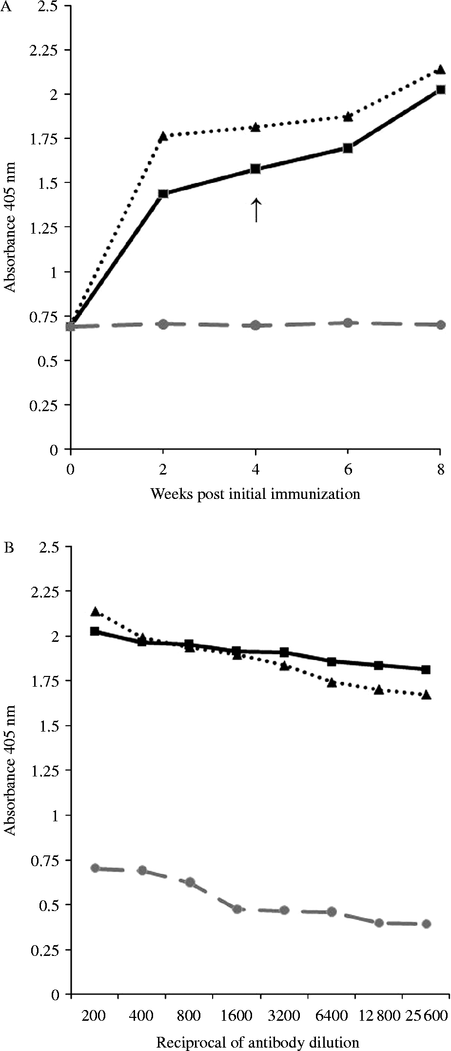
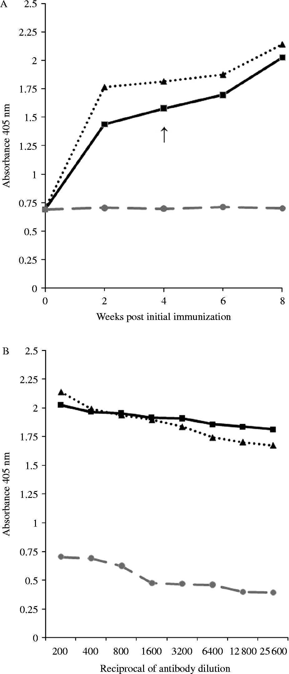
Reactivity of serum from vaccinated mice against selected phage clones was analysed by ELISA (fig. 1A). In vaccinated mice a high humoral response against the homologous phages was observed, showing an elevation after the first immunization until week 8 when they were sacrificed. The response was stronger against clone 1, followed by that to clone 20, the control group showed the lowest response. The titres of specific antibodies were tested by ELISA. Antibody titres in sera against phage clones of immunized mice, after the third immunization, were >1:25,600 at week 8, significantly higher than those of the control group (fig. 1B). Antibody produced by immunization with these phage clones did not react with E/S products in ELISA (data not shown).
(A) Serum IgG levels against selected phage clones from all groups during the experiment (1:200-diluted serum) and (B) collected after the third vaccination at week 8. Mice were given three immunizations at week 0, 2 and 4 with 2.5 × 10¹¹ phage particles without adjuvant. The time of challenge is indicated by the arrow. Results are the means of the ten animals run in duplicate. Group A, clone 1 (▲); group B, clone 20 (■) and group C, control ( ).
Film in situ zymography
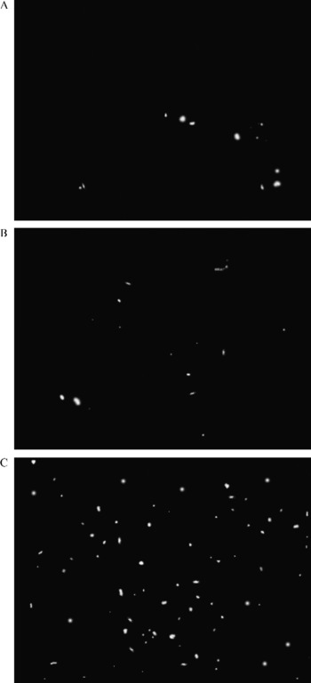
In situ zymography of liver sections demonstrated gelatinolytic activities, represented as unstained spots. Gelatin degradation from mice immunized with clones 1 (fig. 2A) and 20 (fig. 2B) was significantly decreased (80.64% and 72.58%, respectively), as compared to control group animals (P < 0.05) (fig. 2C).
Representative photomicrographs of film in situ zymography for detecting the proteolytic activity of Fasciola hepatica in liver sections collected from mice immunized with clone 1 (A), clone 20 (B) and control group animals (C). Strong gelatinolytic activity was visualized as a transparent pattern on a black background in the latter group, whereas in vaccinated animals low gelatinolytic activity was detected.

Discussion
Reports on the use of purified native or recombinant molecules have shown potential for protection against F. hepatica (Dalton et al., Reference Dalton, O'Neill, Stack, Collins, Walshe, Sekiya, Doyle, Mulcahy, Hoyle, Khaznadji, Moiré, Brennan, Mousley, Kreshchenko, Maule and Donnelly2003; Hillyer, Reference Hillyer2005; McManus & Dalton, Reference McManus and Dalton2006). Amoung these is cathepsin L, which induces significant reduction in both worm burden and egg output. Vaccinating with the epitope only, rather than with the entire organism or the isolated antigen, could be much safer, and may be as effective (De Groot, Reference De Groot2004). Random peptide phage libraries have been successfully used for the identification of immunogenic epitopes or mimotopes, and induce humoral immune responses in animals (Smith & Pretenko, 1997). Phage displaying peptides have several advantages as candidates in the development of vaccines: they are relatively easy and inexpensive to produce and therefore high quantities may be obtained without the necessity for complicated purification procedures.
Our results showed that clones 1 and 20 induced 45.83–66.67% reduction in worm burdens compared to the control group (P < 0.05). Moreover, mice immunized with phage developed high titres of anti-mimotope antibodies. This study confirms previous results obtained in our laboratories (Villa-Mancera et al., Reference Villa-Mancera, Quiroz-Romero, Correa, Ibarra, Reyes-Pérez, Reyes-Vivas, López-Velázquez, Gazarian, Gazarian and Alonso2008), on the protective efficacy of phage clones in sheep immunized without adjuvants and subsequently challenged with F. hepatica metacercariae. Immunological observations in mice, sheep and cattle with infections of F. hepatica metacercariae showed that the immune responses were predominantly Th2 type (Mulcahy et al., Reference Mulcahy, Joyce, Dalton and Dalton1999; O'Neill et al., Reference O'Neill, Brady, Callanan, Mills and Dalton2000; Hoyle et al., Reference Hoyle, Dalton, Chase-Topping and Taylor2002). On the other hand, in cattle vaccinated with cathepsin L a positive correlation was observed between the number of liver fluke recovered and high titres of IgG1 and IgG2, indicating that protection is associated with the induction of a Th1 or a mixed Th1/Th2 response (Mulcahy et al., Reference Mulcahy, O'Connor, McGonigle, Dowd, Clery, Andrews and Dalton1998).
Cathepsin L proteases appear to be involved in activities such as migration of this parasite through liver tissue, egg shedding, immune evasion and nutrition (reviewed by Dalton et al., Reference Dalton, O'Neill, Stack, Collins, Walshe, Sekiya, Doyle, Mulcahy, Hoyle, Khaznadji, Moiré, Brennan, Mousley, Kreshchenko, Maule and Donnelly2003). Two cathepsin L peptidases, cathepsin L1 and cathepsin L2, are the predominant molecules secreted by the parasite in the E/S products and together represent >80% of the protein content (Dowd et al., Reference Dowd, Smith, McGonigle and Dalton1994). In this trial, the cathepsin L activity may have been blocked by the induction of antibodies produced in the groups vaccinated with phage clones. The proteolytic activity of cathepsin L was evaluated with FIZ, which showed that the activity of gelatinase was reduced in these tissues. This result is in accordance with previous reports that detected a significant reduction of liver fluke proteolytic activity using FIZ after intraperitoneal administration of a cysteine protease inhibitor (Alcalá-Canto et al., Reference Alcalá-Canto, Ibarra-Velarde, Gracia-Mora and Sumano-López2005). In addition, reductions of egg count and viability were observed in mice treated with N-[N-(l-3-trans-carboxyoxirane-2-carbonyl)-l-leucyl]-agmatine (E-64) (Alcalá-Canto et al., Reference Alcalá-Canto, Ibarra-Velarde, Sumano-Lopez, Gracia-Mora and Alberti-Navarro2006).
The present study demonstrates that immunization of mice with mimotopes of cathepsins L induced a reduction in fluke burden, and that the proteolysis caused by liver fluke cathepsins in hepatic tissue was reduced after the vaccination with phage clones. The results in sheep and mice confirm that the technology of phage display could be applied to vaccine development against F. hepatica. In the future, field trials are required to evaluate these mimotopes under variable environmental and disease transmission conditions. Further studies focusing on identification of the cellular components will be necessary to explain the mechanism of immune protection of vaccinated animals.
Acknowledgements
This study was supported by the project PAPIIT- IN208703-03 DGAPA-UNAM, and the Consejo Nacional de Ciencia y Tecnología (CONACYT), México.