Introduction
Patulous eustachian tube is postulated to result from a longitudinal, concave defect at the anterolateral aspect of the mucosal valve superiorly.Reference Poe1 It can result in disabling autophony of one's own voice and breathing.
The treatment of patulous eustachian tube presents a challenge to the otolaryngologist. A variety of treatment options have been described, of varying degrees of invasiveness. However, there is currently no ‘gold standard’ treatment, and a proportion of patients fail to achieve therapeutic benefit from conventional treatments.
We describe a novel computed tomography (CT) guided treatment for a patient with patulous eustachian tube related autophony which was refractory to conventional treatment.
All procedures related to this report complied with the Helsinki Declaration of 1975, as revised in 2008.
Case report
A 36-year-old woman presented with a 7-month history of a constant muffled sensation within her right ear. She described associated echoing of her own voice (autophony) and impaired hearing. She also experienced an abnormal sensation of her heart beating. Her symptoms were worse on standing and after taking exercise. There was no history of current or previous ear infection or nasal obstruction. Her past medical history was unremarkable and, with the exception of the oral contraceptive pill, she took no regular medication.
On otoscopic examination, her right tympanic membrane did not move well with the Valsalva manoeuvre. Flexible nasendoscopy was normal, as was her pure tone audiogram. Patulous eustachian tube was suspected clinically, but a high resolution temporal bone CT scan was performed to exclude other pathology. This demonstrated a high-riding right jugular bulb but no dehiscence, and confirmed that the right superior semicircular canal roof was intact.
The patient underwent attempted reconstruction of the right eustachian tube valve with a submucosal silicone elastomer suspension implant (Vox, Uroplasty, Manchester, UK) inserted via a nasendoscopic approach under local anaesthesia. This resulted in partial relief of symptoms but these recurred one week later, probably due to the difficulty in placing biofiller at the lateral aspect of the eustachian tube meatus.
The patient was keen for further intervention. A repeated endoscopic procedure was considered but felt unlikely to be successful due to the patient's anatomy. Thus, external, trans-cutaneous insertion of a silicone elastomer suspension implant to the lateral aspect of the eustachian tube was attempted under CT guidance. The patient gave full, informed, written consent for this procedure, which risked occluding the eustachian tube, causing middle-ear effusion, and also carried a small risk of damage to a branch of the facial nerve.
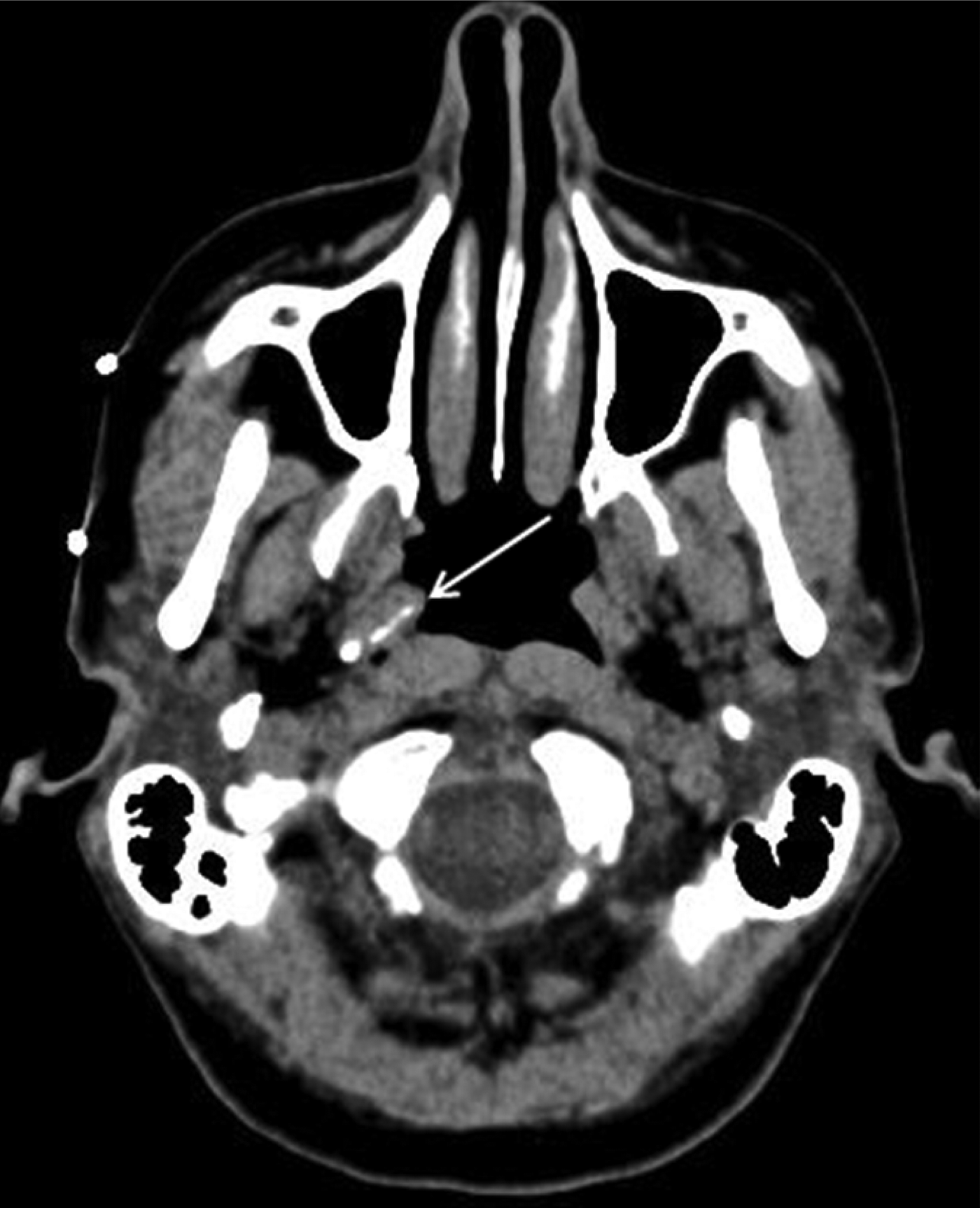
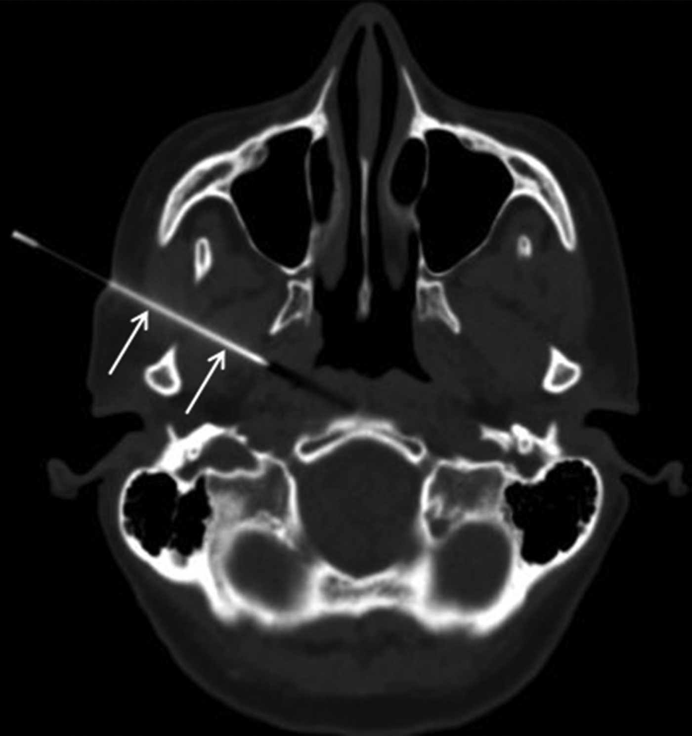
The procedure was carried out under joint radiological and otolaryngological care. The right eustachian tube was identified using a Siemens Somatom Sensation 16 multidetector row CT scanner (Siemens Medical Solutions, Erlangen, Germany) without the use of intravenous contrast medium (Figure 1). Using a radio-opaque skin marker (Figure 1), a site on the cheek which would allow access to the lateral aspect of the eustachian tube was selected, running the needle over the mandibular notch, just posterior to the lateral pterygoid plate. Using aseptic technique, 5 ml of lidocaine 1 per cent was administered to the subcutaneous tissues, then progressively introduced from the skin surface to the deep tissues around the right eustachian tube, using a 21 gauge needle. Under CT guidance, a 70 mm, 18 gauge needle (Medex, Smiths Medical, London, UK) was advanced until its tip was immediately lateral to the right eustachian tube (Figure 2). A 2 ml volume of silicone elastomer suspension implant (Vox, Uroplasty) was injected.
Axial, unenhanced computed tomography (CT) image taken prior to the CT-guided procedure, showing the high density silicone elastomer suspension implant (arrow) medial to the right eustachian tube, which was placed during previous endoscopy but failed to resolve the patient's patulous eustachian tube related symptoms. Radio-opaque skin markers are seen on the skin surface, placed to assist the planning of the subsequent intervention.

Axial, unenhanced computed tomography image showing the position of the 70 mm, 18 gauge needle tip (arrows) at the lateral aspect of the right eustachian tube.
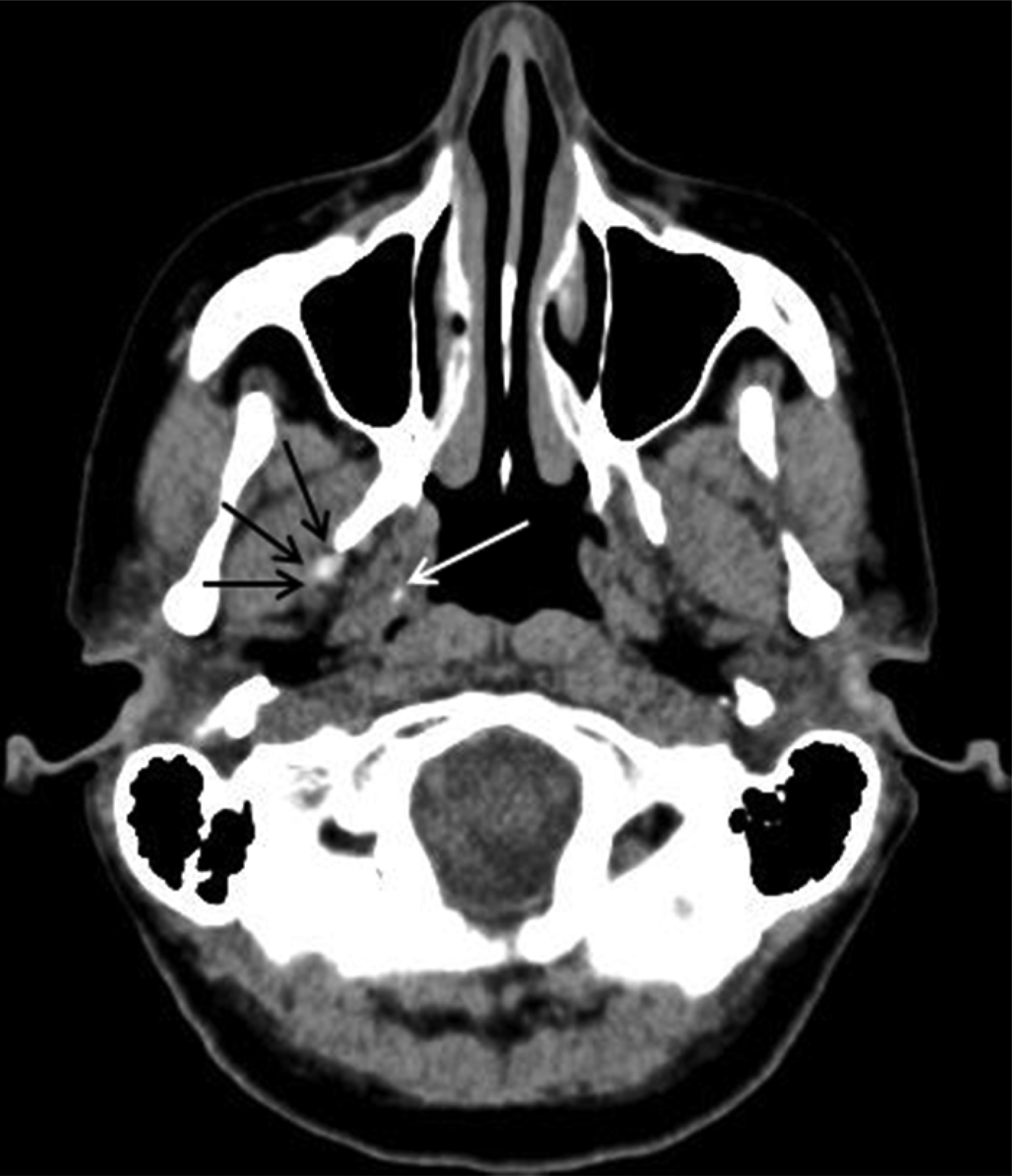
There were no immediate complications (Figure 3). The patient required simple oral analgesia for one week post-procedure. The procedure was successful in completely relieving the patient's patulous eustachian tube related autophony, and she remained asymptomatic at six-month follow up, at which point she was discharged from our care. The following extract is taken from a letter of thanks sent by the patient to the consultant otolaryngologist and radiologist.
I am no longer deafening myself when I speak or stopping mid-sentence because I am so unnerved by the sensation of it [i.e. the eustachian tube] opening and closing all the time. My head feels clearer, I don't spend half the day laying down or with my head between my knees to get some relief, it's fantastic.
Axial, unenhanced computed tomography (CT) image taken after the CT-guided procedure, showing the new high density silicone elastomer suspension implant, lateral to the right eustachian tube (black arrows). The previously placed silicone elastomer suspension implant is also discernible medial to the eustachian tube (white arrow).
Discussion
Patulous eustachian tube is postulated to result from a longitudinal, concave defect at the anterolateral aspect of the mucosal valve superiorly.Reference Poe1 It can result in disabling autophony of one's own voice and breathing. A high resolution CT scan of the temporal bones is commonly performed to investigate patients with autophony. This may detect other causes of autophony such as superior semicircular canal dehiscence, which can be readily treated with surgical plugging.Reference Crane, Lin, Minor and Carey2 Equally, it can lend weight to the diagnosis of patulous eustachian tube, if there is loss of soft tissue in the cartilaginous eustachian tube.Reference Grimmer and Poe3
Once diagnosed, there is much debate about the treatment of patulous eustachian tube.
Simple, conservative methods such as nasal instillation of saline can be effective in relieving symptoms, as Oshima et al. observed in 63.5 per cent of their 52 patients with patulous eustachian tube.Reference Oshima, Kikuchi, Kawase and Kobayashi4 Lower frequencies are preferentially transmitted in the patulous eustachian tube.Reference Bartlett, Pennings, Ho, Kirkpatrick, van Wijhe and Bance5 Mass loading of the eardrum can reduce vibrations at these frequencies, with consequent relief of symptoms, albeit temporary.Reference Bartlett, Pennings, Ho, Kirkpatrick, van Wijhe and Bance5 The relatively high proportion of patients who obtain no benefit from these conservative treatments usually resort to surgical intervention.
There are many potential surgical options. In a study of 14 ears affected by patulous eustachian tube, multilayer ligation (using a combination of fat plugging, endoluminal cauterisation and suture ligation, via a transnasal endoscopic approach) subjectively improved symptoms in 85.7 per cent.Reference Rotenberg, Busato and Agrawal6 Laser-assisted curvature inversion of the medial and lateral lamina of the eustachian tube improved voice autophony in 72.7 per cent of 11 patients studied.Reference Yañez, Pirrón and Mora7 Less invasive endoscopic techniques also exist, and focus on augmentation of the eustachian tube with a variety of different materials. Injection of the eustachian tube valve with autologous cartilage was reported to be effective in a case series of two patients.Reference Kong, Lee, Goh and Park8 In another study, of 14 patients, endoscopic injection of the eustachian tube with hydroxyapatite resulted in significant or complete symptom relief in 57 to 63 per cent.Reference Vaezeafshar, Turner, Li and Hwang9
• Patulous eustachian tube may result from a longitudinal, concave defect at the anterolateral aspect of the mucosal valve
• This condition can cause disabling autophony
• There is a variety of endoscopic treatment options but no current ‘gold standard’
• The presented patient obtained no benefit from conventional endoscopic treatment
• Subsequent trans-cutaneous, computed tomography guided placement of biofiller completely relieved her symptoms
There is no current gold standard treatment for patulous eustachian tube. The majority of the published literature consists of small case series, often with fewer than 20 patients. However, it is clear that, despite treatment, a cohort of patients is left with persistent, disabling patulous eustachian tube symptoms. We describe a novel, minimally invasive method of treating patulous eustachian tube in a patient with previous failed endoscopic biofiller augmentation. To the best of our knowledge, computed tomography guided, trans-cutaneous augmentation of the eustachian tube has not been previously described. Placing biofiller at the lateral aspect of the eustachian tube, which may be hard to access via the endoscopic route, makes anatomical sense, given that patulous eustachian tube is postulated to result from a longitudinal, concave defect at the anterolateral aspect of the mucosal valve superiorly.Reference Poe1 In our patient, it was unclear whether the lateral CT-guided injection alone resulted in the complete symptomatic relief observed, or whether this effect was aided by prior medial augmentation via an endoscopic approach.
Conclusion
Patulous eustachian tube can result in disabling symptoms. There are a variety of available treatments which can help relieve symptoms, often involving an endoscopic approach. However, in a significant minority of patients symptoms persist despite conventional treatment. Otolaryngologists should be aware that CT-guided, trans-cutaneous placement of silicone elastomer suspension implants lateral to the eustachian tube is minimally invasive and may result in complete relief of symptoms in patients who have obtained no benefit from conventional endoscopic treatment, as in our case.