Anomalous aortic origin of coronary artery is a rare congenital heart disease, especially interarterial course, which is the lethal type. The appropriate surgical options depend on individual anatomy. We presented a case of a young boy with presyncopal episode.
A case report
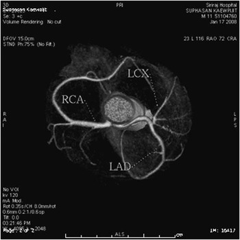
A 12-year-old Thai boy presented with presyncopal symptoms and chest pain that radiated to the neck upon exertion. Investigation showed myocardial ischaemic pattern, that is, ST depression in leads II, III, aVF, V2–V6 in electrocardiogram together with increased levels of cardiac troponin-T; 1.7 (normal 0–0.1) nanogram per milliletre and creatine kinase-MB; 74 (normal 0–4.9) Unit per litre. Echocardiogram and cardiac catheterisation revealed an anomaly, that is, the left main coronary artery that originated from the right aortic coronary sinus of valsalva took an interarterial course between the ascending aorta and the main pulmonary artery. There was neither ostial stenosis nor intramural course. Figure 1 shows the 64-slice computed tomography coronary angiography that the left main coronary artery was compressed by the ascending aorta and the main pulmonary artery, which resulted in luminal narrowing. The narrowest diameter of the left main coronary artery was 1.8 millilitre. There was no intramural course. There was no stenosis of the left anterior descending, left circumflex, and right coronary arteries.
64-slice computed tomography coronary angiography demonstrated a separate origin of the right coronary artery and an anomalous left main coronary artery, that is, an interarterial course originating from the right aortic sinus of valsalva that was compressed by the ascending aorta and the main pulmonary artery. LAD = left anterior descending; LCX = left circumflex; RCA = right coronary artery.

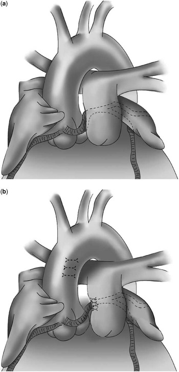
The patient underwent a surgical technique that increased the distance between the aorta and the main pulmonary artery, that is, mobilisation of the pulmonic root, pulmonary bifurcation, suspension of the right pulmonary artery to the left posterior aspect of the ascending aorta, and plication of the pulmonic root juxta to the left main coronary artery (Fig. 2). Intra-operative dobutamine stress echocardiogram revealed no regional wall abnormality and there was no ST–T change on electrocardiographic monitoring.
The (a) picture demonstrated an interarterial course of anomalous left main coronary artery that was compressed between the aorta and the main pulmonary artery. The (b) picture demonstrated a surgical technique that increased the distance between the aorta and the main pulmonary artery by mobilisation of the pulmonic root, pulmonary bifurcation towards the left lateral position, suspension of the right pulmonary artery to the ascending aorta, and plication of the main pulmonary artery juxta to the left main coronary artery.

Post-operative 99mTc-methoxyisobutylisonitrile stress perfusion scan and exercise stress test (BRUCE protocol) were performed at 3 and 9 months, respectively. The results showed no evidence of myocardial ischaemia. No cardiovascular problems were presented during the follow-up visit of 43 months.
Discussion
The prevalence of an anomalous left main coronary artery originating from the right aortic sinus of valsalva is 0.003–0.03% (1.2–7% of coronary anomalies).Reference Tuncer, Gokce and Sokmen1 Anomalous left main coronary artery is classified according to their pathways into five categories, that is, septal course, anterior to the aorta, posterior to the aorta, interarterial course – between the aorta and the pulmonary trunk – and mixed course.Reference Sherif, Zehr, Mookadam, Lorenz and Mookadam2 The interarterial course is considered a rare condition, but it is the most symptomatic and fatal variant with various clinical presentations, for example, sudden death, chest pain, syncope, presyncope, dyspnoea, palpitation, myocardial infarction, and asymptomatic in some cases.Reference Sherif, Zehr, Mookadam, Lorenz and Mookadam2 Coronary blood flow is compromised by several mechanisms, for example, compression of the intramural segment of the coronary artery, stenosis of the intramural segment of the coronary artery, a flap or slit-like orifice, narrowing of the lumen at the acute angle of take-off of the coronary artery, compression of the left main coronary artery by the aortic root and the pulmonary trunk, especially when they are dilated, and increased myocardial oxygen consumption during exercise.Reference Gavrielatos, Letsas, Pappas, Antonellis and Kadaras3–Reference Taylor, Rogan and Virmani5 Multi-slice computed tomography angiography can reveal the exact origin and course of anomalous coronary arteries. The procedure is the first-line imaging technique in the detection of coronary anomalies;Reference Kosar, Ergun, Ozturk and Kosar6 however, the diagnosis is most frequently made by echocardiography in children.
Surgery is indicated in symptomatic patients with anomalous left main coronary artery, but in asymptomatic patients surgery remains controversial.Reference Brothers, Gaynor, Paridon, Lorber and Jacobs7 There are several surgical options for anomalous left main coronary artery, for example the modified unroofed procedure that creates a new coronary ostium in the correct sinus, removes the intramural course, and also corrects the problem of the acute angle of take-off and small orifice of coronary artery. Other procedures include the coronary artery translocation and reimplantation, the coronary artery bypass grafting, the main pulmonary artery translocation to the left pulmonary artery – lateral pulmonary artery translocation – the coronary artery arterioplasty with autologous pericardial patch,Reference Jaggers and Pal8–Reference Gulati, Reddy and Culberson9 and the transcatheter stent placement.Reference Doorey, Pasquale, Lally, Mintz, Marshall and Ramos10 The appropriate surgical selection depends on the anomalous anatomy and the patient's status.
In our case, coronary artery translocation and reimplantation could not be performed because of inadequate tissue between the coronary arteries, as both the right coronary artery and left main coronary artery orifices were adjacent to each other and there was no intramural course to do the unroofed procedure. The patient was too young for the coronary artery bypass grafting procedure because of the possibility of long-term graft problem as a result of competitive blood flow from the native coronary artery, which could decrease the patency of the graft over time especially when vein graft was used. Finally, he underwent a surgical procedure, that is, mobilisation of the pulmonic root, pulmonary bifurcation towards the left lateral position away from the aorta, division of the ligamentum arteriosum, suspension of the right pulmonary artery to the left posterior aspect of the ascending aorta by placing surgical stitches, and plication of the pulmonic root juxta to the left main coronary artery, as shown in Figure 2. The whole procedure increased the distance between the aorta and the main pulmonary artery, which resolved the myocardial ischaemic problem by decreasing the compression on the left main coronary artery.
The advantages of this procedure were as follows: elimination of compression on the left main coronary artery from the aorta and the main pulmonary artery; lowering the risk of myocardial ischaemia during the procedure as it could be performed under a beating heart; no interruption of the aortic root, which might cause aortic insufficiency in the future; no dissection of the pulmonary artery, which may increase the risk of pulmonary artery stenosis; and less bleeding.
Post-operative computed tomography angiography revealed mild kinking of the left main coronary artery. There were no symptoms and no evidence of myocardial ischaemia from electrocardiography and stress tests at 3 and 9 months of follow-up. However, long-term follow-up is warranted.
Conclusion
A surgical procedure, consisting of mobilisation of the pulmonic root, pulmonary bifurcation, and plication of the pulmonic root juxta to the left main coronary artery, was a less-invasive and promising alternative option for the interarterial course of the left main coronary artery originating from the right aortic sinus of valsalva. This operative technique is useful in the case with absent intramural course and no coronary ostial stenosis.
Acknowledgement
We would like to thank the trainee officer of Suratthani hospital for contributing to draw the picture as shown in the figure 2.