INTRODUCTION
Gymnophallid digeneans are parasites of marine and coastal birds; they use marine bivalve molluscs as first intermediate hosts and bivalve or gastropod molluscs as second intermediate hosts. Szidat (1962) was first to record a gymnophallid metacercariae that parthenogenetically produced a subsequent generation of metacercariae. They were from the bivalve Mytilus edulis platensis D'orbigny, 1846 from the coastal waters of Argentina. Later James (1964) described the parthenogenetic metacercariae (the so-called germinal sacs) of Parvatrema homoeotecnumJames, 1964 from the periwinkle Littorina saxatilis (Olivi, 1792) on the Welsh coast of the British Isles and concluded that this mollusc was the only intermediate host for that parasite. This point of view has been called into question many times (Cable, 1965; Ching, 1982, 1995; Pearson, 1992). Ching (1982) described 2 successive generations of parthenogenetic metacercariae (M1 and M2) of Cercaria margaritensis in subtidal gastropods Margarites costalis (Gould, 1841), M. pupillus (Gould, 1841) and M. olivaceous (Brown, 1827) from British Columbia. She proposed that these molluscs were the second intermediate host.
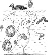
During the course of ongoing research into seabird parasite larvae in invertebrates in the coastal waters of the Barents Sea, a high prevalence of parthenogenetic metacercariae was recorded in the gastropod Margarites helicinus (Phipps, 1774) at one particular site. The bivalves Turtonia minuta (Fabricius, 1780) were collected at this region and some of them shed small gymnophallid cercariae that had reduced tail stems and furcae that were thick at their bases. It was shown that, under experimental conditions, these furcocercariae penetrated the extrapallial cavity of the gastropod M. helicinus, where they lost their tails and transformed into parthenogenetic metacercariae which were identical to those of Cercaria margaritensis. This lead to the proposal of a new life-cycle that was published in Russian (Galaktionov, 1996). That life-cycle, having now been subjected to a much more detailed examination, is presented as Fig. 1. For the present study, adults of the species concerned were obtained from laboratory-infected seabirds. A differential diagnosis confirmed that they were members of an independent species of Parvatrema Cable, 1953. That species is presented here as Parvatrema margaritense (Ching, 1982) n. comb. Its life-cycle supports the proposal of Ching (1982) that the first intermediate host is a bivalve and there are 2 (M1 and M2) generations of metacercariae in a second intermediate host which is a gastropod. Reproduction in the M1 and M2 stages has already been described by Irwin et al. (2003) but the other life-cycle stages will be dealt with in detail in this study. In addition, possible implications of this type of life-cycle for theories of early digenean evolution are proposed and discussed.

Fig. 1. Drawing summarizing the life-cycle of Parvatrema margaritenses. 1, Adult in the common eider (Somateria mollissima) intestine; 2, eggs with miracidia; 3, daughter sporocyst in the first intermediate host – the bivalve Turtonia minuta; 4, cercaria; 5–8, stages of development in the second intermediate host – gastropod Margarites helicinus; 5, young M1 (first parthenogenetic generation metacercaria); 6, fully formed M1 containing developing metacercariae of the second parthenogenetic generation (M2); 7, fully formed M2 containing metacercariae (M3); (8) invasive for the definitive host. (Based on Galaktionov, 1996.)
MATERIALS AND METHODS
Stages of the life-cycle of P. margaritense were studied using sporocysts and cercariae obtained from naturally infected Turtonia minuta (Fabricius, 1780) and metacercariae from naturally infected Margarites helicinus (Phipps, 1774). The molluscs were collected during 1995–2002 from the upper subtidal zone of the Barents Sea at Dal'nezelenetskaya Inlet, Kil'dinskaya Salma Inlet and the Ainovy and Kiiskiye Islands and from the Kandalaksha and Onega Bays on the White Sea.
Initial developmental stages of the parthenogenetic first generation (M1) metacercariae were studied using material obtained by experimentally infecting the second intermediate hosts, M. helicinus. To identify infected bivalves, T. minuta collected from the lower intertidal zone of small islands in the Dal'nezelenetskaya Inlet (69 °07′N, 36 °05′E, on the Barents Sea) were placed individually in small vessels with seawater and exposed for 1–2 h at 10 °C to natural daylight. The vessels were then viewed under a stereomicroscope and molluscs that had shed the P. margaritense cercariae were selected. The infected molluscs were placed in Petri dishes with seawater for 24 h together with individual infection-free Margarites helicinus. The latter were collected at a depth of 5–10 m at a point where the Dal'nezelenetskaya Inlet joins the sea. Dissection of 200 molluscs from this location demonstrated that they were free from trematode infection. Following experimental infection, the M. helicinus were transferred to small vessels with seawater and kept at 7 °C for 43 days. The molluscs were fed on thallus fragments of the kelp Laminaria saccharina (L.). The water in the vessels was regularly changed. Molluscs were dissected on days 1, 5, 14, 20, 30 and 43 post-infection (p.i.).
To assess daily production of cercariae, 10 infected T. minuta molluscs were placed in Petri dishes with seawater (1 mollusc per vessel) and kept at natural daylight (August 1999) at 10 °C for 24 h. The shed cercariae were counted. The experiment was repeated 3 times.
To obtain adults, 4 herring gull (Larus argentatus Pontoppidan, 1763) chicks and 2 eider (Somateria mollissima L.) ducklings caught at islands of the Dal'nezelenetskaya Inlet were experimentally infected. In the former case, 5 to 12-day-old chicks with juvenile plumage were used and, in the latter case, newly hatched eider ducklings that had not yet fed were used. Infection was performed by per os introduction, together with water, of 30–50 parthenogenetic M2 metacercariae that contained M3 larvae that are infective to birds. Dissection of birds was performed 2 and 5 days p.i.
Intramolluscan stages were obtained by dissecting naturally infected M. helicinus under a stereomicroscope. For each infected mollusc the numbers of M1 and M2 parthenogenetic metacercariae and their stages of maturity were recorded. The morphology of sporocysts, cercariae and metacercariae was studied in vivo using an Olympus CH40 microscope. Photographs were taken with a SC-35 Olympus photomicrography camera. Measurements were made on heat-killed specimens in seawater, under light cover-glass pressure. At least 20 specimens of daughter sporocysts, cercariae and metacercariae were measured. Adults obtained from experimentally infected eider chicks were fixed in 70% ethanol, stained with aluminous carmine, cleared in isobutyl alcohol and xylene, and mounted in Canada balsam. Fourteen adults mounted in this manner were measured and all measurements (length×width) presented in the paper are in micrometres, with the mean in parenthesis. The drawings were made with the aid of a camera lucida.
M2 and M3 individuals were fixed for scanning electron microscopy in 3% glutaraldehyde made up in filtered seawater. They were washed in distilled water before further fixation in 1% osmium tetroxide. Next they were dehydrated through an ascending series of ethanols, washed in hexamethyldisilazane before being allowed to dry in a fume hood. Finally they were mounted on ‘stubs’ with double-sided tape and sputter-coated with gold. They were viewed in a Jeol 840S scanning electron microscope.
RESULTS
Daughter sporocysts
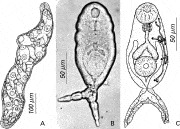
Daughter sporocysts are located in the haemolymph of the digestive gland, gills and gonad tissues of T. minuta. They are elongate worm-like organisms (250–800×70–180) (Fig. 2A). The birth pore is terminal at the anterior body end. Release of fully formed cercariae is via the birth pore in living sporocysts. The anterior end of each sporocyst is motile, and it can extend and retract. The body cavity of the sporocyst is filled with embryos and cercariae at different developmental stages. The germinal mass is located in the posterior end of the sporocyst.

Fig. 2. Drawings and light micrograph (phase contrast) of daughter sporocyst (A) and cercaria (B, C).
Infected T. minuta usually possess only 3–7 daughter sporocysts. The number of cercariae shed by each infected mollusc is also only 10–300 per 24 h.
Cercariae
Cercariae (Fig. 2B,C) are furcocercous, being 130–185 (148) long and 60–80 (73) wide at the mid-body. Their surface is covered with small spines decreasing in size from the anterior to the posterior. The tail stem is very short, 3–8 (5)×10–18 (13). The furcae are 30–50 (44) long and 15 wide at the base. Oral and ventral sucker diameters are 30–43 (35) and 35–45 (38) respectively. The pre-pharynx is not pronounced and the pharynx is more or less round at 12–20 (16)×15–20 (17). Gut caeca may reach the level of the middle of the ventral sucker. Glands in the cercarial body are not revealed during in vivo observations. The excretory formula is 2[(2+2)+(2+2)]=16. The branches of a V-shaped excretory bladder extend to the level of the middle of the oesophagus. The main collecting tube starts subterminally from the bladder branch. It proceeds forwards, then turns abruptly backwards, making a loop. There are 5–6 tufts of cilia inside the main collecting tube. A short unpaired caudal duct runs from the bladder before branching into 2 ducts that lead into each of the furcae. Proximal areas of these ducts, located at the furcal bases, are dilate. The distal parts open through an excretory pore located subterminally at the internal surface of the furcae. Sucker-like structures are present on the ends of the furcae.
Newly shed cercariae do not swim. They bring their furcae together and attach to the substrate by the sucker-like structures. The larval body rotates, as if scanning the surrounding space (as indicated on Fig. 1). The cercariae can crawl by alternating attachment to the substrate by the now adjacent ends of the furcae and the oral sucker. If a prospective second intermediate host, M. helicinus, comes close to a cercaria, it immediately detaches from the substrate, moves rapidly towards the gastropod and migrates into its extrapallial cavity. As a similar behavioural pattern was referred to as ‘ambuscade behaviour’ by Prokofiev (1994) and Galaktionov and Dobrovolskij (2003), who described it for a number of short-tailed cercariae such as Levinseniella brachysoma (Creplin, 1837) (Microphallidae) and Podocotyle atomon (Rudolphi, 1802) (Opecoelidae), the term ‘ambuscade behaviour’ is adopted in this study.
Metacercariae of the first parthenogenetic generation – M1
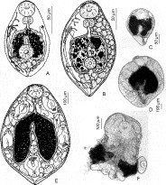
Inside the extrapallial cavity of Margarites helicinus, each cercaria drops its tail to become an M1 metacercaria. They feed on molluscan extrapallial fluid and desquamated pallial epithelium. Black pigment contained in the ingested cells of the pallial epithelium can already be seen in the gut of metacercariae shed by the molluscs 1 day post-infection (p.i.). M1 grow slowly, at least under experimental conditions. For the first 2 weeks p.i. their body size remains roughly the same as that of cercariae, the only visible change being an enlargement of the gut caeca. The reproductive system organs are not differentiated. The first germinal balls (embryos of the second metacercarial generation, the M2) appear 3–4 weeks p.i. at the posterior end of the M1 body (Fig. 3A). Later they became dispersed throughout the metacercarial body (Fig. 3B,C). These young M1 measure 190–320 (240)×120–220 (180). The oral sucker (38–63 (45)×45–52 (48)) and the pharynx (22–30 (26)×25–40 (33)) have increased in size, whereas the diameter of the ventral sucker remains the same as that of cercariae (30–38). Dorsally, along the edge of the oral sucker, 12–20 external pores from glandular ducts become noticeable. The excretory system also develops, the excretory formula becoming 2[(2+2+2)+(2+2)]=20. Branches of the excretory bladder reach the level of the middle of the oesophagus. The number of flame cells is increased, as is the number (8–12) of cilia tufts in the main collecting tube. As the number of M2 embryos increases inside the M1 the posterior end of its body enlarges. As a result, the ventral sucker, located in the posterior third of the body in cercariae and young M1, shifts forward so that it is finally located almost centrally in mature M1.

Fig. 3. Drawings and light micrographs of M1. (A) Young M1 with the first germinal balls at the posterior end of the body. (B, C) Young M1 with the germinal balls dispersed throughout the body. (D, E) Mature M1 with developing M2. (F) Old M1, having lost the posterior part of the body.
Since experimentally infected M1 did not reach maturity during the 43-day-long experiment, mature M1 were studied using material from naturally infected M. helicinus. Mature M1 are large oval organisms (Fig. 3D,E) with a widely varying size range being 450–1000 (790)×320–630 (530). The oral sucker diameter reaches 100–130 and the ventral sucker is 40–60. The pharynx is round with a 56–65 diameter. The gut caeca, filled with black pigment, occupy a considerable part of the M1 body. The rest of the body cavity is filled with numerous embryos and developing M2. Young M2 inside M1 are motile. Eventually they rupture the body wall at the posterior part of the mother organism and escape to start an independent existence in the extrapallial cavity of the molluscan host. Old M1, having lost the posterior part of their bodies, are occasionally observed. These individuals continue to function; they are motile and contain viable developing M2 (Fig. 3F).
Most infected M. helicinus, taken from populations at the Barents and the White Sea, contain only 1 M1 individual. However, some of the molluscs investigated contained 2–5 M1, these presumably being the result of multiple infections. A number, varying from 1–5 to 10–15, of young M2 that have recently left the mother organism are often observed alongside a seemingly intact mature M1 in the extrapallial cavity. No evidence of ruptures or perforations through which the first few M2 would have made their escape is seen in the M1 body wall. M1 that have lost the posterior portions of their bodies are observed only in M2 infrapopulations and, in these cases, some of the M2 contain M3 that are almost fully developed.
Metacercariae of the second parthenogenetic generation – M2
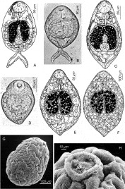
Initial stages of M2 development occur inside M1. M2 embryos that are transferred prematurely along with a mass of fully formed M2 into the extrapallial cavity of M. helicinus, do not survive. M2 pass through a furcocercarian stage during their development. Their tail structure (a short stem with broad furcae) is similar to that of the cercariae that develop inside daughter sporocysts (see above). The tail is eventually dropped, although it is sometimes still present in M2 that have taken up an independent existence in the molluscan host (Fig. 4A,B). In these cases, sucker-like structures, characteristic of the free-living cercariae of this species, are observed at the furcal ends. The gut caeca of the M2 that reside in the M1 brood sac are transparent. Dark pigment only appears in them following their release, i.e. when they start feeding on extrapallial fluid. M3 embryos appear inside very young M2 (for details see Irwin et al. 2003), filling almost the entire body cavity of individuals that are starting an independent existence.

Fig. 4. Progressive developmental stages of M2. (A, B) Young M2 that have taken up an independent existence in the molluscan host and retained or dropped their tails (C, D). (E) Older M2 with heterogeneous brood sac content, varying from embryonic balls to almost fully formed M3. (F) Mature M2 containing fully formed M3. SEM micrographs of mature M2 showing the entire individual (G) and the region of the oral sucker (H). OS, oral sucker.
The youngest M2 found in the molluscan extrapallial cavity measure 140–230 (180)×80–150 (120). They are motile oval organisms, the brood sacs of which contain embryonic balls and M2 embryos at early developmental stages (Fig. 4C,D). The oral sucker, 37–43 in diameter, is larger than the ventral (25–30). There is no pre-pharynx and the pharynx is 15–30 (26)×13–28 (24). Large gut caeca have thickened walls and are filled with black contents. Glandular cells, the ducts of which circumflex the oral sucker and open dorsally at its anterior edge, are found in the anterior end of the body, approximately at pharyneal level. The excretory formula of the youngest M2 is 2[(2+2)+(2+2)]=16, becoming 2[(2+2+2)+(2+2)]=20 in more advanced individuals. The number of the cilia tufts in the loop of the main conducting duct varies from 6 to 12. The excretory bladder branches extend to the level of the middle of the oesophagus.
In larger developing M2 the content of the brood sac is heterogeneous, varying from embryonic balls to almost fully formed M3 (Fig. 4E). The body of these M2 is more rounded than it was in the previous developmental stage (see above), reaching 220–510 (340)×200–370 (250). The size of the oral sucker is increased significantly (40–78 (63)), whereas the ventral sucker remains relatively small (28–42 (34)). The pharynx is now 28–50 (41)×23–50 (38). The digestive system appears to be active as the gut caeca have also increased in size and are filled with black, brown, orange and yellow granular contents in which crystalloid inclusions can be discerned. Glands are no longer visible. Due to closely packed embryos in the body cavity of these parthenogenetic metacercariae, it is impossible to determine the excretory formula.
Mature M2 containing fully formed M3 are less motile than younger individuals but they continue to feed actively (Fig. 4F). Their size varies greatly (430–1400×370–960), correlating with the number of M3 found in the brood sac. The number of these larvae in small mature M2 is 40–80, reaching 200–250 in the largest M2. The SEM confirmed that these organisms are sac-like and, due to their relatively thin body walls (see Irwin et al. (2003) for details) and perhaps as a result of the fixation process, their surface follows the contours of the developing M3 inside (Fig. 4G). It also showed that the organisms are covered by small spines that are arranged in numerous rows around the body. The vast majority of the spines occur in pairs, and frequently each pair is composed of individuals with adjacent parallel sides and lateral contoured sides. The rows of spines extend anteriorly into the raised folds of the oral sucker (Fig. 4H). In some individuals, the oral sucker appears to be filled by a plug of ill-defined material that could be of host origin. The ventral sucker is poorly developed, making it difficult to identify amongst the contours of the underlying developing M3. Light microscopy confirmed that the ventral sucker size (37–42) remains almost the same as that of younger M2 whereas the oral sucker of mature M2 reaches 75–133 (100) in diameter. At light level all parts of the digestive system are distinct. The pharynx is 42–73 (60)×40–65 (52), large gut caeca fill a considerable part of the M2 body cavity. The contents of the caeca have the same appearance as those in younger individuals.
Infected M. helicinus usually contain 10–20 M2, but this number may reach 50–60 in large specimens (shell height 4–6 mm). The composition of M2 groups is heterogeneous. In the youngest M2 infrapopulations M1 can still be observed (see above). More advanced populations contain 1–5 large M2, the others present being much smaller. In such groups it is quite apparent that the larger M2 contain the more advanced M3 in their body cavities. It would appear that these individuals were the first M2 to leave M1 and therefore the first to start an independent existence in the gastropod host.
The size structure in mature groups of M2 remains heterogenous and in these groups each M2, regardless of its size, contains only fully formed M3. Observations would suggest that there are competitive interactions between the M2 individuals in an infrapopulation that suppress the growth of those M2 that were last to leave the M1. Although development of M3 inside small M2 individuals proceeds normally, their M3 are fewer and smaller than those in M2 individuals that were shed from an M1 before them. Similarly, evidence exists to suggest that the life-span of M2 inside the gastropod host is limited. Dead M2 were often observed embedded in calcium salts in the ostracum of the host's shell. Their positions in the ostracum indicated that the largest individuals were the first to die. Rounded bulges of the ostracum covering dead M2 were often observed during the examination of non-infected M. helicinus shells. This would support the contention that the life-span of M2 generations is less than the life-span of their gastropod hosts.
During the duration of an infection of M. helicinus by P. margaritense, the M2 infrapopulation changes its location inside the gastropod. As a rule, mature groups tend to concentrate in the area of the shell apex. These M2 feed on debris accumulated there and the contents of their gut ceaca acquire different shades of brown, yellow and orange.
Infective metacercariae – M3
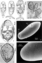
M3 development takes place entirely inside M2. In embryos with a differentiated tail primordium and the excretory formula 2[(1+1)+(1)]=6, primordia of the oral and ventral sucker and those of the digestive system organs are already clearly visible (Fig. 5A,D). Proximal parts of the main collecting tubes are expanded and form a loop, whereas the distal ones open through excretory pores located at the tips of the developing furcae of the tail primordium. In more advanced embryos with the excretory formula 2[(2+1)+(1+1)]=10, all primordia are developed further (Fig. 5B). The lumina in the primordia of the gut caeca, the oesophagus and the pharynx become visible; the walls of the latter develop muscular bands. The loop in the proximal part of the main collecting tubes becomes more distinct than in the previous developmental stage. The component parts of the main collecting tubes in the body-tail junction area merge. A duct is formed, the proximal part of which (located in the larval body) will eventually become the non-paired part of the bladder. Its distal region (located in the tail) will eventually give rise to the non-paired area of the caudal duct.

Fig. 5. M3. Successive stages (A–C) of M3 development. (D) M3 embryos at various developmental stages liberated from an M2. Drawing (E) and light micrograph (F) of a fully formed M3. SEM image of a fully formed M3 (G) and the anterior end (dorsal side) of its body (H). C, caeca; EB, branches of the excretory bladder; OS, oral sucker; VS, ventral sucker.
At the stage with the excretory formula 2[(2+2)+(2+1)]=14, the oral and ventral suckers are, in fact, fully formed (Fig. 5C). The lumina in the digestive system organs and the musculature of the pharynx are clearly visible. Groups of large glandular cells are found on either side of the pharynx. Primordia of the testes and the ovary appear. The common primordium of the genital atrium and the distal parts of the reproductive system are situated to the anterior of the ventral sucker. The first indication of material that will form the ducts of the female reproductive system has the appearance of a string of cells that extends from the primordial ovary around the posterior of the ventral sucker. The excretory bladder becomes V-shaped. Its branches arise as modifications of the embryo's main collecting tubes situated between the merged parts and the proximal loop. The area of the contraflexure and the descending parts of the loops become definitive main collecting tubes. Tufts of cilia appear in the lumens of these. The tail remains underdeveloped, having a short stem similar to tails of cercariae inside sporocysts in T. minuta. It is dropped in the later stages of the M3 morphogenesis.
Fully-formed M3 are small (115–150 (130)×60–93 (76)) ovoid organisms (Figs 5E,F) and this is confirmed by SEM studies (Fig. 5G). There are a number of well-developed sensory papillae situated in a regular pattern around both the oral and ventral suckers. The oral sucker (30–45 (38) in diameter) is approximately 1·3 times larger than the ventral sucker (27–35 (30) in diameter). The tegument is covered with alternating rows of conical spines (Fig. 5H). There is no pre-pharynx. A spherical pharynx, 13–20 (17) in diameter, leads into a short oesophagus from which the gut caeca originate. The bifurcation of the gut lies slightly anterior of the middle of the body. In the anterior end of the body, approximately at the level of the pharynx, 2 paired groups of glands are situated symmetrically on either side of the oesophagus. There are 5 cells in each of the lateral groups and 2 cells in each of the median groups. The ducts of each group of glands circumflex the oral sucker dorsally and open at its anterior edge. The excretory formula is 2[(2+2)+(2+2)]=16. The excretory bladder is V-shaped and its branches extend forward to the level of the posterior edge of the pharynx. There are 6–10 tufts of cilia along the length of each main collecting tube.
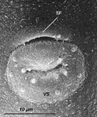
Although the reproductive system of metacercariae appears to be fully formed, there is no evidence that it is functional. The testes lie at the level of the posterior half of the ventral sucker. Vas efferens from each of the testes proceed forward to merge at the level of the anterior end of the ventral sucker to form a short vas deferens that leads to an undivided seminal vesicle. A short ejaculatory duct runs from the seminal vesicle into the genital atrium. An unpaired ovary is situated anteriorly to the right or the left testicle roughly at the level of the middle of the ventral sucker. The oviduct circumflexes the ventral sucker posteriorly and it is joined at its distal end by the seminal receptacle and the common vitelloduct. The vitelloducts, which run from the paired compact vitelline glands lying posteriorly to the ventral sucker, merge to form the common vitelloduct. The common vitelloduct is relatively thick and yolk drops can be seen in its lumen. A short oötype is connected to the uterus which proceeds forward along the edge of the ventral sucker and then forms a lateral loop before discharging into the genital atrium. The latter is tubular, short and opens through a slit-like genital pore which is elongated in transverse section. This is located close to the anterior edge of ventral sucker and its size depends on the state of contraction of the worm. SEM studies show that the genital pore is protected by an overhanging transverse fold of the ventral tegument (Fig. 6).

Fig. 6. SEM image of the M3 ventral surface in the region of the ventral sucker. TF, transverse fold of the ventral tegument; VS, ventral sucker.
Adult
No adults of P. margaritense were found in any of the herring gull chicks dissected in the experiment. In the eider duckling dissected 2 days p.i. numerous young worms were observed with single eggs in the uterus. Fully formed adults were obtained from the first third of the gut of the eider duckling dissected 5 days p.i.
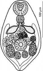
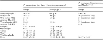
The adults are small and oval (Fig. 7, Table 1). A pre-pharynx is absent. An ovoid pharynx is well developed. Relatively small gut caeca arise from a short oesophagus approximately at the level of the posterior border of the first third of the body. The excretory bladder is V-shaped and its branches reach pharyngeal level. The structure of the reproductive system is essentially the same as in the metacercariae. The testes are oval and lie in the posterior third of the body laterally to the ventral sucker. Anteriorly to the latter the seminal vesicle filled with sperm is located. Posteriorly, between the testes, paired vitelline glands are observed. The position of the ovary varies: it may be located anteriorly to the right or to the left testis. The slit-like genital pore opens at the anterior margin of the ventral sucker. Loops of the uterus occupy the posterior two thirds of the worm's body and may reach into the forebody to the level of the pharynx. Eggs are small numbering 41–108 (87) per worm.

Fig. 7. Adult of Parvatrema margaritense (Ching, 1982) n. comb.

Adults are deposited in the Helminthological Collection of the Zoological Institute RAS (St Petersburg, Russia).
Differential diagnosis
Chubrik (1966) discovered in the bivalves Turtonia minuta from the Barents Sea, sporocysts with cercariae that she identified as Cercaria dichotoma Pelseneer, 1906. These cercariae are very different from the larvae that we have studied in a number of characters, the most significant being a long tail stem (50) which is almost as long as the furcae (70) (Chubrik, 1966). C. dichotoma-infected molluscs have often been observed in the course of our research. Observations upon the cercariae shed by the molluscs showed that they swim actively with the aid of their furcae and never penetrate Margarites helicinus.
In the descriptions of gymnophallid cercariae available in the literature we have not found any mention of larvae with a reduced tail stem, furcae broad at the basis and sucker-like structures at the furcal ends. Ambuscade behaviour has never been noted in gymnophallid cercariae, either. All the above makes us think that the cercaria described in the present paper has never previously been recorded.
At the same time, an almost non-existent tail stem and furcae broad at the basis have been noted in the furcate forms of Cercaria margaritensis from the large germinal sacs (M2, following the terminology of the present paper) in the molluscs Margarites spp. from British Columbia (Ching, 1982). Morphological and morphometric characters of these furcate forms are almost identical to those given above both for the cercariae from T. minuta and for the analogous developmental stage of M3 in the M2 in M. helicinus. The same holds true for the description of germinal sacs presented by Ching (1982). They correspond to various stages of M2 development: small germinal sacs are young M2 with germinal balls; medium and large germinal sacs are developing M2 with a varying number of M3 in them. The forms described by Ching (1982) and those investigated by us are clearly the same species.
Bearing in mind the distinguishing features defined by James (1964) and Scholz (2002), the combination of the morphological characters of the metacercariae and adults of our species indicate that it belongs to Parvatrema Cable, 1953. The most similar representative of Parvatrema appears to be P. ovoplenum (Jameson and Nicoll, 1913), which was described from the intestine of the scoter duck (Melanitta nigra L.). A comparison of the morphometric details of the two species is provided in Table 1 and it shows that the BL[ratio ]OS and OS[ratio ]VS ratios of both species are very similar but the body of P. ovoplenum is much smaller and has slightly larger eggs. Like the species described in this study, P. homoeotecnumJames, 1964 also has parthenogenetic metacercariae (termed germinal sacs by James, 1964) in its life-cycle. However, it differs substantially from P. margaritense by the facts that its BL[ratio ]OS and OS[ratio ]VS ratios are 11[ratio ]5 and 3[ratio ]1 respectively and its surface spines are more well developed. In addition early developmental stages of P. homoeotecnum M2 (termed daughter germinal sacs by James, 1964) and M3 (termed cercariae by James, 1964) bore tails with a distinctive stem which was of a similar length to that of the furcae.
The above differential diagnosis confirms that P. margaritense (Ching, 1982) n. comb. described in the present article is an independent species. Its life-cycle is realized in the coastal waters of Holartic seas. Upper-subtidal molluscs act as intermediate hosts. The sporocysts develop in the bivalve Turtonia minuta, which has a circumboreal distribution. The cercariae invade gastropods such as Margarites helicinus, M. costalis, M. pupillus and M. olivaceous inside which 2 generations of parthenogenetic metacercariae (M1 and M2) successively develop. Metacercariae (M3) formed in M2 are infective for the definitive hosts, the eider and, apparently, other species of marine diving ducks. Adults of P. margaritense have been found in the intestine of the Steller's eiders (Polysticta stelleri Pallas, 1769) caught in the Varanger-fjord of the Barents Sea (Galaktionov and Bustnes, unpublished data).
DISCUSSION
Several species of gymnophallids that possess parthenogenetic metacercariae in their life-cycles are currently known. Among them are Parvatrema homoeotecnumJames, 1964, P. margaritenese (Ching, 1982) n. comb., Cercaria quadriramisChubrik, 1966 from Littorina spp. from the Barents Sea (Chubrik, 1966; Podlipaev, 1979) and Gymnophallus australisSzidat, 1962 from Mytilus edulis platensis (see Szidat, 1962). Tsimbaljuk, Kulikov and Baranova (1978) recorded Parvatrema homoeotecnum (?) forma A from Collisella cassis (Eschscholtz, 1833) and P. homoeotecnum (?) forma B from Falsicingula kurilensis (Pilsbry, 1905) (Sea of Okhotsk). Tsimbaljuk et al. (1978) emphasized that their species identifications were only ‘presumed’ and analysis of the morphological descriptions quoted in their article indicate that P. homoeotecnum (?) forma A and P. homoeotecnum (?) forma B are 2 separate species that are distinct from P. homoeotecnumJames, 1964. Gymnophallid parthenogenetic metacercariae were also found in Epheria vincta (Montagu, 1803) and E. porrecta (Carpenter, 1864) (Sea of Okhotsk) (Galaktionov, unpublished observations). It seems very likely that the life-cycles of all these species are trixenous and follow a similar scenario to the one described here for P. margaritenese.
Recently, the parthenogenetic metacercariae (PM) of a gymnophallid species were recorded in the intertidal gastropods Falsicingula spp. on the coast of the Sakhalin and Kuril islands (Galaktionov et al. 2004, a detailed description is in preparation). This species appears to differ distinctly from the others. Rather than producing M2 or M3, the PM of this species produced typical gymnophallid furcocerous cercariae. The furcocercariae were shed from their gastropod hosts, actively swam in water and penetrated new individuals of Falsicingula spp. On contacting the molluscs, they dropped their tails and migrated into the extrapallial cavity. The fate of these larvae remains unclear. Perhaps they give rise to new parthenogenetic metacercariae or alternatively develop into metacercariae infective for the definitive host. However, we have found neither gymnophallid metacercariae with a developing hermaphrodite reproductive system (as in ‘ordinary’ gymnophallids) nor PM containing metacercariae (like the M2 of P. homoeotecnum and P. margaritense) in dissected Falsicingula spp. Possibly, this gymnophallid's infective metacercariae develop from furcocercariae inside PM. This process may be seasonally dependent, as in the case of sporocysts of Gymnophallus choledochus Odhner, 1900 (see Loos-Frank, 1969).
It is clear that gymnophallids with parthenogenetic metacercariae in their life-cycles are widespread in coastal ecosystems of very different geographical regions. The transmission success of these parasites can be attributed to their acquisition of additional life-cycle stages (parthenogenetic metacercariae) for clonal multiplication of infective stages. This is especially important for parasites using small littoral bivalves as first intermediate hosts. For instance, as noted above, a single P. margaritense-infected Turtonia minuta mollusc (shell length, 1–3 mm) sheds only 10–300 cercariae a day. However, if 1 cercaria infects Margarites helicinus, it gives rise to about 1500–2500 invasive metacercariae (clones) as a result of 2 successive stages of parthenogenetic reproduction.
It may be concluded that, when compared to other trematodes, gymnophallids have made a significant evolutionary advance by colonizing and multiplying in a second intermediate host. The latter have started to play a role in the gymnophallid life-cycle that is similar to the one played by the first intermediate host, i.e. multiple replication. Irwin et al. (2003) suggested that a possible prerequisite for the formation of this adaptation was the fact that gymnophallid metacercariae do not encyst and, unlike most digenean metacercariae, are able to make use of their oral suckers and gut caeca to actively ingest and digest their molluscan host tissues. This contrasts with the majority of digenean metacercariae that depend on their molluscan hosts for provision of their energy resources which are acquired via their teguments. The period during which gymnophallid metacercariae may feed on their second intermediate host's tissue is not limited and that could be a reason why they have adopted parthenogenetic reproduction.
The presence of parthenogenetic metacercariae of P. margaritense in the extrapallial cavity of M. helicinus does not result in parasitic castration. All the infected molluscs that we investigated had normally developed gonads with germinal products. Ching (1982) noted that when large numbers of M2 were present, they caused increased amounts of extrapallial fluid to collect between the shell and body of snail and only occasionally was other damage to the molluscan host tissues observed. The presence of the gymnophallid larvae does not appear to cause the mollusc any serious harm and its life-span is not reduced. This contention is supported by the fact that the molluscs can survive an infection episode by P. margaritense and this was confirmed in the present study by the observation of rounded bulges enclosing dead M2 in the ostracum of shells of non-infected individuals. The evidence all points to the conclusion that P. margaritense metacercariae can be more accurately described as commensals rather than parasites.
It has been conjectured that, in the early stages of digenean evolution, the first intimate relationship between free-living turbellaria-like proto-digeneans and molluscs took the form of commensalism (Cable, 1965, 1974; Pearson, 1972; Ginetsinskaya, 1968; Rohde, 1971, 1994; Gibson, 1987; Gibson and Bray, 1994; Galaktionov and Dobrovolskij, 2003). According to this hypothesis, proto-digeneans adopted commensalism in the extrapallial cavities of molluscs before becoming true tissue parasites. This is exactly the mode of life adopted by the parthenogenetic metacercariae of P. margaritense and the gymnophallid species from Falsicingula spp. (see above). They are essentially commensal in the extrapallial cavity and feed by ingestion of extrapallial fluid. This method of feeding provides these larvae with enough energy resources for early maturation and a rapid passage to parthenogenesis. This mode of life is accompanied by a distinct retardation and eventual cessation of reproductive organ formation (Irwin et al. 2003). The individuals of the next generation develop from germinal cells that appear to originate from the undifferentiated cells of the metacercarial genital primordium. In the course of M2 morphogenesis, some traces of retardation can be observed in the development of some somatic organs. The tail does not reach the developmental stage characteristic of cercariae formed inside sporocysts, the ventral sucker also remains underdeveloped. As a result, the structure of the parthenogenetic metacercariae develops similarities to the organization seen in rediae. Significantly, rediae are considered to be morphologically more archaic than sporocysts (for review see: Galaktionov and Dobrovolskij, 2003).
It should be noted that, whereas the development of P. margaritense M1 and M2 is not accompanied by differentiation of the reproductive system organs, this is not the case in the morphogenesis of the M3. It is only in the M3 that a distinct heterochrony is displayed in that, by the stage when developing individuals have suckers, tails and excretory systems equating to those of fully developed cercariae, the primordia of the ovary and the testes, as well as other parts of the reproductive system, are already developed.
When all the species of gymnophallid parthenogenetic metacercariae are considered, it is apparent that they include species that demonstrate many of the successive stages of transition to tissue parasitism that have been postulated for proto-digeneans. The parthenogenetic metacercariae of P. margaritense and the gymnophallid species from Falsicingula spp. are commensals in the extrapallial fluid of their gastropod hosts. M1 and M2 of Cercaria quadriramis are parasitic in seminal vesicles of Littorina spp. These cercariae probably penetrate the mollusc's male reproductive ducts from the extrapallial cavity via the genital pore. Finally Parvatrema homoeotecnum M1 localize in female Littorina saxatilis gonoducts and the M2 penetrate the haemocoelic space of the digestive gland and gonad, destroying these organs and feeding on tissue debris (James, 1964, 1965). Thus, the parthenogenetic metacercariae of P. homoeotecnum can be considered as true tissue parasites in exactly the same way as can digenean rediae.
Some authors (Heyneman, 1960; Cable, 1965; Ginetsinskaya, 1968; Pearson, 1972) have suggested that larvae of proto-trematodes, rather than reproductively mature forms, were first to parasitize gastropods. James and Bowers (1967) and Galaktionov and Dobrovolskij (2003) contend, on the other hand, that it was reproductively mature proto-digeneans that took the first step to parasitism. Significant to this argument is the fact that the organization of digenean rediae and sporocysts is characterized by the presence of a distinct structure, the germinal mass, in which the reproduction and ‘maturation’ of reproductive cells and initial stages of embryonic development proceed. (It is suggested that the germinal mass was formed on the basis of the ovary (germarium) as it retains some features in common with the gonad).
If the relationship between gymnophallid parthenogenic metacercariae and their gastropod hosts parallels the evolution into parasitism by proto-digeneans, one might expect that they would have gone through a similar developmental stage. This is not the case. No equivalent to a germinal mass has been observed in parthenogenetic metacercariae of gymnophallids. In these organisms it is the larva (cercaria) that penetrates the gastropod and it possesses only an undifferentiated genital primordium. That is why there is no morphological basis for the formation of a germinal mass-like structure. Irwin et al. (2003) demonstrated that in P. margaritense, as progenesis occurs, undifferentiated cells in the genital primordium proliferate before dispersing throughout the body of the developing metacercaria and giving rise to embryos of the next generation.
One can assume that the state of the reproductive system of gymnophallid parthenogenic metacercariae (devoid of germinal mass or any gonad-like structure) and rediae/sporocysts (possessing a germinal mass) illustrates that distinctly different developmental stages acquired the ability to reproduce parthenogenetically. In gymnophallids the cercaria made use of its undifferentiated genital primordium whereas proto-digeneans, probably having already developed gonads, transformed the ovary into the germinal mass in the course of evolution. Thus, the absence of a germinal mass in gymnophallid parthenogenetic metacercariae supports the contention of James and Bowers (1967) and Galaktionov and Dobrovolskij (2003) that adult proto-digeneans, rather than larval forms, were first to parasitize gastropods.
The authors wish to acknowledge support from INTAS (project 01-210) and a RFBR grant (N 04-04-49439) and to thank Dr D. I. Gibson (The Natural History Museum, London) for his valuable advice on taxonomy and the two anonymous referees for their constructive criticism and valuable comments.