Introduction
Most epidemiological studies of the incidence and prevalence of dementia report that Alzheimer's disease (AD) is the most common dementia, with vascular dementia (VaD) in second place.Reference Lobo, Launer and Fratiglioni1–Reference Jellinger4 However, prospective pathological-based studies support the view that dementia due to both vascular-based pathology and AD pathology (mixed dementia) is in fact a far more common underlying cause for dementia in elderly people than indicated by the figures based upon epidemiological evidence.Reference Snowdon, Greiner and Mortimer5–14
Early clinicopathological studies,Reference Erkinjuntti, Halatia, Palo, Sulkava and Paetau15, Reference Esiri, Wilcock and Morris16 later confirmed by neuroimaging, identified cortical and subcortical infarcts, assumed to be the result of small diameter blood vessel (arteriole) disease (SVD), were important factors in dementia. More recent evidence has shown that large vessel atherosclerosis could also be important in the aetiology of these changes in mainly elderly people.Reference Markus17–Reference Pantoni19 Intracranial atherosclerosis (involving large diameter arteries) was regarded as the most likely explanation for multi-infarct dementiaReference Hachinski, Lassen and Marshall20 and recently intracranial atherosclerosis has been identified as a risk factor for dementia that is independent of cerebral infarction and other risk factors, such as peripheral atherosclerosis, systemic hypertension and age.Reference Dolan, Crain, Troncoso, Resnick, Zonderman and O'Brien18, Reference Hachinski, Lassen and Marshall20–Reference Troncosco, Zonderman, Resnick, Crain, Pletnikova and O'Brien23 Vascular pathology, including SVD, co-existing in the same brain as individuals with only moderate AD pathology, unexpectedly increased the severity of dementia, raising the possibility of a common set of risk factors shared by VaD and AD.Reference Snowdon, Greiner and Mortimer5, Reference Esiri, Nagy, Smith, Barnetson and Smith25, Reference Petrovitch, Ross, Steinhorn, Abbott, Markesbery, Davis, Nelson, Hardman, Masaki, Vogt, Launer and White26 White matter lesions detected by imaging have also been shown to have a strong influence on progression of cognitive decline and are regarded a further surrogate marker of SVD.Reference Fazekas, Kleinert, Offenbacher, Schmidt, Kleinert, Payer, Radner and Lechner27–Reference Patel and Markus32
Despite these detailed clinicopathological studies, there remains a lack of accepted neuropathological criteria to validate clinically identified VaD.Reference Gold, Giannakopoulos, Herrmann, Bouras and Kovari33, Reference Wiederkehr, Simard, Fortin and van Reekum34 Similar problems have been encountered in the interpretation of neuroimaging, and the relationship between radiological images and neuropathological findings remain undecided, adding further uncertainty to the efforts to provide an agreed diagnostic criteria (clinical and pathological) to assist with the diagnosis of VaD.Reference Bronge and Wahlund35
This review will outline the evidence collected from pathological studies showing that both large and small diameter blood vessels contribute to VaD, not only through focal brain infarction, but as the result of microbleeds, venous changes and also through the endothelium expressing inflammatory cytokines and neurotoxic proteins that cross a damaged blood–brain barrier (BBB) to activate adjacent microglia.Reference Pantoni19, Reference Pantoni and Gorelick36 The emerging concept of injury to the neurovascular unit (endothelium, BBB and neuron) will be discussed with reference to potential biomarkers of SVD pathology in AD and VaD.Reference Hassan, Hunt, O'Sullivan, Parmar, Bamford, Briley, Brown, Thomas and Markus37–Reference Grammas39 Furthermore, many of the risk factors associated with ischaemic vascular pathology are reversible, making the cerebral circulation an important new target for the treatment and prevention of dementia of both AD and VaD varieties.Reference Marchesi40
Clinicopathological studies rediscover the importance of vascular pathology in dementia
Post-mortem examination of elderly individuals with dementiaReference Tomlinson, Blessed and Roth41, Reference Tomlinson, Blessed and Roth42 found that a significant proportion of cases were the result of a combination of both AD pathology (neurodegeneration) and arteriosclerosis (ischaemic pathology), hence the probability that multiple pathological processes, rather than a single pathology, were responsible for the cognitive decline.Reference Hachinski, Lassen and Marshall20
Hachinski et al. did not regard cerebral atherosclerosis as an explanation for mental deterioration in elderly people because it did not produce ‘a relentless strangulation of the brain's blood supply’ as previously considered.Reference Hachinski, Lassen and Marshall20 In his opinion, both small and large acute ischaemic cortical infarcts contributed to the entity known as ‘multi-infarct dementia’. The majority of cerebral infarcts were due to thromboembolic events originating from extracranial vessels, with a minority due to in situ thrombosis of medium/large diameter cerebral vessels. Autopsy studies found that multi-infarct dementia was, however, rare and VaD was not entirely explicable by large vessel atheroma and thromboembolic-related events.Reference Hachinski43, Reference Hachinski44
The importance of pathologically identified subcortical infarcts for VaD
Autopsy examination of VaD cases (as defined clinically by abrupt onset and stepwise deterioration of cognitive function) found that the vast majority showed multiple infarcts (micro- and macroscopic) with mean loss of brain volume of 39 ml (range 1–229 ml). The location of the infarct(s) with regard to dementia was significant; in those individuals with dementia, 96% had bilateral temporal infarcts and over 80% had subcortical infarcts (basal ganglia, globus pallidus, caudate nucleus).Reference Erkinjuntti, Halatia, Palo, Sulkava and Paetau15
A more detailed clinicopathological study compared elderly people with VaD (based upon the criteria of insignificant numbers of neurofibrillary tangles (NFT) and neuritic plaques (NP) to satisfy Consortium to Establish Registry for Alzheimer's Disease (CERAD) criteriaReference Mirra, Heyman and McKeel45 with those with either no dementia or cerebrovascular disease.Reference Esiri, Wilcock and Morris16 The main difference between vascular and non-vascular groups was the frequency of microscopic vascular change, described as SVD. This is not regarded as a single pathological entity in this study, but included cribriform, perivascular, subcortical white matter damage, microinfarction and congophilic material in vessel walls. Ninety per cent of dementia cases had bilateral, multiple, temporal lobe infarcts with subcortical infarction (no infarct diameter dimensions provided). The prevalence of atheroma in the internal carotid arteries and Circle of Willis, and macroscopic infarction in all groups (VaD, non-demented with stroke and control groups) was similar, indicating that large diameter intracranial vessel disease is not necessarily the most important factor producing subcortical infarction and dementia. The emphasis on SVD-associated infarction in subcortical sites as an important underlying cause for VaD was confirmed.Reference Erkinjuntti, Halatia, Palo, Sulkava and Paetau15, Reference Esiri, Wilcock and Morris16, Reference Chui46
Interaction between vascular disease and moderate AD pathology results in more severe dementia
The co-existence of subcortical lacunar infarction (LI) and moderate AD pathology results in a more severe dementia than predicted from simple additive effects of ischaemic and neurodegenerative pathology. The Nun study investigated cognitive function in women, with and without AD pathology (based on Khachaturian criteriaReference Khachaturian47), in the presence of brain infarction. The prevalence of dementia with subcortical LI (defined as lesions <1.5 mm in diameter) due to SVD was 88%, whereas dementia was present in only 57% without either lacunes or infarcts.Reference Snowdon, Greiner and Mortimer5 This study concluded that ‘a few small infarcts in strategic locations may be sufficient to produce dementia in those individuals made vulnerable by abundant neuropathological lesion of AD in the neocortex’.Reference Snowdon, Greiner and Mortimer5, Reference Esiri, Nagy, Smith, Barnetson and Smith25 The effect of subcortical LI upon cognition was explained as a disconnection of the already damaged AD neocortex from the subcortical structures by single or multiple subcortical LI. The prevalence of infarction did not increase with severity of AD pathology, and neither lacunes nor large infarcts were associated with APOEɛ4 status.Reference Snowdon, Greiner and Mortimer5
The Honolulu-Asia Aging (HAA) Study is a longitudinal, prospective, community study of over 6000 cases with documented cognitive function and 220 autopsies, involving elderly Japanese American men on the island of Oahu. It showed that co-existing vascular-related factors, especially LI (infarcts not exceeding 1 cm), determined the severity of dementia.Reference Ross, Petrovitch and White6, Reference White, Petrovich and Hardmann8, Reference Petrovitch, Ross, Steinhorn, Abbott, Markesbery, Davis, Nelson, Hardman, Masaki, Vogt, Launer and White26 Of 68 men with VaD, 50% had small vessel infarcts, 23% had large vessel infarcts and 16% mixed (small and large vessel infarcts).Reference Ross, Petrovitch and White6 The data showed that vascular lesions increase the likelihood and severity of dementia in men who would otherwise have normal cognition and only moderate AD pathology (CERAD criteria). Similar findings were seen in early clinical AD cases.Reference Schneider, Arvanitakis, Bang and Bennet11, Reference Esiri, Nagy, Smith, Barnetson and Smith25, Reference Schneider, Boyle, Arvanitakis, Bienias and Bennet48 The importance of stroke prevention in early stages of AD was therefore confirmed and it was estimated that 24% of cases of dementia in men of Japanese ancestry would be prevented if risk factors such as systemic hypertension were eliminated.Reference Petrovitch, Ross, Steinhorn, Abbott, Markesbery, Davis, Nelson, Hardman, Masaki, Vogt, Launer and White26 The Rush study confirmed the synergistic interaction between subcortical infarcts due to SVD and AD pathology, concluding ‘the presence of subcortical infarction implies that subcortical infarcts do not simply add to the deleterious effects of AD pathology (NFT and NP counts), but they somehow lead to a greater than expected cognitive loss’.Reference Schneider, Boyle, Arvanitakis, Bienias and Bennet48
A note of caution regarding the significance of subcortical infarcts, SVD and dementia
The clinical significance of SVD and the associated subcortical LIs is not as well established as perhaps implied from the above studies. In the Cardiovascular Health Study, over 20% of individuals (aged over 65 years) had lacunes, of which approximately 90% were clinically silent.Reference Lopez, Jagust, Dulberg, Becker, DeKosky, Fitzpatrick, Breitner, Lyketsos, Jones, Kawas, Carlson and Kuller49 Neuropathological studies also found lacunes in 25% of elderly people apparently without evidence of dementia or neurological disability.Reference Jellinger and Attems14 LI of 1.5 cm in diameter or less, as defined by occlusion or in situ SVD of a single deep perforating artery of the middle cerebral artery, cannot be identified radiologically and can only be confirmed at autopsy. Several arterial systems (not all of them end arteries) supply the subcortical structures, so a subcortical infarct cannot be assumed to represent a LI due to SVD unless this is identified microscopically.Reference Pantoni19 Furthermore, SVD represents a range of possible pathologies and should not be regarded as a single pathological entity.Reference Esiri, Wilcock and Morris16, Reference Pantoni19 This is discussed in more detail below.
The prospective Baltimore Longitudinal Study of Aging (BLSA) examined 179 brains of 579 participantsReference Dolan, Crain, Troncoso, Resnick, Zonderman and O'Brien18, Reference Troncosco, Zonderman, Resnick, Crain, Pletnikova and O'Brien23 and showed that the number of macroscopic hemispheric infarcts, either obvious or clinically silent, contributed to 35% of the dementia cases in the study. It was the presence of a cerebral infarct and not any associated risk factors, such as coronary artery disease and hypertension, that determined the risk of dementia. The BLSA study found the effect of deep subcortical infarcts on dementia was no longer significant after adjusting for the number of hemispheral infarcts and that the ‘presence of hemisphere infarcts remained highly related to the odds of dementia after controlling for number of deep subcortical infarcts’. This evidence does not support the implied major contribution of SVD-related subcortical infarcts to dementia, contradicting the findings of others.Reference Snowdon, Greiner and Mortimer5, Reference Esiri, Wilcock and Morris16, Reference Gold, Giannakopoulos, Herrmann, Bouras and Kovari33, Reference Schneider, Boyle, Arvanitakis, Bienias and Bennet48 Furthermore, another large retrospective study found no association between small vessel arteriosclerosis, plaques and tangles.Reference Braak and Braak50 Conversely, large vessel atherosclerosis was strongly associated with increased frequency of NPs and, by inference, dementia.Reference Honig, Kukull and Mayeux51
The BLSA data also raise the possibility that large diameter vessel intracranial atherosclerosis has a local effect upon the brain to produce dementia that is independent of the presence of cerebral infarction, systemic atherosclerosis (carotid disease) and AD pathology.Reference Dolan, Crain, Troncoso, Resnick, Zonderman and O'Brien18 According to these authors, these data support the view of ‘an association between atherosclerosis and cognition that is independent of cerebral infarction’. An explanation for this would be a common mechanism that produces both intracranial atherosclerosis and neurodegeneration (dementia), such as oxidative stress, disruption of the BBB, or dysfunction of communication between local blood flow and neuron metabolism (neurovascular unit).Reference Zlokovic38 This conclusion was supported by Roher et al.Reference Roher, Esh, Kokjohn, Kalback, Luehrs, Seward, Sue and Beach21 and Beach et al.,Reference Beach, Wilson and Sue22 who proposed that hypoperfusion due to narrowing of large diameter intracranial arteries resulted in a breakdown of brain energy metabolism. However, these findings disagree with the Rotterdam study, where extracranial carotid artery atherosclerosis was positively associated with risk of developing AD and VaD and had an inconsistent relationship with apolipoprotein E genotype.Reference Hofman, Ott, Breteler, Bots, Slooter, van Harskamp, van Duijn, Van Broeckhoven and Grobbee52, Reference van Oijen, de Jong, Witteman, Hofman, Koudstaal and Breteler53
Neuroimaging and SVD
The limitations of neuroimaging for identifying silent (lacunar) infarcts
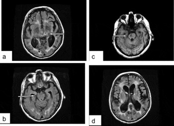
The variation in scanning techniques and criteria adopted for identifying silent (non-clinically apparent) LI presents problems for estimating their incidence and prevalence, but some general conclusions can be drawn. It should emphasized that MRI cannot detect actual SVD, but detects evidence of surrogate markers of SVD, namely LI and white matter hyperintense lesions (WML).Reference Pantoni19 MRI detects more silent infarcts than CT, because LI are only visible on T2-weighted imaging.Reference Vermeer, Longstreth and Koudstaal31 It is generally accepted that focal lesions greater than 3 mm in diameter on imaging are regarded as LI, whereas autopsy studies generally agree on 1.0–1.5 cm.Reference Vermeer, Prins, den Heijer, Hofman, Koudstaal and Breteler54, Reference Zhu, Dufoil, Tzourio and Chabriat55 A recent review of MRI studies noted that the distinction between dilated perivascular space and LI is especially difficult when the lesion is less than 3 mm in diameter.Reference Zhu, Dufoil, Tzourio and Chabriat55 To add to the difficulty, a recent MRI study has identified dilated perivascular space as a surrogate marker of brain inflammation and not LI (Figure 1).Reference Wuerfel, Haertle, Waiczies, Tysiak, Bechmann, Wernecke, Zipp and Paul56
(a) MRI-FLAIR scan image of a lesion (arrow) in the left thalamus corresponding to a lacunar subcortical infarct; (b) MRI-FLAIR scan image of a lacunar infarct in the brainstem (pons); (c) same brain but with T1 weighted MRI scan showing the same lesion in pons (arrow); (d) MRI-FLAIR scan demonstrating perivenular stippling corresponding to Virchoff Robin space dilatation in the thalamus.

Neuroimaging identifies surrogate markers for SVD and their relationship with dementia
The Rotterdam Prospective study, using MRI to initially scan non-demented elderly individuals, found that increasing numbers of so-called silent strokes (subcortical LI > 3 mm) and WML correlated with increasing risk of dementia.Reference Vermeer, Longstreth and Koudstaal31, Reference Vermeer, Prins, den Heijer, Hofman, Koudstaal and Breteler54, Reference Van Djik, Prins and Vrooman57
The prevalence of silent infarcts (8–28% in the general population) is five times greater than clinically obvious stroke and higher in women and in individuals with cardiovascular disease and depression.Reference Vermeer, Longstreth and Koudstaal31 Silent infarcts also have a five times higher incidence for all age groups than clinically obvious stroke in the same populations.Reference Vermeer, Longstreth and Koudstaal31, Reference Vermeer, Prins, den Heijer, Hofman, Koudstaal and Breteler54 The presence of silent infarcts dramatically increases the risk of clinically obvious stroke, especially in those individuals with co-existing WML, providing evidence that SVD is responsible for both silent infarcts and WML.Reference Vermeer, Longstreth and Koudstaal31, Reference Kobayashi, Okada, Koide, Bokura and Yamaguchi58 The risk factors associated with silent infarcts are age, systemic hypertension, retinal microvascular changes, severe white matter changes and a family history of stroke, and these are shared with SVD. Other risk factors, some of which are associated with atheroma formation such as smoking, cholesterol level and surprisingly diabetes, have been found not to be associated consistently with silent infarcts and by inference not to be risk factors for SVD.Reference Vermeer, Longstreth and Koudstaal31, Reference Vermeer, Prins, den Heijer, Hofman, Koudstaal and Breteler54
The Rotterdam study found that the presence of silent infarcts in elderly individuals more than doubles the risk of developing dementia, including AD, and mild cognitive impairment.Reference Vermeer, Longstreth and Koudstaal31, Reference Lopez, Jagust, Dulberg, Becker, DeKosky, Fitzpatrick, Breitner, Lyketsos, Jones, Kawas, Carlson and Kuller49 In the same study, the presence of silent infarcts due to SVD reduced information processing and executive memory function.Reference Prins, van Dijk, den Heijer, Vermeer, Jolles, Koudstaal, Hofman and Breteler59 Evidence that SVD is responsible for the majority of silent strokes is also provided by radiological evidence showing that silent infarcts are mainly LI (<3 mm) located in the same subcortical areas (thalamus and putamen)Reference Gold, Kövari, Herrmann, Canuto, Hof, Michel, Bouras and Giannakopoulos60 as those assumed to be responsible for dementia in pathological studies.Reference Snowdon, Greiner and Mortimer5, Reference Erkinjuntti, Halatia, Palo, Sulkava and Paetau15, Reference Esiri, Wilcock and Morris16, Reference Chui46 These strategically located subcortical infarcts disturbed urinary retention, motor control and executive function,Reference Tatemichi, Steinke, Duncan, Bello, Odel, Behrens, Hilal and Mohr61 whereas LIs in the anterior thalamus and putamen inhibited short-term memory.Reference Benisty, Gouw, Porcher, Madureira, Hernandez, Poggesi, van der Flier, Van Straaten, Verdelho, Ferro, Pantoni, Inzitari, Barkhof, Fazekas and Chabriat62
The mechanism responsible for a silent infarct due to SVD increasing the severity of dementia is not clear. Several possible explanations exist: (1) the accelerated formation of NFT and NP in already ‘vulnerable brains’,Reference Vermeer, Longstreth and Koudstaal31 (but this is unlikely in view of Honig et al.Reference Honig, Kukull and Mayeux51); (2) white matter damage, especially of periventricular distribution; and (3) a LI resulting in an increased loss of synapses and neurons, even in cases with only moderate numbers of NFT and NP, increasing the severity of dementia.Reference Snowdon, Greiner and Mortimer5, 7
Neuroimaging identified leukoaraiosis in old age and the demented – what does it represent?
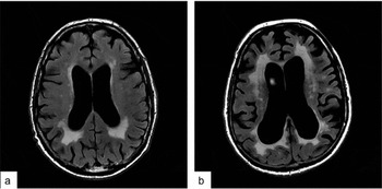
The term leukoaraiosis (LA) was a descriptive term, first employed by Hachinski in 1987,Reference Hachinski44 to describe diffuse WML in the periventricular or subcortical (centrum semiovale) areas.Reference O'Sullivan63, Reference Pantoni64 On scans, LA corresponded to poorly defined lesions that were either hypodense on CT or hyperintense (‘holes’) on T2-weighted or FLAIR (fluid-attenuated inversion recovery) MRI scans (Figure 2); these lesions can be described as white matter hyperintensities (WMH).Reference Patel and Markus32, Reference Bronge and Wahlund35 Initially, LA was regarded as of doubtful significance, but with more widespread use of imaging studies it was found to be present in 30–40% of cases of dementia with lacunar strokes and in over 60% of individuals with clinical VaD. The ischaemic aetiology for LA is supported by evidence from the Leukoaraiosis and Disability in the Elderly Study (LADIS),Reference van der Flier, van Straaten, Barkhof, Verdelho, Madureira, Pantoni, Inzitari, Erkinjuntti, Crisby, Waldemar, Schmidt, Fazekas and Scheltens65, Reference Pantoni, Basile, Pracucci, Asplund, Bogousslavsky, Chabriat, Erkinjuntti, Fazekas, Ferro, Hennerici, O'Brien, Scheltens, Visser, Wahlund, Waldemar, Wallin and Inzitari66 showing that age, systemic hypertension, diabetes and lacunar stroke are the major determinants of age-related white matter changes (LA) seen with neuroimaging.Reference van der Flier, van Straaten, Barkhof, Verdelho, Madureira, Pantoni, Inzitari, Erkinjuntti, Crisby, Waldemar, Schmidt, Fazekas and Scheltens65–Reference Debette and Markus68 Neurological features associated with increasing LA load, as for SVD, include bladder control, gait disturbance and information processing, not all of which are necessarily related to cognitive decline.Reference Vermeer, Prins, den Heijer, Hofman, Koudstaal and Breteler54 However, as the number of LA lesions from the first to subsequent scans increased, so too did the clinical progression to dementia.Reference Prins, van Dijk, den Heijer, Vermeer, Jolles, Koudstaal, Hofman and Breteler59, Reference O'Sullivan63, Reference van der Flier, van Straaten, Barkhof, Verdelho, Madureira, Pantoni, Inzitari, Erkinjuntti, Crisby, Waldemar, Schmidt, Fazekas and Scheltens65, Reference Pantoni, Basile, Pracucci, Asplund, Bogousslavsky, Chabriat, Erkinjuntti, Fazekas, Ferro, Hennerici, O'Brien, Scheltens, Visser, Wahlund, Waldemar, Wallin and Inzitari66
MRI-FLAIR scans showing varying degrees of periventricular leukoaraiosis (LA). In (a) there are moderate periventricular changes of LA, whereas in (b) the extent of periventricular changes is more extensive.

The correlation between radiologically identified WML with their pathology and their significance for dementia
As LA lesion size increases, from small punctuate hyperintense lesions to large confluent lesions, they are associated with myelin loss, gliosis and microinfarction, contributing to a decline in cognitive performance. By contrast, the periventricular lesions have a non-ischaemic appearance and are associated with the loss of normal ependymal lining and subependymal gliosis.Reference Fazekas, Kleinert, Offenbacher, Schmidt, Kleinert, Payer, Radner and Lechner27, Reference O'Sullivan63, Reference Frisioni, Gallizzi, Pantoni and Filippi69 In MRI identified areas of brain containing LA, tissue markers of hypoxia are increased in activated microglia, pointing to hypoperfusion as a contributory factor to myelin damage in LA. However, evidence for hypoperfusion as defined by radiological techniques in the same areas is not convincingReference O'Sullivan, Lythgoe and Pereria70 and as yet any initiating factor triggering hypoxia has not been identified.Reference Fernando, Simpson, Matthews, Brayne, Lewis, Barber, Kalaria, Forster, Esteves, Wharton, Shaw, O'Brien and Ince71
The Rotterdam scan study identified that both periventricular and subcortical WML were associated with dementia, but the association was stronger for periventricular changes than for subcortical ones.Reference Prins, van Dijk, den Heijer, Vermeer, Jolles, Koudstaal, Hofman and Breteler59, Reference Pantoni64, Reference Schmidt, Petrovic, Ropele, Enzinger and Fazekas72 The mechanism for this association could be related to the potential ischaemic aetiology of large WML, but the effect of the periventricular lesions was independent of cerebral infarcts and could be the result of interrupting cortico-cortical association fibres. It therefore represents an independent risk for dementia.Reference Prins, van Dijk, den Heijer, Vermeer, Koudstaal, Oudkerk, Hofman and Breteler30 Neuropathological studies have confirmed MRI identified WML lesions are the result of SVD, including histologically identified arteriosclerosis.7, Reference van Swieten, van den Hout, van Ketel, Hijdra, Wokke and van Gijn73
The strong correlation between periventricular, and to a lesser extent diffuse WML (both identified by histological staining for myelin) with cognitive decline in individuals with dementia, but no co-existing AD pathology was confirmed by Kovari et al., raising the possibility that periventricular WML identified by MRI is a surrogate marker for non-AD dementia.Reference Prins, van Dijk, den Heijer, Vermeer, Koudstaal, Oudkerk, Hofman and Breteler30, Reference Vermeer, Prins, den Heijer, Hofman, Koudstaal and Breteler54,Reference Kovari, Gold and Herrmann74
By contrast, the significance of WML (identified by histological staining for myelin) and LIs (that co-existed in 95% of cases) for dementia was investigated prospectively in brains from individuals with no evidence of co-existing macrovascular or AD pathology. The presence of grey matter LIs (1 mm – 1.5 cm) correlated with cognitive decline, but the presence of co-existing WML identified by myelin staining did not correlate with dementia and therefore questions the use of histologically and radiologically identified WML as a surrogate marker for SVD.Reference Gold, Kövari, Herrmann, Canuto, Hof, Michel, Bouras and Giannakopoulos75
White matter vascular changes due to SVD and AD pathology – a possible causal relationship
The Vantaa study found only frontal periventricular and diffuse WML on MRI scan correlated with high Braak stage, raising the possibility of a link between AD pathology in the frontal lobe (NFT formation) and periventricular WML as a consequence of underlying SVD. Large vessel macroscopic infarcts did not correlate with severity of AD pathology in this study, in contrast to several others.Reference Roher, Esh, Kokjohn, Kalback, Luehrs, Seward, Sue and Beach21, Reference Troncosco, Zonderman, Resnick, Crain, Pletnikova and O'Brien23 However, the presence of SVD was proposed as an explanation for both frontal lobe NFT formation and white matter changes because of (1) retrograde axonal degeneration with subsequent NFT formation in frontal lobe neurons and/or (2) neuron loss due to SVD-related ischaemia in the grey matter resulting in anterograde axonal loss in adjacent areas of white matter. On this basis, radiologically detected frontal WMLs are regarded as a surrogate marker of SVD.Reference Polvikoski, van Straaten, Barkhof, Sulkava, Aronen, Niinistö, Oinas, Scheltens, Erkinjuntti and Kalaria76
Some conclusions?
The evidence for WML influencing cognitive decline is confusing, but the most recent meta-analysis of 46 MRI studies concluded that WML are correlated with increased risk of stroke, dementia, cognitive decline and death.Reference Debette and Markus68 The data from this review were not able to identify the specific vascular basis for white matter disease in stroke and dementia, but suggested that the WML could result from several pathways: (1) disruption of subcortical neural networks, supported by diffusion tensor MRI scan evidence; (2) the same aetiological factors responsible for AD pathology; and (3) the same SVD that is also responsible for LI, reflecting underlying vascular injury.Reference Debette and Markus68
SVD – what is the histopathology?
The effects of ageing on cerebral blood vessels
Ageing produces structural changes in small diameter blood vessels, including basal membrane thickening and endothelial changes, both reducing blood flow and preventing efficient transport across the BBB.Reference Brown and Thorne77 The change in the density of cerebral capillaries with age has been a controversial topic, but it is now accepted that capillary density is reduced in ageing and AD.Reference Riddle, Sonntag and Lichtenwalner78 Elderly capillaries are unable to respond as efficiently as younger capillaries when exposed to hypoxia, preventing efficient autoregulation of cerebral blood flow. In young animals, tissue hypoxia increases hypoxic inducible factor (HIF), promoting vascular endothelial growth factor (VEGF) expression and endothelial changes. This results in new vessel formation, or angiogenesis, thus restoring blood flow. The HIF-1 response to hypoxia is lost in elderly blood vessels, as is cerebral angiogenesis, preventing new vessel formation after ischaemic conditions.Reference Black, Polinsky and Greenough79, Reference Rivard, Fabre, Silver, Chen, Murohara, Kearney, Magner, Asahara and Isner80, Reference Frenkle–Denkberg, Gershon and Levy81 A combination of age-related changes (capillary density decline) and systemic hypertension prevent normal autoregulation of cerebral blood flow, placing the elderly brain at risk from the effects of hypoperfusion and resulting in ischaemic changes, microbleeds, white matter ischaemia and cognitive decline.Reference Van Beek, Claasen, Rikkert and Jansen82 Microvascular changes, including endothelial damage, are present in AD, with reduced endothelial CD34 and CD31 demonstrating degeneration of the vessel wall.Reference Van Beek, Claasen, Rikkert and Jansen82 Accumulation of cerebral arteriole wall β amyloid has also been proposed as a factor preventing efflux of β amyloid from the ageing brain, with neurotoxic consequences.Reference Weller, Subash, Preston, Mazanti and Carare83
Vessel pathology, large and small
Atheroma involves the wall of large diameter arteries (e.g. carotid artery, Circle of Willis, coronary arteries) and is responsible for ischaemic infarction through occlusion of the lumen with thrombus formation.Reference Ince and Kalimo84, Reference Ogata, Yamanishi and Ishibashi-Ueda85 By contrast, SVD is restricted by definition to small diameter vessels and is associated with pathological and radiological evidence of LIs and white matter changes variously called LA and WML (Figure 3). Whether or not LI and WML are the result of different types of SVD remains to be established.Reference Lammie and Kalimo86
(a), Lacunar infarct (arrow) in the basal ganglia stained with haematoxylin & eosin (H&E). The periphery of the infarct contains macrophages with loss of brain tissue from the centre, forming a so-called lacune. (b), transverse section through a small diameter arteriole in the basal ganglia, stained with H&E with the endothelial, intimal and media layers replaced by pathological changes amounting to arteriosclerosis (note the narrowing of the vessel lumen); (c), the brain surrounding an arteriole with arteriosclerosis contains numerous reactive glial cells (arrows) stained with H&E, some of which are capable of expressing inflammatory cytokines.

Arteriosclerosis (thickening), or age-related vascular risk factor type 1, is found in arterioles.Reference Pantoni19 This group also includes Fisher's segmental arteriolar disorganization (lipohyalinosis), fibrinoid necrosis, microatheroma (plaque-like proteins, plasma cells and macrophages) and microaneurysm formation.Reference Grinberg and Thal87 The pathology is characterized by age-related loss of smooth muscle cells and replacement by deposits of fibro-lipohyalinosis material producing lumen occlusion and vessel wall thickening, including basement membrane structural changes.Reference Lammie and Kalimo86 Type 1 vessel wall changes have been described in basal ganglia, brainstem (in the same distribution as LIs) and in white matter lesions, but not in cortex.Reference Grinberg and Thal87
Cerebral amyloid angiopathy (CAA) type 2Reference Pantoni19 involves arteries, veins, arterioles and capillaries (β amyloid in the capillary wall is type 1).Reference Grinberg and Thal87 This results in vessel wall dilation, disruption and fragmentation, microscopic haemorrhage, haematoma formation and parenchymal damage, as found in AD and β amyloid-related angiitis. CAA does not involve white matter.
Venous collagenosis is a pathological thickening of veins closely located to the lateral ventricles.Reference Pantoni19 Thickening of the vessel wall can result in occlusion of the lumen, with a reduction of local venous return increasing the likelihood of WML in periventricular areas.
Atheroma and SVD are different
A systematic review of 25 studies found no difference between risk factors for developing SVD-related LI and non-LI, embolic/atheromanous-related stroke. This included as risk factors high systemic blood pressure and diabetes.Reference Jackson and Sudlow88 One problem with this meta-analysis was the variation in the definition of ischaemic stroke between different studies producing a classification bias. When these data were re-analysed, hypertension and diabetes were found not to be associated with LI and the individual stroke subtypes cannot be distinguished on the basis of these risk factors alone.Reference Jackson and Sudlow88, Reference Jackson, Hutchison, Dennis, Wardlaw, Lewis and Sudlow89
However, although unable to discriminate SVD from atheroma on the basis of related risk factors, SVD-related LIs are also apparently less likely to be associated with coronary thrombosis, carotid artery atheroma and a previous myocardial infarction than non-LIs due to atheroma.Reference Jackson and Sudlow88 This suggests that SVD pathology does not share the same aetiology as peripheral atheroma and thrombo-embolic disease. In a large prospective study,Reference Jackson, Hutchison, Dennis, Wardlaw, Lewis and Sudlow89 cerebral non-atheromatous LIs were usually followed by subsequent LIs, rather than atheroma-related ischaemic infarction elsewhere in the brain or heart. This supports the conclusion that SVD and LIs are independent of the disease process responsible for atheroma and thrombus formation.
Causes and consequences of SVD
The lacunar hypothesis and systemic hypertension
Fisher defined clinically significant LIs as ‘infarcts caused by occlusion of small penetrating cerebral arteries’ (200–800 μm diameter) and he attributed the underlying pathology to narrowing of the lumen of deep perforating arteries. This produced a lacune, more strictly defined as a healing stage of a deep infarct, and these were increasingly found in the brains of hypertensive individuals.Reference Fisher90 The LIs are the basis for radiologically identified subcortical infarcts located in basal ganglia and thalamus, as described in dementia. The pathology of the SVD assumed to be responsible for LIs was initially described as fibrinoid necrosis, lipohyalinosis and miliary aneurysms.Reference Fisher90 The fibrinoid necrosis and lipohyalinosis were regarded as a consequence of hypertension, but a recent study of hypertensive individuals with SVD found these changes are uncommon and the vessel wall pathology is better described by the term hyaline arteriosclerois.Reference Lammie, Brannan, Slatery and Wardlaw91 In fact, the author recommends that the generic term ‘SVD’ is a more accurate term, because it does not imply any pathological understanding, but does localize responsibility for LI at the level of the arteriole.
The early explanations for LI involved two possibilities. Embolic occlusion proved to be unlikely because occluded arterioles have been rarely found in autopsy material and emboli would account for only 10–15% of LIs.Reference Jackson, Hutchison, Dennis, Wardlaw, Lewis and Sudlow89, Reference Wardlaw, Sandercock, Dennis and Starr92, Reference Wardlaw93 Early studies by Fisher proposed hypertension produced either fibrinoid necrosis or vasospasm, which in turn initiated degenerative changes (fibrinoid necrosis lipohyalinosis) in the vessel wall.Reference Wardlaw, Sandercock, Dennis and Starr92 The Rotterdam silent infarct study provided support for this explanation, identifying hypertension as a risk factor, as did animal models of experimental hypertension.Reference Wardlaw, Sandercock, Dennis and Starr92, Reference Wardlaw93 However, in humans, typical SVD (hyaline arteriosclerosis) changes were found to be equally prevalent in hypertensive and normotensive non-diabetic brains.Reference Lammie, Brannan, Slatery and Wardlaw91 This study supports the findings that systemic hypertension in the general population is not a risk factor for LI, because it is unlikely to initiate SVD.Reference Jackson and Sudlow88 Furthermore, neither subcortical infarcts due to SVD nor systemic hypertension were identified as risk factors for dementia in the BLSA cohort.Reference Dolan, Crain, Troncoso, Resnick, Zonderman and O'Brien18, Reference Troncosco, Zonderman, Resnick, Crain, Pletnikova and O'Brien23
The leaky vessel or disrupted BBB hypothesis
An alternative explanation for SVD is that lipohyalinosis and type I arteriole damage are in fact secondary effects, due to injury to the vessel wall endothelial lining resulting in BBB disruption, with leakage into the neuropil of serum proteins.Reference Wardlaw, Sandercock, Dennis and Starr92 These serum proteins include IgG, coagulation and complement proteins and are capable of activating local microglia in white matter, with secondary small vessel wall changes including perivascular oedema resulting in an ‘incomplete’ LI (type b).Reference Ince and Kalimo84, Reference Lammie and Kalimo86 Although the leaky hypothesis is plausible, the initiating event(s) provoking prolonged disruption of the endothelium with increasing BBB permeability is not yet known and this could represent a secondary effect following primary local neuron ischaemia. A systemic stimulus, such as oxidative stress, would potentially involve all cerebral arterioles and provide a better explanation for SVD, rather than individual small vessels being occluded by chance embolic events.Reference Wardlaw, Sandercock, Dennis and Starr92, Reference Bogousslavsky94, Reference Thompson and Hakim97 This possibility, rather than the effects of systemic hypertension, has been discussed by Dolan et al. as an underlying cause for both vascular and AD pathology.Reference Dolan, Crain, Troncoso, Resnick, Zonderman and O'Brien18
Small vein pathology, white matter lesions and cognitive decline
Periventricular, non-necrotic, white matter hyperintensities (PVH), as detected by MRI scanning, are present adjacent to the ventricle wall and are generally silent. However, if extensive, they can be associated with gait disturbance and cognitive decline.Reference Black, Gao and Bilbao98 More detailed clinicopathological analysis found that PVH correlated with decline of executive dysfunction as a consequence of interrupting cortico-cortical axons. Pathological analysis of PVH found myelin pallor, astrocytosis and reduced numbers of oligodendroglia due to apoptosis.Reference Black, Gao and Bilbao98 One explanation for these findings was periventricular intraparenchymal venular disease, due to collagen thickening of vein walls, resulting in stenosis and occlusion considered sufficient to produce vasogenic oedema and eventually chronic hypoperfusion of adjacent periventricular white matter.Reference Black, Gao and Bilbao98 This is supported by studies showing that glia in post-mortem tissue from MRI-identified PVH regions expressed higher levels of HIF-1a and HIF-2a than in WM controls. This provides evidence that WM tissue hypoxia with activation of microglia, expression of pro-inflammatory cytokines, metalloproteinases and oligodendroglial apoptosis would contribute to the disruption of the BBB.Reference Rosenberg, Sullivan and Esiri99, Reference Rosenberg100 The identity of the stimuli initiating venular disease is regarded as ischaemic, but the evidence for localized white matter hypoperfusion is undecided.Reference O'Sullivan, Lythgoe and Pereria70
Consequences of SVD, microbleeds and cognitive performance
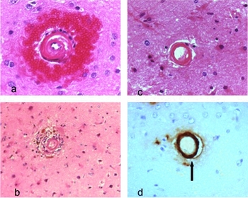
Type 2 vessel wall rupture due to sporadic amyloid deposition results in haemorrhage in the surrounding brain (Figure 4), with an MRI appearance of micro-hyperintense lesions in associated cerebral white matter and micro-infarcts.Reference Pantoni19 Approximately 40% of elderly individuals over the age of 80 years will have MRI evidence of microbleedsReference Fisher, Rench, Ji and Kim101 and there was high association with LA, LIs, systemic hypertension and cognitive decline due to caudate and frontal lobe involvement.Reference Viswanathan, Guichard, Gschwendtner, Buffon, Cumurcuic, Boutron, Vicaut, Holtmannspötter, Pachai, Bousser, Dichgans and Chabriat102
(a), Small diameter arteriole in the basal ganglia surrounded by perivascular haemorrhage – a so-called ‘microbleed’ stained with H&E; (b), higher magnification shows a small diameter arteriole with vessel wall changes surrounding reactive glia and macrophages containing haemosiderin pigment (iron) originating from the breakdown products of haemoglobin in a perivascular microbleed; (c), cerebral capillary stained with H&E showing a thickened hyalinized wall; (d), the same capillary stained with anti-Aβ4 antibody showing the vessel wall contains Aβ4 amyloid.

As the result of either acute or chronic ischaemia and vessel wall rupture, serum proteins such as thrombin, a serine protease vital for blood coagulation, will gain access to brain parenchyma.Reference Xi, Greiser and Keep103 Under physiological conditions, thrombin is synthesized by neurons and astrocytes at low concentrations and this is important for brain repair.Reference Gingrich and Traynelis104, Reference Striggow, Riek, Breder, Henrich-Noack, Reymann and Reiser105 However, at high concentrations (500 nM) following BBB disruption, thrombin activates microglia via the JAK2-STAT3 signalling pathway, promoting inflammatory cytokine production, myelin damage, apoptosis and axonal injury.Reference Huang, Ma, Sun, Wei, Fang, Liu and Liu106
Future prospects and topics for research
Neuropathological criteria – an urgent requirement
At present there is no accepted histological criteria to provide assessment of clinically diagnosed VaD.Reference Wiederkehr, Simard, Fortin and van Reekum34 By analogy with the CERAD, any proposed histological criteria should include small diameter vessel wall appearance, white matter changes, infarct localization (subcortical, cortical, thalamic) and take into consideration age, neuroimaging, cognitive status and the presence or absence of risk factors (hypertension, diabetes), in order to provide a qualitative score of possible, probable and definite VaD.
The neurovascular functional unit – a possible explanation for SVD and the co-existence of vascular and amyloid-related dementias?
There is a growing amount of evidence to show that vascular dysfunction is potentially a primary event in both AD and VaD pathology. Neuron metabolism is closely linked with blood flow through cerebral capillaries and the close relationship between neurons and capillaries, as well as the BBB, is regarded as a functional neurovascular unit.Reference Zlokovic38 Following primary vascular changes due to endothelial injury, initiated by risk factors such as oxidative stress, the capillary endothelium expresses inflammatory cytokines that activate local microglia, disrupt the BBB and inhibit normal neuron metabolism. Oxidative stress is a risk factor for both atheroma and AD pathology and it is possible it has its primary effect upon the cerebral vessel wall, resulting in local neuronal dysfunction, leaky BBB (SVD pathology) and β amyloid deposition (AD pathology), exposure of the brain to potentially neurotoxic systemic proteins and activation of microglia.Reference Craft107–Reference Nunomura, Castellani and Zhu109
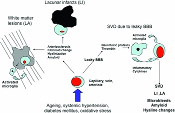
One serum protease, thrombin, crosses the disrupted BBB and has several potentially neurotoxic effects mediated by thrombin receptor protease activated receptor-1 (PAR-1) distributed on astrocytes and neurons. Thrombin also stimulates the endothelial release of pro-inflammatory cytokines (MCP-1 VEGF), tau aggregation and neuron death, as the result of increasing synthesis of pro-apoptotic proteins triggering NADPH oxidase-mediated oxidative stress and activation of microglia, with release of nitric oxide.Reference Nunomura, Castellani and Zhu109In vitro thrombin can stimulate amyloid precursor protein production and subsequent cleavage into β amyloid peptides, which in turn increase neurovascular pro-inflammatory cytokine synthesis and endothelial damage.Reference Ciallela, Figueiredo, Smith-Swintosky and McGillis110 This is summarized in Figure 5.
A schematic diagram to summarize the possible pathways producing SVD and associated pathological changes. Various pathological stimuli (systemic hypertension, diabetes mellitus, oxidative stress) combined with ageing produce primary changes in the wall of small calibre cerebral vessels resulting in arteriosclerosis, atheroma, hyalinization, fibrinoid necrosis and amyloid deposition (as documented in SVD). These changes in vessel wall lumen produce white matter pathology (LA) with activation of local microglia; the other main pathology, ischaemic lacunar infarction (LI) found in subcortical sites, is the result of small vessel occlusion. The alternative explanation is that the same initial pathological stimuli, including ageing, produce a ‘leaky blood–brain barrier’ with systemic neurotoxic proteins entering the brain parenchyma to activate microglia. Microglia express inflammatory cytokines, resulting in secondary pathological changes in small calibre blood vessel walls that amount to SVD.

Endothelial inflammation and potential biomarkers for SVD
Inflammatory molecules synthesized by the endothelium can act as biomarkers of endothelial and BBB dysfunction and provide insight into the evolution of SVD before clinical consequences become apparent.Reference Markus, Hunt, Palmer, Enzinger, Schmidt and Schmidt111 The endothelial cells express a range of proteins present in the serum and associated with coagulation and fibrinolysis, including inter-cellular adhesion molecule 1 (ICAM-1), thrombomodulin (TM), activation of anticoagulant protein C, and tissue factor pathway inhibitor (TFPI). Serum levels of C-reactive protein (CRP), fibrinogen and IL-6 represent biomarkers of tissue inflammation and have been associated with brain infarction and white matter changes.Reference Schmidt, Schmidt, Curb, Masaki, White and Launer112 Hassan et al. found that serum levels of ICAM-1, TM and TFPI were all elevated in individuals with radiologically detected LIs and LA, supporting the concept of a leaky BBB reflecting endothelial dysfunction.Reference Hassan, Hunt, O'Sullivan, Parmar, Bamford, Briley, Brown, Thomas and Markus37 However, a similar studyReference Knottnerus, Govers-Riemslag, Hamulyak, Rouhl, Staals, Spronk, van Oerle, van Raak, Lodder, ten Cate and van Oostenbrugge113 found there was an increase of serum tissue plasminogen activator (tPA) in WML but no difference between LI and non-LI groups for tPa, TFPI or TM, in contrast to Hassan, who found these serum markers increased with severity of LA. TM is also distributed throughout the systemic circulation, so it is unlikely these proteins are suitable specific serum biomarkers for SVD, although they do indicate activation of endothelium in LI.Reference Schmidt, Schmidt, Curb, Masaki, White and Launer112, Reference Knottnerus, Govers-Riemslag, Hamulyak, Rouhl, Staals, Spronk, van Oerle, van Raak, Lodder, ten Cate and van Oostenbrugge113 More general biomarkers of inflammation, such as CRP and IL-6, have been found to be significantly associated with radiologically detected WML and LIs,Reference Benchenane, Berezowski, Ali, Fernández-Monreal, López-Atalaya, Brillault, Chuquet, Nouvelot, MacKenzie, Bu, Cecchelli, Touzani and Vivien114 as well as cerebral infarcts due to large vessel atherosclerosis.Reference Fornage, Chiang, O'Meara, Psaty, Reiner, Siscovick, Tracy and Longstreth115
Therapeutic interventions to target inflammation and coagulation
The role of antihypertensive treatment reducing the risk of dementia as the result of preventing subcortical LI due to SVD is well established. Anti-inflammatory drugs, such as inhibitors of cyclo-oxygenase-2, nitric oxide, tumour necrosis factor (TNF) and prostaglandins, have been tried without success in dementia, but this is probably the result of them being administered in the more advanced stages of AD and VaD rather than at the outset.Reference Hoshi, Kitagawa, Yamagami, Furukado, Hougaku and Hori116 Similarly, anticoagulant agents to prevent the neurotoxic effects of thrombin, TM and TF could be of potential benefit if started sufficiently early. Reliable serum and radiological biomarkers to identify at-risk-of-dementia groups should mean that these drugs can be given at an earlier stage in the evolution of SVD and will have a potentially greater chance of success.
Conclusions
Clinicopathological studies identified small infarcts in subcortical structures and white matter changes were major contributory factors in VaD. This has been confirmed by MRI scanning of individuals with VaD, showing that many have LIs and white matter changes. These radiological lesions are regarded as surrogate markers for SVD, despite disagreement about the radiological criteria with which they are identified.
Although subcortical LIs were easily identified at autopsy, their aetiology was assumed to be the result of hypertension damaging the wall of small calibre arterioles supplying these subcortical structures. Evidence to confirm this explanation has been less than conclusive. Similarly, pathological changes in white matter tracts have been assumed to be the result of ischaemic changes due to arteriole obstruction, but evidence for local hypoperfusion is inconclusive. SVD is not a single pathological process, as it includes arteriosclerosis, amyloid deposition, perivascular space and venous wall thickening, implying that a range of different aetiologies are responsible. The wide range of pathology included in SVD has contributed to the failure to construct a universally agreed scoring system to allow documentation of SVD, making comparisons across different studies difficult and inconclusive. To date the vascular pathology of SVD in AD and VaD is not thought to differ, although this could be a reflection of the failure to adopt a unified classification of SVD, preventing meaningful comparisons.
The favoured explanation for SVD includes primary endothelial injury due to systemic hypertension, metabolic changes and oxidative stress. Injury of capillary wall endothelium will contribute to BBB disruption, with exposure of neurons, capillaries/arterioles and white matter changes due to inflammatory cytokines expressed by activated microglia. Many of the risk factors for SVD are also shared with AD, raising the possibility that AD and VaD share a common aetiology, based upon primary vessel wall changes.
Therapeutic agents designed to reduce endothelial wall inflammation, maintain BBB integrity and inhibit the neurotoxic effects of serum proteins such as thrombin should be developed and prescribed at an early stage to those at high risk of developing VaD, in order to gain maximum benefit. Biomakers of SVD are currently available and, once these have been refined, will allow more accurate identification of ‘at-risk groups’ to be treated prophylactically, before clinical signs and radiological markers of VaD become apparent.
Perhaps it is now an opportune time to accept that the majority of dementias result from different proportions of both neurodegeneration and ischaemia, because these two apparently different pathologies share many common underlying and reversible vascular-related aetiologies.
Conflicts of interest
The author declared no conflicts of interest
Acknowledgements
I am extremely grateful to Dr M. O'Sullivan, Consultant Senior Lecturer and MRC Research Scientist in Cognitive Neuroscience, Deparment of Neurology and CUBRIC (Department of Psychology, Cardiff University) for providing the MRI and MRI-FLAIR images.