Hypertrophic cardiomyopathy is a kind of primary hereditary cardiomyopathy that is characterised by left ventricular hypertrophy without ventricular enlargement and excludes other cardiovascular or systemic diseases that cause left ventricular hypertrophy. Reference Maron, Towbin and Thiene1 Hypertrophic cardiomyopathy is the second most common cardiomyopathy in children, accounting for 42% of all cardiomyopathy cases. Reference McKenna, Maron and Thiene2 Clinical manifestations of hypertrophic cardiomyopathy include sudden cardiac death, syncope, progressive heart failure, mild to moderate symptoms or even no clinical symptoms. Reference Elliott, Andersson and Arbustini3,Reference Gersh, Maron and Bonow4 The mortality rate of hypertrophic cardiomyopathy is about 1%, Reference Hershberger, Cowan, Morales and Siegfried5 , and it is one of the causes of sudden death in older children and adolescents. Therefore, early and accurate diagnosis of the cause of the disease is very important. Genetic gene defects encoding sarcomere and myofibrillar proteins are the main cause of hypertrophic cardiomyopathy. Reference Maron6 Common non-sarcomere pathogeny of hypertrophic cardiomyopathy in children includes metabolic defects, malformation syndrome, and neuromuscular diseases. Reference Gersh, Maron and Bonow7 More than 1500 variants have been identified in more than 21 genes to date. Reference Maron, Ommen, Semsarian, Spirito, Olivotto and Maron8 Screening pathogenic genes and identifying variants are helpful to clarify the cause of hypertrophic cardiomyopathy in children and conduct early diagnosis and screening of their family members. The latest American College of Cardiology Foundation/American Heart Association guidelines recommend comprehensive detection of pathogenic genes in patients with hypertrophic cardiomyopathy. Reference Gersh, Maron and Bonow4 However, traditional screening methods, such as Sanger sequencing, are time consuming and expensive due to the large number of pathogenic genes to be tested. Thus, these methods are difficult to use for rapid and comprehensive screening. Individual genome sequencing has become possible with the development of next-generation sequencing technology; in this regard, whole-exome sequencing is an important technology that can be used to detect the genetic causes of hypertrophic cardiomyopathy. Reference Bos, Will, Gersh, Kruisselbrink, Ommen and Ackerman9 In the present study, we used whole-exome sequencing to detect pathogenic genes in a Han Chinese family with hypertrophic cardiomyopathy. We also evaluated the feasibility of whole-exome sequencing in genetic detection of hypertrophic cardiomyopathy in children. We analysed the correlation between the pathogenic gene variant and their phenotype. This study will increase clinical experience to establish the application of whole-exome sequencing detection in judging the prognosis and stratifying the risk of hypertrophic cardiomyopathy.
Objects and methods
Objectives and clinical data collection
Seven people in three generations (four males and three females) from a Han Chinese pedigree with hypertrophic cardiomyopathy were enrolled in the study in the Department of Pediatrics, Qilu Hospital of Shandong University in November 2019. The proband is a 4-year-old boy who suffered from syncope during running and was diagnosed with hypertrophic cardiomyopathy by echocardiography. Detailed clinical data of all family members, including clinical symptoms, physical examination, electrocardiogram and echocardiography, mobile electrocardiogram, and/(or) cardiac magnetic resonance examination as needed, were collected. Information of the patient with hypertrophic cardiomyopathy, including sex, age, age of onset, and main symptoms, were collected. A total of 200 healthy volunteers were selected as controls, including 97 males and 103 females, with an average age of (35.1 ± 10.7) years. The effect of gene variant on the phenotype of hypertrophic cardiomyopathy was analysed. All the candidates signed the informed consent form. This study was approved by the Ethics Committee of Qilu Hospital of Shandong University.
Diagnosis and exclusion criteria of hypertrophic cardiomyopathy
According to the diagnostic criteria of hypertrophic cardiomyopathy established by the European Society of Cardiology in 2014, Reference Elliott, Anastasakis and Borger10 the wall thickness of one or more segments of the left ventricle measured by cardiac imaging techniques (echocardiography, CMR, or CT) is more than 15 mm in adults and ≥ mean + 2 SD in children. Hypertrophic cardiomyopathy can be diagnosed if cardiac imaging technology of the first-degree relative of the patient with hypertrophic cardiomyopathy reveals that the thickness of one or more segments of the left ventricular wall is ≥ 13 mm without other known causes. The exclusion criteria include ventricular wall hypertrophy caused by hypertension, coronary heart diseases, coarctation of aortic arch, valvular diseases, CHDs, metabolic diseases, and heart hypertrophy of athletes.
Whole-exome sequencing and analysis of gene variant
First, 5 ml of peripheral blood of the children and all family members was collected and stored in a refrigerator at −80°C after adding anticoagulant with ethylenediaminetetraacetic acid. Genomic deoxyribonucleic acid was then extracted and randomly broken into 180–280 bp fragments by a Covaris fragmentation instrument. After end repair and A-tail addition, the two ends of the fragment were connected with a connector to prepare a deoxyribonucleic acid library. The exon-coding region of the sample gene and its upstream and downstream 20 bp regions were captured and amplified to build a library. After quality inspection, the NextSeq500 high-throughput sequencing platform of Illumina Company was used for sequencing. Each sample produced paired-end sequencing data of 15 bp. The average depth of the sample sequenced was 100X. The sequencing depth of the core target sequence region was not less than 20X.
The data were analysed and filtered after sequencing. The sequencing reads were aligned to the National Center for Biotechnology Information human reference genome (Hg19) by using a Burrows-Wheeler alignment tool (bamtools-2.4.0). GATK software (GenomeAnalysisTK-4.0.8.1) was used to identify and filter the single-base variant and the insertion–deletion variant. ExAC (http://gnomad.broadinstitute.org/), gnomAD (http://gnomad.broadinstitute.org/), HGMD (http://www.hgmd.org), ClinVar (http://www.ncbi.nlm.nih.gov/clinvar), and OMIM databases (http://www.omim.org) were used to annotate the data. The pathogenicity of candidate gene variant sites was predicted by SIFT (http://sift.jcvi.org), Polyphen2 (http://genetics.bwh.harvard.edu/pph2/), MutationTaster (http://www.mutationtaster.org/), GERP++ (http://mendel.stanford.edu/SidowLab/downloads/gerp/), and REVEL software (https://sites.google.com/site/revelgenomics/). The genetic variant detected was explained according to the guidelines of the genetic variant classification standard of the American College of Medical Genetics and Genomics.
Five steps were used to select potential pathogenic variants in downstream analysis by variant reads should be more than 5. Variant ration should be not less than 30%; in removing the variants, the frequency should be more than 5% in 1000 Genomes, gnomAD, ESP6500, and Inhouse database; if the variants exist in the InNormal database (MyGenostics), then they are dropped; synonymous reads were removed; and after (i), (ii), and (iii), if the variants are synonymous and reported in HGMD, then they are retained.
Verification of gene variant by Sanger sequencing
The region of the gene variant was amplified by polymerase chain reaction in the proband, family members, and 200 healthy controls. The polymerase chain reaction products were sent to MyGenomics (Beijing, China) for Sanger sequencing (ABI3730XL) to verify the gene variant.
Amino acid conservation and conformational analysis of variation sites
The amino acid sequence homology and protein conformation analysis of gene variant were compared. The protein sequences of the wild genes and variant genes were downloaded from the UniProt database (https://www.uniprot.org/). Swissmodelserver (https://swissmodel.expasy.org/) was used to construct the 3D conformation of the wild protein and the variant protein. If no complete structural protein was observed, then the protein domain of the variant site was truncated for 3D conformation construction. The protein data bank file was used to visualise the 3D conformation in SAVESv5.0 (https://servicesn.mbi.ucla.edu/SAVES/).
Results
Results of clinical data analysis
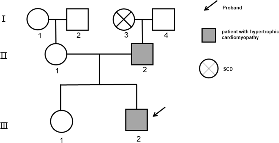
The pedigree of the hypertrophic cardiomyopathy is shown in Fig 1. The detailed clinical data are as follows: the proband (III 2) is a 4-year-old boy who was admitted to a hospital for syncope while running. The electrocardiogram showed sinus bradycardia, abnormal Q wave (I, aVL, and V4-V6 lead), and ST-T changes. The echocardiography result showed the thickening of the interventricular septum and left ventricular posterior wall, mainly above the middle part of the interventricular septum, which was 23 mm at the thickest part. The systolic anterior motion sign was positive (complete). The pressure difference in the left ventricular outflow tract was 78 mmHg. The left ventricular end-diastolic diameter was 4.5 cm. The left ventricular end-systolic diameter was 2.8 cm. The left ventricular ejection fraction was 60%. At present, the proband is being treated with β-blocker (bisoprolol) and is being followed up regularly; the father of the proband (II 2) is 41 years old. He had dyspnoea, palpitation, and chest pain after strenuous exercise a year ago. These clinical symptoms were relieved after rest, and no syncope occurred. The electrocardiogram showed atrial fibrillation rhythm, complete left bundle branch block, ST-T changes, and left ventricular high voltage. The echocardiography showed thickening of the interventricular septal, and the thickest part was 21 mm. The systolic anterior motion sign was negative, the left ventricular ejection fraction was 58%, and no left ventricular outflow tract obstruction was observed; the grandmother of the proband (I 3) died suddenly at the age of 58 years, and no relevant blood samples were collected. Thus, whether she died due to sudden cardiac death induced by hypertrophic cardiomyopathy was impossible to speculate. Her blood was not also collected for genetic testing; and the mother (II 1), sister (III 1), grandmother (I 1), grandfather (I 2), and grandfather (I 4) of the proband had neither clinical symptoms nor substantial genetic variation.
Pedigree of the family with hypertrophic cardiomyopathy (I, II, and III refer to the first, second, and third generations of the family, respectively). Gray shadow represents clinically diagnosed patient with hypertrophic cardiomyopathy. III-2 and = 2 \* ROMAN II-2 are the carriers of MYBPC3: c.2731G>T (+/-).

Gene detection and bioinformatics analysis
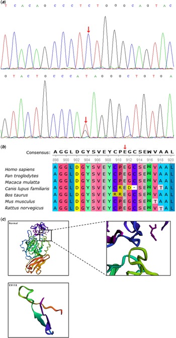
According to the OMIM database and clinical phenotypic analysis, only the MYBPC3 gene variant was found to be associated with the clinical phenotype. The proband carried a novel heterozygous nonsense variant of MYBPC3(NM_000256):c.2731G > T (p.E911X), which is a kind of protein-truncated variation (Fig 2). The variant was further confirmed using Sanger sequencing (Fig 2a). The variant was not present in the 1000 Genomes, gnomAD, Inhouse and ESP6500 databases and was located in the conservative functional domain. This variant was predicted to be harmful by bioinformatics protein function prediction software SIFT, PolyPhen-2, Mutation Taster, and REVEL. According to the American College of Medical Genetics guidelines, the variant was identified as pathogenic: PVS1 + PM2. No report about the variant was found in the ClinVar database. The father of the proband carried the same heterozygous variant at this site, whereas his mother did not. Therefore, the variant of the proband was inherited from his father. ClustalX analysis showed that the amino acid at the variant site is highly conserved among different species (Fig 2b). The 3D visualisation conformation of the protein is shown in Fig 2c. No variant was detected in the rest of the family members and the healthy control group.
Images of deoxyribonucleic acid sequence, amino acid conservation, and three-dimensional protein structures of variant (c. 2731G > T) in MYBPC3. (a) Variant deoxyribonucleic acid sequence analysis: The mutated sequence of MYBPC3(c. 2731G > T) from the proband is shown in the lower panel(arrow). The wild sequence is shown in the upper panel (arrow). (b) Conservation analysis of amino acids: amino acid variation site c. 2731G > T (p.E911X) is highly conserved in many species. (c) Three-dimensional protein structure of variant (p.E911X): The wild protein structure is shown in the upper panel. The variant protein structure is shown in the lower panel: The 911 amino acid mutated to termination codon (p.E911X), resulting in the incompleteness of Fibronectintype-III2 (p872–967) domain, deletion of domain Ig-like C2-type 6 (p971–1065), Fibronectin type-III 3, and Ig-like C2-type 7 (p1181–1274), in which p1253–1268 is predicted to have transmembrane helix structure.

Discussion
Hypertrophic cardiomyopathy is an autosomal dominant genetic disease with an annual incidence of 0.5–1%. Reference Semsarian, Ingles, Maron and Maron11 MYBPC3 encoding cardiac myosin-binding protein C is the gene with the highest variant frequency in hypertrophic cardiomyopathy. Reference Alfares, Kelly and McDermott12 This gene is a member of the immunoglobulin superfamily and a key component of sarcomere. The myosin-binding site of MYBPC3 is mainly located at the C-terminal and directly affects the assembly of cardiac muscle fibres by binding to myosin and actin, which is necessary for myocardial contraction. The regulatory domain of MYBPC3 located at the N-terminal binds to the myosin S2 domain, which participates in the occurrence of hypertrophic cardiomyopathy through interaction with β-myosin. Three β-adrenergic phosphorylation sites exist at the junction of C1 and C2 domains of MYBPC3 and are involved in the regulation of myocardial contraction. Most variants in MYBPC3 lead to a decreased level of functional cMyBP-C, resulting in the acceleration of sarcomere cross-bridge circulation and increased contractile dynamics in animal models. MYBPC3-knocked mice have been widely used as models of hypertrophic cardiomyopathy. Reference Farrell, Grimes, de Lange, Armstrong and Ralphe13 According to statistics, more than 64% of MYBPC3 variants are truncated and produce an unstable mutant peptide. However, nonsense variants are rarely reported in Chinese families with hypertrophic cardiomyopathy. Reference Chen, Jiang, Zhu, Wu and Su14,Reference Tong, Liu and Guo15 Elucidating the clinical characteristics of hypertrophic cardiomyopathy caused by MYBPC3 variations is of great significance for accurately judging the prognosis and stratifying the risk of hypertrophic cardiomyopathy.
In this study, comprehensive screening of pathogenic genes was carried out in a Han Chinese family with hypertrophic cardiomyopathy. We found a novel heterozygous nonsense variant of MYBPC3(NM_000256):c.2731G > T (p.E911X). According to the American College of Medical Genetics guidelines, the variant is a pathogenic protein-truncated variant (PVS1 + PM2). Conservative analysis of amino acids suggested that the variant is highly conserved in many species. Three-dimensional protein structures showed the variant results in the incompleteness of the fibronectintype-III2 (p872–967) domain and the deletion of Ig-like C2-type 6 (p971–1065) and fibronectin type-III 3 and Ig-like C2-type 7 (p1181–1274) domains, in which p1253–1268 is predicted to have transmembrane helix structure. Hence, the variant may change the original structure and cause the functional abnormalities of MYBPC3. Clinical data analysis showed that the phenotypes of the variant carriers with hypertrophic cardiomyopathy were of variable severity and clinical manifestations. Previous studies showed that haploid deficiency is the main pathogenesis of truncation variants. Premature termination of codons usually leads to the degradation of abnormal mRNA through nonsense-mediated decay, which results in haploidy deficiency. Reference Seeger, Shrestha and Lam16 Therefore, patients with MYBPC3 variations need comprehensive clinical risk analysis. Mearini et al Reference Mearini, Stimpel and Geertz17 reported the successful long-term gene therapy of hypertrophic cardiomyopathy in mice for the first time. Moreover, haploid insufficiency and production of toxic peptide were corrected by gene therapy. Gene therapy may become a realistic treatment option for patients with severe hypertrophic cardiomyopathy with MYBPC3 variant given that no other treatment options are available except for heart transplantation.
About 3–5% of adult patients with hypertrophic cardiomyopathy carry the compound or double heterozygotes of two pathogenic variants in the same or different sarcomere genes. Reference van Spaendonck-Zwarts, van Rijsingen and van den Berg18,Reference Fourey, Care and Siminovitch19 Whole-exome sequencing technology can detect gene sequence information and help identify hereditary diseases quickly and accurately. The identification of these pathogenic gene variants not only explains the incidence of childhood probands with hypertrophic cardiomyopathy but also allows screening of other adult relatives at risk of hypertrophic cardiomyopathy. Reference Alsters, Wong and Peferoen20 Identifying the genetic factors of the disease is of great significance for the early diagnosis of family members and is helpful for targeted early intervention and treatment of variant carriers. Therefore, members of families with hypertrophic cardiomyopathy and pathogenic gene variations can be informed of the potential risk. Doctors can also focus on prevention and regular follow-up to provide timely intervention considering the abnormal cardiac function. However, the diagnosis rate, cost-effectiveness, and economic evaluation of whole-exome sequencing should be further improved. Reference Mak, Lee and Tang21,Reference Schwarze, Buchanan, Taylor and Wordsworth22 Proper genetic testing should be achieved in the near future. The detection of new variations in hypertrophic cardiomyopathy not only provides a new understanding of the pathophysiology of hypertrophic cardiomyopathy but also plays an important role in the clinical treatment and prognosis of patients with hypertrophic cardiomyopathy and their families by early and even pre-symptomatic intervention. Reference Williams, Marion and McDonald23,Reference Janin, Chanavat and Rollat-Farnier24 MYBPC3 polymorphisms have also been implicated in sudden infant death syndrome. Reference Brion, Allegue and Gil25 However, further research is needed to explore the functional analysis of the detected MYBPC3 variants by using induced pluripotent stem cells to elucidate phenotypical differences.
Conclusion
In this study, a Han Chinese family with hypertrophic cardiomyopathy was screened using whole-exome sequencing. The proband carried a novel heterozygous nonsense variant of MYBPC3:c.2731G > T (p.E911X). According to the American College of Medical Genetics guidelines, the variant was identified as a pathogenic protein-truncated variant. The phenotypes of the variant carriers with hypertrophic cardiomyopathy were of variable severity and clinical manifestations. Whole-exome sequencing technology is a reliable method for screening hypertrophic cardiomyopathy gene variations and has great significance for early treatment, follow up, and prognosis of hypertrophic cardiomyopathy.
Acknowledgements
We express thanks to the MyGenostics Inc., Beijing, P. R. China.
Financial support
This research was supported by the Projects of Medical and Health Technology Development Plan (2018WS070 and 2018WS072) and the Key Research and Development Plan (2019GSF108186 and 2014GSF118066) of Shandong province in China.
Conflicts of interest
None.
Ethical standards
This study was approved by the ethics committee of Qilu Hospital of Shandong University.