Stenosis within the pulmonary arterial tree can be produced by a heterogeneous group of malformations, including obstructions in both small and large vessels due to congenital or acquired lesions. Although surgical repair is possible in many cases, relief of pulmonary arterial stenosis is the most common indication for therapeutic intervention in our laboratory for paediatric catheterization. Improvements in the technology for manufacture of balloons and stents have resulted, over the past decade, in an increase in the number of tools available to the interventionist. Angioplasty balloons now come in many sizes, are capable of inflation at low or high pressures, and can be equipped with blades for cutting the wall of the vessel. Stents, used to support the wall of the vessel, are also offered in different designs. Recent advances have resulted in a significant reduction in the technical challenges associated with their placement. In this review, we will discuss the unique as opposed to the complementary roles of these therapeutic tools in the trans-catheter management of pulmonary arterial obstructions.
History and evolution of options for treatment of pulmonary arterial stenosis
Trans-catheter management of pulmonary arterial had its genesis in the 1980s. Validation of the technique in animal models1 was followed by clinical application of balloon angioplasty in children with obstructions of the proximal pulmonary arteries in the setting of congenital cardiac disease as an alternative to surgical intervention.2 Improvement in the diameter of the involved vessel, combined with a reduction in right ventricular pressure and normalization of pulmonary perfusion, in most cases with minimal complications, led to the rapid acceptance and application of the technique.3 Initial attempts to use balloons to dilate more distal obstructions, however, were limited due to the size and stiffness of the available balloons. In consequence, a substantial subset of vessels remained resistant to therapy.4 In the next decade, rapid development of balloons with lower profiles, and in wide ranges of size, allowed entry and dilation of virtually all segments of the pulmonary arterial tree. Despite this, it proved that some lesions remained refractory to simple angioplasty.
Innovative application of new technologies, originally developed for use in adults with coronary arterial and peripheral vascular disease, proved necessary to overcome the problem of unresponsive lesions. Lesions fail to respond to simple balloon angioplasty for three distinct reasons. The first is a resistance to tearing or disruption of the wall, as evidenced by a persistent waist in the balloon. The second reason is that compliant lesions are able to expand with angioplasty, but recoiling to their original diameter. Finally, in some instances, the narrowing is caused by asymmetric compression from adjacent structures.
In the late 1980s, therefore, Mullins et al.,5 using a canine model, assessed the potential role of stents in the relief of obstructed pulmonary arteries. This led to multi-institutional efforts to evaluate the safety and efficacy of placement of such stents in children with congenital vascular obstructions.6, 7 Placement of stents also resulted in significant increases in the diameter of the vessels, reduction in pressure gradients across the lesions, and improvements in differential pulmonary perfusion. Most importantly, lesions previously unresponsive to simple angioplasty because of recoil or compression could be kept open by the supporting structure of the rigid metal stent. Experience has now shown that stents are particularly effective for large, compliant, proximal vessels, as well as those compressed by adjacent structures.
Albeit not supported by evidence, it was clear from the beginning that stenotic lesions in pulmonary arteries were unpredictable, and variable in their response to balloon dilation. In our experience, low-pressure angioplasty succeeds in only three-fifths of such dilations. Most of the involved vessels demonstrate resistance to the tearing required for a successful procedure, again as evidenced by failure to eliminate the waist with a balloon. Although placement of stents has proven useful for compliant lesions, their use in vessels resistant to balloon angioplasty is only partially successful. Furthermore, many of these resistant lesions are found distally in the arterial tree, where it may be less desirable to place a stent because of restenosis within the device, and jailing of side branches, with or without occlusion. While the introduction of high-pressure balloons in the 1990s improved the results of simple angioplasty, a subset of vessels remained resistant to therapy. Thus, experience has grown with the use of angioplasty balloons with blades imbedded in the material of the balloon. These so-called cutting balloons have been used successfully in lesions previously resistant to simple angioplasty. In this review, we will compare and contrast the three techniques of angioplasty, placement of stents, and use of cutting balloons.
General concepts for the treatment of pulmonary arterial stenosis
Population involved
Pulmonary arterial obstructions occur in patients with both congenital and acquired disease, and are found as isolated or multiple lesions. Genetic syndromes, such as those of Williams or Alagille, may be associated with such obstructions. They may also be complicating features of simple or complex intra-cardiac malformations, such as septal defects, pulmonary valvar stenosis, or tetralogy of Fallot. Acquired obstructions most commonly occur after surgical interventions for complex congenital cardiac malformations, including anastomoses between a pulmonary artery and either a shunt, a collateral artery, or a conduit. Pulmonary arterial obstructions may also result from stretching, as occurs subsequent to use of the LeCompte manoeuvre for repair of transposed arterial trunks, or compression after surgical augmentation of the aortic arch in palliation of patients with functionally single ventricles. Since the underlying aetiology, and thus the pathology and mechanism of obstruction, is quite different in these conditions, it is not surprising that optimal therapeutic strategies will also vary.
Indications for intervention
Obstruction within the pulmonary arterial tree can cause cyanosis due to limited flow with intra-cardiac shunting, or result in elevated right ventricular pressures. This can lead to ventricular dysfunction, or cause pulmonary hypertension in unaffected pulmonary segments, resulting in irreversible vascular disease. Ideally, interventions should take place prior to development of signs or symptoms from chronic pulmonary obstruction. Universally accepted indications for intervention include symptoms, near or higher than systemic right ventricular pressure or dysfunction, and significant differential pulmonary perfusion. Non-invasive imaging, such as echocardiograms and lung scans, are useful for monitoring patients with known or suspected disease, and can be used to determine the optimal timing for interventions in the absence of symptoms.
Procedural cardio-respiratory support
Patients undergoing pulmonary angioplasty may have isolated proximal obstruction or multiple distal obstructions, with variable haemodynamic consequences. Many cases can be performed with conscious sedation and spontaneous breathing, particularly in cases of isolated obstruction. Management under general anaesthesia, nonetheless, can be life-saving for patients with suprasystemic right ventricular pressures or right ventricular dysfunction, and should be considered for infants and patients requiring treatment of multiple lesions. Inotropic support may also be necessary during the procedure. Patients with suprasystemic right ventricular pressure and an intact atrial septum usually benefit from deliberate creation of an atrial septal defect, thereby decompressing the right heart to maintain cardiac output during the dilations.8
Pulmonary oedema
Pulmonary oedema is common following successful angioplasty or stenting. Oedema due to high flow has been shown to occur when the mean distal pressure exceeds 20 millimetres of mercury, or when acute changes exceed more than 150 percent.9 Clinically significant pulmonary oedema can be successfully managed with diuretics, oxygen, and mechanical ventilatory support, and in our experience has resolved in all patients within 72 hours of the procedure.
Treatment and imaging
Wires placed in the distal pulmonary vessels facilitate delivery of catheters and sheaths. Imaging with hand injections assures placement of stiff wires in safe positions, as wires are a recognized cause of trauma. Wires should be placed in the largest branch of the involved vessel distal to the obstruction to prevent the balloons from being forced out proximally by small distal vessels, or causing aneurysms in vessels stretched excessively. In most cases, the most severe stenosis affecting the greatest proportion of lung is dilated first. When multiple distal interventions are required, proximal lesions are dilated after distal interventions, thus avoiding manipulating and crossing the region after intervention. The use of torque wires is preferred when crossing previously dilated regions. It is important to relieve all possible obstructions, distributing the flow of blood as evenly as possible to avoid excessive flow in unobstructed segments, which can result in pulmonary vascular disease. Angiography can be performed after the intervention by exchanging an angiography catheter over a wire, by positioning of an angiography catheter through a second venous line, or by injecting through a long sheath over a wire. Utilizing a long sheath positioned in a pulmonary artery has the advantage of security, simultaneous measurements of pressure, efficient angiography, and ensures rapid delivery of stents or coils in the event of trauma.
Simple angioplasty
Results
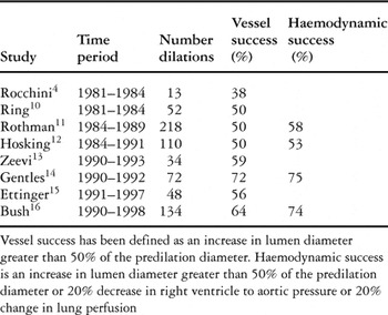
Beginning in the early 1980s, low-pressure balloons were shown to be successful in increasing lumen diameter by at least half in up to three-fifths of cases. Dilations not meeting the change in criterions were considered successful if they resulted in an increase of greater than one-fifth increase in pulmonary perfusion, and a decrease by greater than one-fifth in the ration of right ventricular to aortic pressures. Initial experience included mostly large proximal arteries in young children and adults, with rarely more than one site of dilation per procedure. In the late 1980s, the development of stiff, non-compliant balloons capable of inflation at high pressures improved the proportion of successful dilations in pulmonary angioplasty from 59 to 72 percent (Table 1).4, 10–16 The development of noncompliant balloons on softer catheters with a lower profile led to broader applications, including more distal obstructions, younger patients, and procedures involving multiple dilations.8, 15–20 Despite these improvements in balloon technology, approximately one-third of vessels remain resistant to simple angioplasty.
Table 1. Success of simple balloon angioplasty.

Predictors of success
Vessel tears.
Successful balloon dilations result in tears in the intima and media of the arterial wall, as observed in histological studies and by intravascular ultrasound.21–23 Following successful angioplasty, angiography frequently demonstrates non-obstructive intra-luminal filling defects, or “therapeutic tears”. Initially, investigators observed an association between failed dilations and a persistent waist in the balloon, presumably resulting from failure to tear the stenotic portion of the vessel.11 Subsequently, elimination of the waist with high-pressure balloons, and the presence of a tear, was found to predict acutely successful procedures.14, 16
Size of balloons.
Early experience, confirmed in subsequent series, showed that a larger ratio of the balloon to the minimal luminal diameter was associated with successful procedures.3, 11, 14, 15 Based on these data, a ratio of 3 to 1, but not exceeding twice the distal diameter, became the suggested size of the balloon for pulmonary arterial dilations.24 In a recent review comparing proximal and distal sites of dilation, the initial, and generally conservative, choice of balloon was frequently at a ratio of less than 3 to 1. The size differed by according to the site of dilation, being 2.0 for proximal sites as opposed to 2.7 for distal ones, and increased with subsequent dilations if there was no or minimal waist on the balloon.17 Interventionists must consider multiple characteristics of the morphology, including location, size, and length of the obstruction, as well as features of the adjacent normal arterial segments, when choosing the initial size for the balloon.
Site of angioplasty: post-surgical compared to congenital.
Despite early suggestions that sites of previous surgery may be less amenable to simple angioplasty, a history of surgical intervention at the site of dilation was found to be predictive of success following multivariate analysis.14 In the first report of high-pressure angioplasty results, success was achieved in two-thirds of congenital lesions, compared to over nine-tenths of surgical lesions not previously dilated. In vessels that had previously failed low-pressure dilation, high-pressure angioplasty was successful at almost nine-tenths of surgical sites, compare to less than half of congenital lesions. Such statistically significant differences in success for congenital and acquired lesions have not been confirmed or denied in other series.
Restenosis
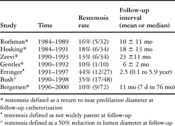
Lasting benefits of simple balloon angioplasty for pulmonary arterial obstructions have been achieved in from two-thirds to nine-tenths of procedures (Table 2).11–17 In series describing restenosis as a return to the diameter prior to dilation, regression has been observed in from one-tenth to one-fifth of dilations. Bush et al.16 provided a more stringent definition, categorizing all vessels with a loss in luminal diameter of more than half as restenotic, resulting in one-third being positive. In a series of proximal dilations reported by Ettinger et al.,15 late failures were treated with stents or surgery, as vessels with recoil characteristics are more common at proximal sites of dilation.17 Although many investigators have sought to identify a cause for restenosis, an association with procedural or arterial characteristics has not been identified.
Table 2. Restenosis rates for simple balloon angioplasty.

Complications
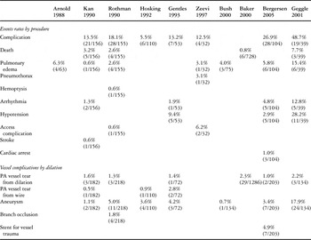
Complications from pulmonary angioplasty can result from excessive tearing of the arterial wall. This can lead to development of obstructive intimal flaps, self-limited extravasation, or castastrophic, life-threatening extravasation (Table 3).3, 8, 9, 11–14, 16, 17, 25 Although the risk of significant pulmonary arterial trauma has remained stable at 1 to 2 percent of procedures, recognition of life-threatening tears, and rapid trans-catheter management with coils, has reduced the rate of mortality to less than 1 percent (Table 4).11, 17, 25 The rate of aneurysmal formation, or focal enlargement of vessels caused by stretching of normal segments distal to the site of obstruction, has also decreased with the recognition of the importance of placing the balloon in the largest possible distal arterial branch.
Table 3. Complications with simple angioplasty.

Table 4. Balloon angioplasty: changes in major complication rates reported over time.

Angioplasty with cutting balloons
A Cutting Balloon™ (Boston Scientific Inc., San Diego CA) has 4 longitudinal microtome blades bonded to a non-compliant balloon. Folded between the material of the balloon, the blades are exposed when the balloon is slowly inflated, achieving a profile of 0.007 inches from the surface (Fig. 1). This device was developed to cut plaques in coronary atherosclerotic lesions at lower pressures of inflation, with smaller ratios of balloon to lesion, thereby reducing trauma to the tissues, and achieving lower rates of restenosis. Results of the global randomized trial showed comparable initial acute gains, rates of dissection, and late loss in luminal diameter for treatment with cutting balloons compared to conventional percutaneous trans-catheter angioplastic techniques.26 Until recently, cutting balloons were only available in the United States of America in 0.25 millimetre sizes from 2.0 to 4.0 millimetres. In June of 2004, the Federal Drug Administration approved the Peripheral Cutting Balloon for use in dialysis grafts, similar in design to the coronary arterial cutting balloon. These balloons are available in 1 millimetre sizes from 5.0 to 8.0 millimetres, with blades of either 1 or 2 centimetres in length.

Figure 1. A cutting balloon.
Use in treating resistant pulmonary arterial stenosis
The novel design of the tool did not go unrecognized by paediatric cardiologists around the world, some of whom were quick to use the device in congenital cardiac malformations which previously had been difficult to manage. Using animal models, the balloon was shown to be capable of creating an atrial septal defect and treating resistant pulmonary arterial obstructions.27–29 Simultaneously, many groups started exploring the effectiveness of the tool in clinical applications, including vessels resistant to high-pressure angioplasty.
Results and technique
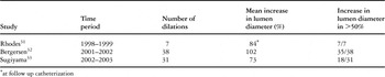
In 2002, Rhodes et al.31 published a small series of dilations for resistant pulmonary arterial stenosis. In all vessels, including four unifocalized collateral arteries, the waist was eliminated with the cutting balloon, and the vessel was subsequently dilated at high pressure with a 5 or 6 millimetre balloon. In these vessels, the mean minimal luminal diameter increased significantly, with follow-up showing maintenance of the initial gains, with an increase in luminal diameter of 84 percent from initial to follow up measurement, which occurred from between 3 and 24 months after the procedure, with a median of 14 months.
At Boston Childrens Hospital, pulmonary arteries resistant to conventional angioplasty have been treated in this fashion since 2001. We use balloons between 0.5 and 1.0 millimetres larger than the waist in the involved vessel persisting at pressures greater than 15 Atmospheres, and more recently greater than 8 Atmospheres. The balloon is slowly inflated to 8 Atmospheres, with inflation maintained for 20 to 60 seconds. It is then slowly deflated to allow proper exposure of the blades and subsequent inward folding of the balloon. A follow-up low-pressure inflation is performed with a slightly larger angioplasty balloon. In our series, we included small children with tetralogy of Fallot undergoing combined catheter and surgical repair, half of whom weighed less than 10 kilograms. Using the technique, we successfully dilated in nearly all cases small pulmonary vessels with a mean minimal luminal diameter of 1.5, ranging from 0.4 to 4.0 millimetres, achieving increases of greater than half in 35 of 38 vessels previously resistant to therapy. We failed with one lesion in a larger vessel requiring a catheter-augmented technique. Prior to the availability of larger sized peripheral cutting balloons, vessels with resistant waists greater than 4 millimetres were treated with a 4.0 millimetre cutting balloon alongside a rigid catheter. This technique was successful in at least 5 other vessels, with the only serious complication in the initial report resulting from distal perforation, requiring coil occlusion after augmented therapy.
The larger peripheral cutting balloons were first used for treatment of pulmonary arterial stenosis at the Hospital for Sick Children in Canada,33 proximal lesions being involved in 12 of 31 dilations. Their series also extended the experience in children with syndromes such as Alagille's and Williams', including multiple peripheral obstructions. In this series,33 the cutting balloon was sized 1 to 2 millimetres larger than the diameter of the normal vessel adjacent to the stenosis. The analysis revealed a relationship between successful increase in luminal diameter and a minimal ratio of the lumen to the balloon of greater than 200 percent. In the 26 vessels with a persistent waist following the initial dilation, subsequent use of cutting balloons resulted in a gain of at least half in luminal diameter in 18 of 26 dilations. In addition to these findings, the investigators demonstrated that diffusely hypoplastic vessels, and vessels in which a waist did not form with simple angioplasty, did not change significantly following use of cutting balloons.
Restenosis
Cutting balloons appear to produce a tear when the radial strength of simple angioplasty is insufficient, producing evidence of therapeutic tears in at least half dilations. Despite initial concerns about restenosis, the angiographic appearance of both coronary and pulmonary arteries have shown rates of restenosis comparable to simple angioplasty.34
Complications
To date, no significant concerns have resulted from use of cutting balloons as described to treat native pulmonary arterial stenosis. The majority of patients requiring such interventions are at high risk, requiring multiple dilations often resistant to high pressure techniques. In this population, the procedures are often lengthy, with patients frequently requiring anaesthesia and haemodynamic support. Signs of transient pulmonary oedema due to high flow often occur following successful dilations. We do not yet know whether the risk of rupture of 1 percent observed with simple angioplasty will change with the use of cutting balloons.
Current and future indications
Currently, the balloons are being used to treat pulmonary arterial obstructions in vessels resistant to simple angioplasty (Table 5). Successful therapy has not been observed in compliant vessels. Based on encouraging initial experiences using cutting balloons for resistant pulmonary arterial stenosis, Boston Children's Hospital is sponsoring a prospective, randomized, multi-centric trial to evaluate their safety and efficacy. In this trial, approved by the Federal Drug Administration, lesions resistant to dilation at pressures lower than 8 Atmospheres as evidenced by a persistent waist are randomized to either high-pressure angioplasty or dilation with cutting balloons. Results of this trial will allow a direct comparison of the efficacy and safety of these techniques.
Table 5. Results of cutting balloon angioplasty for resistant peripheral pulmonary arterial stenosis.

Placement of stents
Indications
Compliant obstructions with recoil characteristics, and obstructions caused by external compression, kinking, or tension on a vessel, require the rigid framework of a stent to maintain their patency. Special consideration should be given to placement of stents at sites of obstruction in the early post-operative period to avoid the need for therapeutic tears in fresh surgical sites, or in cases of recurrent obstruction after angioplasty.35–37 In addition, stents are sometimes necessary for the management of vascular trauma when either an obstructive intimal flap develops after angioplasty, or a tear with extravasation, can be managed with a covered stent. Stents are often optimal treatment for proximal pulmonary arterial obstructions with recoil characteristics, and appear to have excellent short and long-term results in relief of obstruction to flow. In nearly all cases, it is possible further to expand these stents at follow-up to account for somatic growth. There is, however, one major drawback to their implantation, since an expanded stent is nearly impossible to retrieve or reposition accurately.
Advantages and disadvantages of their design
In the 1990s, The Palmaz™ stent (Johnson and Johnson, Sommerville, NJ) was the most commonly used stent for congenital cardiac malformations (Fig. 2).38 Clinical application in systemic, pulmonary, arterial and venous obstruction has been reported with extended follow-up. In proximal pulmonary arterial obstructions, investigators have found significant and consistent increases in the diameter of the involved artery, reduction in pressure gradient, and improved pulmonary perfusion.6, 7, 39–41 Although a one-millimetre reduction in luminal diameter has been observed due to intimal growth, significant re-obstruction, thrombosis, or aneurysms are rare when stents are placed in proximal pulmonary arteries. Those placed in the relatively smaller vessels of the distal vasculature, in contrast, have a higher rate of re-obstruction and incidence of jailing of side branches.42 Although many studies have shown that redilation up to 2 years after placement is possible to account for somatic growth, the extent of expansion is limited by the foreshortening that occurs with the closed cell design.43–45 In small infants and children, placement of renal stents in the proximal pulmonary arteries was often palliative, but they eventually required excision due to the foreshortening and inadequate final diameter needed for adults.

Figure 2. The closed cell design of the Palmaz stent.
Some of the technical problems with Palmaz stents lead to investigations of alternative technology. Palmaz stents require hand crimping onto a balloon, with a final profile necessitating large sheaths, frequently exceeding 8 French. The size of the sheath is an obvious limitation when trying to treat infants and children with congenital malformations. This problem was partially overcome by innovative front-loading techniques.46 In many cases, front loading was necessary to minimize displacement of the stent from the balloon during delivery, particularly when accessing pulmonary arteries via a tortuous course. Rupture of balloons on the edges of the stent during placement is a serious problem that may result in permanent malposition. Balloon-in-balloon angioplasty catheters, therefore, were developed to overcome such rupture and slippage. Despite technical modifications, however, misplacement requiring removal or expansion of a stent in a non-obstructive site still occurred in at least 2 percent of procedures.39 Clinical evaluation has revealed that flexible and self-expanding stents are less effective for initial procedures, with a higher rate of restenosis.47, 48
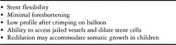
Availability of recent new designs, nonetheless, has led to widespread application in paediatric interventional cardiology. Limited publications suggest impressive advantages in the closed cell design with interposed sigma hinges of the Genesis™ stent (Cordis, Warren NJ – Fig. 3), and open cell stent design of the IntraStent™ (ev3 Inc., Plymouth MN) for the treatment of congenital vascular obstructions. In vitro testing of the Genesis XD 19/29 stent demonstrates more flexibility, less foreshortening, lower profile when crimped on a balloon, and superior radial strength compared to the Palmaz 308.49 Clinical use has further revealed two important clinical advantages, first that stent cells can be crossed and dilated to access and restore flow to jailed vessels, and second, that redilation is possible with minimal foreshortening, expanding to adult size (Table 6). With respect to over-expansion and redilation, however, the open cell design appears to be limited by asymmetric expansion and recoil.50–51 Although not yet shown in a large clinical series, it is our impression that complications such as balloon rupture, malposition, and occlusion of side branches have all been reduced with the use of premounted and non-premounted Genesis stents. While experimental testing of the Genesis stent has suggested superior radial strength, follow-up data is needed to determine if this design can withstand the external forces of compression encountered in many congenital and surgical lesions requiring stents. The clinical advantages and disadvantages of this technology in pulmonary arteries, and other sites of implantation, must be critically evaluated in the near future.

Figure 3. The Genesis stent has closed cells with interposed sigma joints.
Table 6. Advantages of new stent designs.

Summary
The trans-catheter treatment for pulmonary arterial stenosis cannot be encompassed by one ideal technology (Fig. 4). Instead, the complementary and unique roles of available tools must be understood and exploited to treat a disease characterized by variable characteristics of the involved arteries, and mechanisms of their obstruction. Simple angioplasty is therapeutic in many cases, and is critical for the evaluation of vessel compliance as evidenced by a balloon waist in non-compliant lesions, or recoil, as seen with compression, stretching, and kinking of vessels. In the case of resistant obstructions, growing experience with cutting balloons has shown effective relief of non-compliant obstructions in distal pulmonary vessels. For compliant obstructions, the rigid support provided by stents is required for the effective management of vessels with recoil characteristics or compression by adjacent structures. Additionally, stents are often crucial in the management of vascular trauma caused by angioplasty. New designs appear to offer many advantages over previously available stents, and may result in fewer procedural complications. These innovations in stent and angioplasty technology have resulted in more effective treatment of congenital and surgical vascular obstructions in both proximal and distal pulmonary arteries.

Figure 4. Therapeutic choices for pulmonary arterial obstructions: a single institutional experience in 2004.