Left ventricle to right atrium communication which is a rare defect, may be either congenital or acquired.Reference Gerbode, Hultgren, Melrose and Osborn1–Reference Sinisalo, Sreeram and Jokinen3
After the first successful device closure of acquired LV-RA communication in 2006, interventional therapy with devices as well as surgical treatment have started to be used in the management of Gerbode defects.Reference Trehan, Ramakrishnan and Goyal4 Different devices such as Amplatzer Duct Occluder (St Jude Medical, St Paul, Minnesota, United States of America) ADO 2, ADO 1, Amplatzer Vascular Plug II, and muscular VSD device have been used to close this defect.Reference Yuan5
Herein, we present a patient in whom the ADO 2 device was successfully used for the closure of a supravalvar type LV-to-RA communication.
Case
A 7-year-old girl was referred to our hospital because of right heart failure and supraventricular tachycardia. The patient was receiving amiodarone (IV, 5 mcg/kg/min), spironolactone, enalapril, and propranolol at admission. The patient had undergone transitional atrioventricular septal defect and left atrioventricular valve repair, and right pulmonary artery reconstruction in another hospital 1 year before.
At the time of presentation, the patient had signs of right heart failure. Nonsustained atrial tachycardia episodes were detected by 24-hour Holter monitoring.
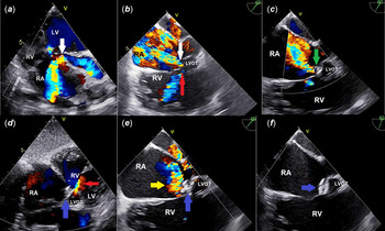
Transthoracic echocardiography revealed iatrogenic Gerbode defect (direct LV-RA communication) measuring 3.7 mm at a distance of 9 mm and 5 mm, respectively, from the hinge point of the aortic valve and tricuspid leaflets and also 2.5 mm residual perimembranous VSD and moderate tricuspid regurgitation (Fig 1a). Peak gradient from TR was 40 mmHg and right atrial dilatation was determined.
TTE: Modified apical four-chamber view showing LV-RA communication (white arrowhead) with the left-to-right shunt. ( a ) TEE: Modified short-axis view showing LV-RA communication (white arrowhead) and residual ventricular septal defect (red arrowhead) with the left-to-right shunt. ( b ) TEE: Modified short-axis view showing important residual shunt, after AVP 2 device (green arrowhead) defect, which is still attached to the delivery cable positioned in the defect. ( c ) TTE: Subcostal modified apical four-chamber view ADO 2 device (blue arrowhead) in situ with no residual LV-RA shunt and no residual shunt of the ventricular septal defect (red arrowhead). ( d ) TEE: Modified short-axis view showing ADO 2 device (blue arrowhead) in situ with no residual LV-RA shunt, tricuspid regurgitation jet (yellow arrowhead). ( e ). TEE: Modified short-axis view showing the final position of the ADO 2 device being released ( f ). ADO = Amplatzer Duct Occluder; AVP = Amplatzer Vascular Plug; LV = Left Ventricle; LVOT = Left Ventricular Outflow Tract; RA = Right Atrium; RV = Right ventricle; TEE = Transesophageal echocardiogram; TTE = Transthoracic echocardiogram.

Informed consent was obtained from the parents. Cardiac catheterisation was performed under general anaesthesia.
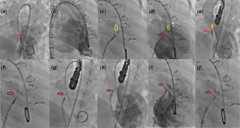
The size of the defect was measured 3.5 mm by transesophageal echocardiography (TEE, modified short- axis window) and LV angiogram (RAO 30° , LAO 45, and cranial 25° positions) (Fig 1b and Fig 2a and b). LV-RA communication was crossed from the LV using a 5F Judkins right coronary catheter (Cordis Corporation, Bridgewater, NJ, USA) along with an 0.035'' Terumo wire (Terumo Corporation, Tokyo, Japan). An arteriovenous loop was established. AVP II 6 mm device was placed in the defect from the venous side via the 5F Amplatzer TorqVue LP Delivery System. AVP II device was positioned in the defect, but an important residual shunt was detected by echocardiography and LV angiography before release and the device was pulled back into the long sheath (Fig 1c and Fig 2c–e). Thereafter, an ADO 2 device 6/4 was placed in the defect from the venous side using the antegrade route, and the distal disc of the device was deployed in the LV pulling in to the level of the defect and finally releasing the body and proximal disc of the device into the defect and in the RA side, respectively, (Fig 2f and g). Venous access was used to shorten the procedure time and to avoid complications related to the arteriovenous loop. In addition, the wire passed easily and quickly from the right atrium to the left ventricle. It was confirmed by TEE that the wire was not associated with any intracardiac structure.
Left ventriculography LAO 40°, cranial 25° ( a ) and RAO 30° ( b ) view showing post-operative Gerbode defect (red arrowhead) and LV-to-RA shunt diameter 3.5 mm (red line) ( a and b ). After AVP 2 device (yellow arrowhead), which is still attached to the delivery cable, positioned in the defect, left ventricular angiogram RAO 30°, LAO 40°, and cranial 25° showing important residual shunt (red arrowhead) ( c , d , e ). ADO 2 device (red arrowhead), which is still attached to the delivery cable, positioned in the defect ( f , g ). Left ventriculography and LAO 40°, cranial 25° ( h ) and RAO 30° ( i ) view showing complete disappearance of LV-to-RA shunt ( h and i ). Final position of the ADO 2 device being released ( j ). ADO = Amplatzer Duct Occluder; AVP = Amplatzer Vascular Plug; LAO= Left Anterior Oblique; LV = Left Ventricle; RA = Right Atrium; RAO = Right Anterior Oblique.
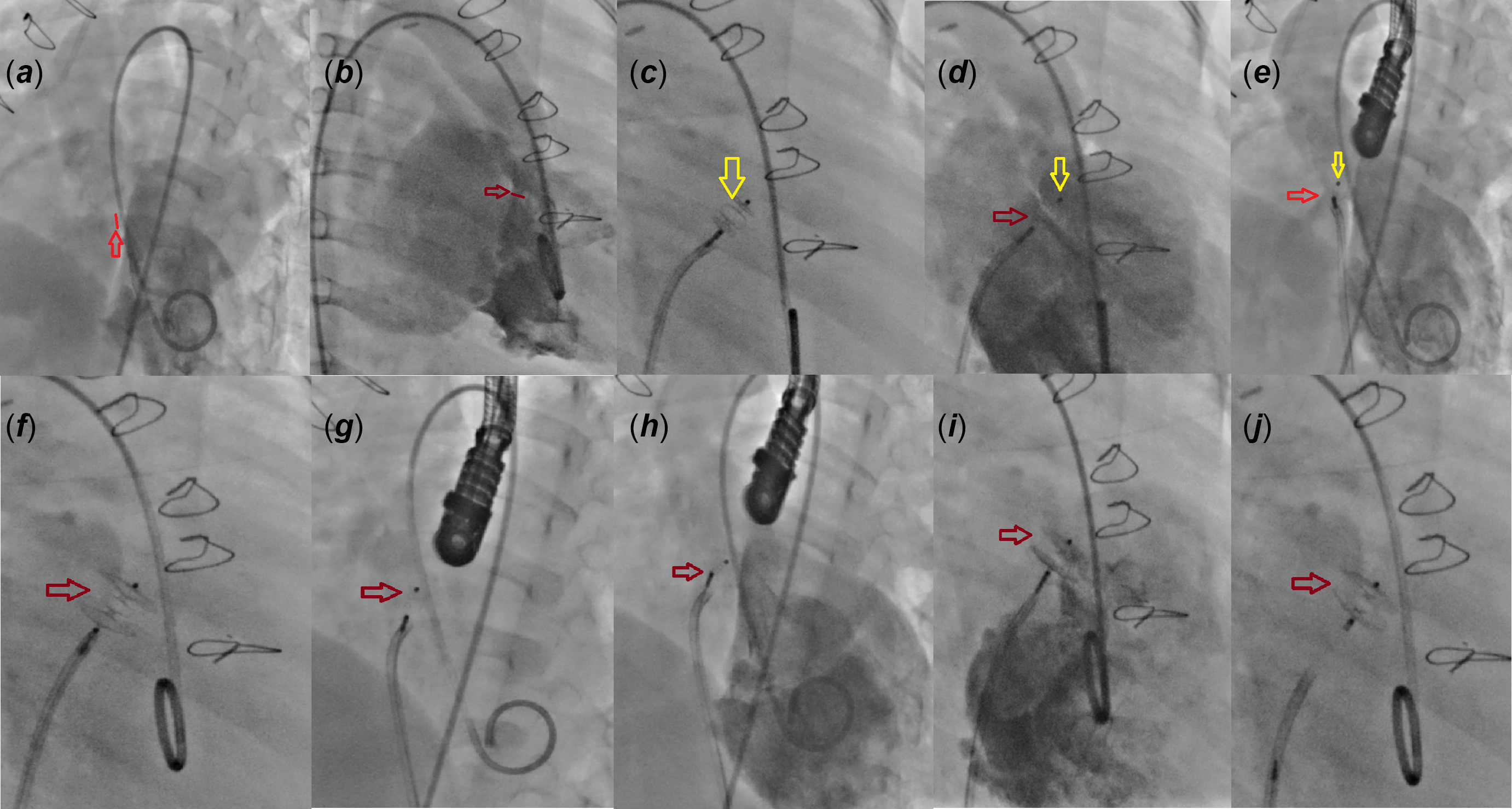
Left ventriculography was performed showing a proper position of the device with no residual shunt (Fig 2h and i). TEE and TTE showed no residual flow through the device without any progression of TR, and no aortic regurgitation (Fig 1d and e). A brief period of sinus bradycardia and junctional escape rhythm was recorded. The patient was given steroids and atropine, and then sinus rhythm was recorded by electrocardiogram and the device was released (Figs 1f and Fig 2j).
After the procedure, the signs of right heart failure improved, Holter monitoring was normal, and antiarrhythmic treatment was discontinued. She was discharged on the 7th day after the procedure of 5 mg/kg/day aspirin and enalapril.
Discussion
We reported successful closure of iatrogenic type 1 Gerbode defect with ADO 2 after attempting to close with an AVP II device. Different devices such as ADO 1–2, AVP II, muscular VSD devices have been used to close Gerbode defect.Reference Yuan5,Reference Vijayalakshmi, Setty and Narasimhan6 Although the ADO 2 device is designed for the patent ductus arteriosus, it is also used properly for LV-RA communication. Successful transcatheter closure of this defect with the ADO 2 device has been reported previously.Reference Vijayalakshmi, Setty and Narasimhan6,Reference Shi, Wang and Li7
Advantages of using the ADO 2 device in closing Gerbode defects are anterograde or retrograde placement without the need for an arteriovenous loop, having a soft low profile, minimising embolisation, and residual shunt risk by having two retention discs.
We chose the ADO 2 device because we thought that the retention discs would better cover the defect. When using the ADO 2 device in type 1 Gerbode defects, although it has soft retention discs, we think that a distance of more than 3 mm between the defect and the aortic valve and tricuspid valve will be better.
Transient junctional rhythm in 2 patients and transient complete heart block requiring temporary pacemaker implantation in one patient was reported in a case series including 12 patients with Gerbode defect closed with ADO 2.Reference Vijayalakshmi, Setty and Narasimhan6 In the present case, although a brief period of junctional escape rhythm was recorded during the procedure, atrioventricular block and junctional rhythm were not detected by 24-hour Holter monitoring.
Although the AVP II device was first chosen in this case because of its low profile and inert effect on the AV node, it was unsuccessful due to lack of retention discs. AVP II failure on Gerbode defect closure was described previously.Reference Ganesan, Paul and Mahadevan8 However, successful transcatheter closure iatrogenic Gerbode defect with AVP II was reported by Vázquez et al.Reference Vázquez, Reyes and Jiménez9
Conclusion
This report supports the view that transcatheter closure of post-operative Gerbode defect with the ADO 2 device is safe, effective, and alternative to surgical treatment. Also in our opinion, ADO 2 device is a better choice than AVP II in this procedure. However, this needs to be validated in a large cohort of patients.
Acknowledgements
None.
Financial support
This research received no specific grant from any funding agency or commercial or not-for-profit sectors.
Conflicts of interest
None.
Ethical standards
The authors assert that all procedures contributing to this work comply with the ethical standards of the relevant national guidelines on human experimentation (ethical guidelines for biomedical research on human participants 2006, India) and with the Helsinki Declaration of 1975, as revised in 2008, and has been approved by the institutional committees.