Burn injuries are an important source of morbidity and mortality in the United States; an estimated 486,000 people required medical treatment and ~40,000 required hospitalization for a burn in 2016 alone. 1 , Reference Mason, Esselman, Fraser, Schomer, Truitt and Johnson 2 Burn patients are particularly vulnerable to infection due to the nature of their injury, their prolonged hospitalizations, hypermetabolic and hypercatabolic conditions, inhalational injuries, and frequent use of invasive devices.Reference Mayhall 3 , Reference Rafla and Tredget 4 In addition to these factors that impact local immunity, systemic immunity is also altered in the burn setting.Reference Cairns, Maile, Barnes, Frelinger and Meyer 5 – Reference Neely, Maile, Wang, Vadlamudi, Meyer and Cairns 7
The evolution of burn care management has led to significant improvements in morbidity and mortality rates. Early excision and grafting, enhanced antimicrobial options, and a better understanding of the pathophysiology of burns have significantly improved outcomes for burn survivors: survival is the rule, no longer the exception. In addition to the baseline risk for healthcare-associated infection (HAI) in hospitalized patients, burn patients are at increased risk of infection until their wounds are grafted and/or healed. Those patients with comorbid conditions may be at even higher risk of infection. This risk is a devastating consequence of burn trauma. It has been estimated that infections cause half of all burn deaths.Reference D’Avignon, Hogan and Murray 8
Despite the tremendous impact of infections in the burn patient population, risk factors beyond burn size and inhalational injury are unclear, and whether burn size impacts the effect of risk factors on HAI incidence remains unknown. Thus, the goal of this study was to estimate the association between patient demographics, comorbidities, and burn characteristics with time to HAI among adult patients hospitalized for burn injuries. To the best of our knowledge, this is the first study to assess potential risk factors for HAIs irrespective of site and pathogen.
METHODS
Study Population
Patients admitted between January 1, 2004, and December 31, 2013, to the North Carolina Jaycee Burn Center at the University of North Carolina (UNC) Hospitals were identified using the burn center registry, which includes data collected for reporting to the National Burn Repository. Adult patients (ie, ≥18 years old) admitted with burn injury (including inhalation injury alone) were eligible for inclusion. Only the first hospitalization for a patient’s first burn within the period was included in the analyses (n=5,576). Patients were excluded if the discharge date could not be determined (n=37; 0.7%; these patients were all discharged alive) or if they were hospitalized for <2 days (n=1,113; 20.0%).
Measures
Registry data were linked to the Carolina Data Warehouse for Health, a central repository for clinical and administrative data from the UNC Healthcare System and the UNC Hospital Epidemiology database, which captures healthcare-associated infections (HAIs) through real-time, comprehensive, hospital-wide surveillance in accordance with the Centers for Disease Control and Prevention (CDC) criteria. 9 , Reference Weber, Sickbert-Bennett, Brown and Rutala 10 Age was categorized as <30 years old, 30–39 years old, 40–49 years old, 50–59 years old, or ≥60 years old, and total burn surface area (TBSA) was categorized as <5% TBSA, 5–10% TBSA, 11–20% TBSA, or >20% TBSA. Revised Baux scores were calculated as described by Osler et al.Reference Osler, Glance and Hosmer 11 Comorbidities were identified using International Classification of Disease, Ninth Revision, Clinical Modification (ICD-9-CM) diagnostic codes in the administrative records, and included diabetes (250–250.9), chronic pulmonary disease (490–496.9, 500–505.9, and 506.4), congestive heart failure (428–428.9), prior myocardial infarction (412–412.9), pre-existing renal disease (582– 583.7, 585 – 586.9, and 588 – 588.9), peripheral vascular disease (443.9, 441–441.9, 785.4, and V43.4), and cerebrovascular disease (430–438.9). These coding schemes were all validated by Deyo et al.Reference Deyo, Cherkin and Ciol 12
Statistical Analyses
Bivariate analyses comparing demographics, comorbidities, and burn characteristics between patients with and without an HAI within the first 60 days of hospitalization were performed using 2-tailed, Fisher’s exact, and Wilcoxon-Mann-Whitney tests, where appropriate. Kaplan-Meier survival curves were used to estimate the 60-day cumulative incidence of HAI, stratified by each factor of interest; inpatient mortality was treated as a competing risk.Reference Lau, Cole and Gange 13 A 2-sided P<.05 was considered statistically significant.
Multivariable Cox proportional hazards regression was used to estimate the direct effect of demographics, comorbidities, and burn characteristics (after adjusting for all other potential risk factors) on the time to HAI. The multivariable model included patient age, sex, race/ethnicity, comorbidities (ie, diabetes, chronic pulmonary disease, congestive heart failure, prior myocardial infarction, pre-existing renal disease, peripheral vascular disease, and cerebrovascular disease), burn mechanism, TBSA, and inhalational injury. Patients contributed time from first day of hospitalization until their first HAI, death, discharge, or administrative censoring at 60 days postadmission. Radiation was reclassified as an “other” mechanism due to low incidence. To assess whether effect measure modification by TBSA of any risk factor on time to HAI, the multivariable Cox model described above (plus additional interaction terms between TBSA and each risk factor) was also fit. Wald χ2 tests were used to identify significant modification. Variables with a P<.10 were considered to have statistically significant modification.
To account for potentially informative censoring (ie, being discharged alive <60 days after admission), inverse probabilities of censor weights were calculated.Reference Cole and Hernan 14 Briefly, among patients censored (ie, did not have an infection or died), length of stay was partitioned into quintiles. Using those cut points, a pooled logistic regression model was used to estimate the probability of each patient being censored in each period, given the patient had not been discharged and had not been infected or died during the prior period, adjusting for patient age, sex, race/ethnicity, comorbidities, burn mechanism, TBSA, inhalational injury, and admit year. Weights were scaled by the marginal (ie, overall) probability of being censored in each period and truncated at the 5th and 95th percentiles. Robust sandwich estimators were used to account for the weighting in the Cox regression models.
All analyses were performed using SAS version 9.4 software (SAS Institute, Cary, NC). Approval for this study was obtained from the Institutional Review Board (IRB) of the University of North Carolina at Chapel Hill.
RESULTS
Overall, 4,426 patients were hospitalized for at least 2 days and were included in the analysis. The median length of stay was 10 days (interquartile range 5–17). Only 120 patients (2.7%) had hospitalizations longer than 60 days without an HAI and were censored; of those patients, 29 (24.2%) had HAIs after 60 days. Moreover, 172 patients (3.9%) died during hospitalization and within 60 days of admission; among them, 68 (39.5%) died occurred after an HAI.
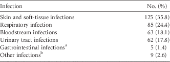
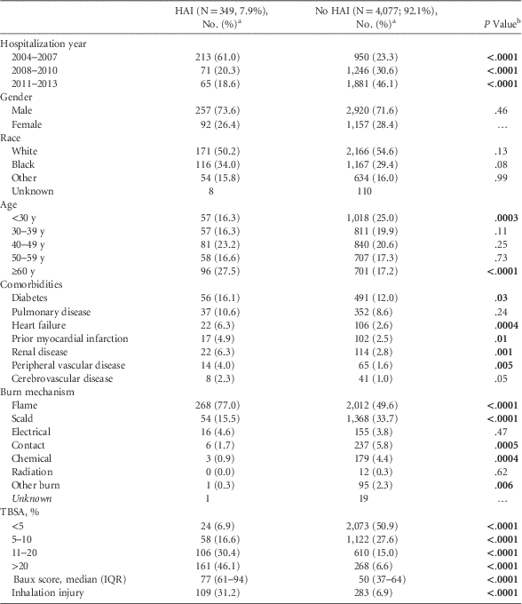
Within 60 days of admission, 349 (7.9%) patients had at least 1 HAI. Skin and soft-tissue infections (n=125; 35.8%) and respiratory infections (n=85; 24.4%) were the most common infection types (Table 1). In addition, 50 respiratory infections (52.6%) were ventilator associated and 45 urinary tract infections (67.2%) were catheter associated. Patients with HAIs were more likely to be older (median age, 48 years vs 43 years; P<.0001), to have diabetes (16.1% vs 12.0%; P=.03), to have congestive heart failure (6.3% vs 2.6%; P=.004), to have a prior myocardial infarction (4.9% vs 2.5%; P=.01), to have pre-existing renal disease (6.3% vs 2.8%, P=.001), and to have peripheral vascular disease (4.0% vs 1.6%; P=.005) (Table 2). Additionally, patients with HAIs were more likely to have flame burns (77.0% vs 49.6%; P<.0001), larger burns (median TBSA, 19% vs 5%; P<.0001), and inhalational injuries (31.2% vs 6.9%; P<.0001). No significant differences were detected in the distribution of gender, race, or pulmonary disease.
Breakdown of First HAI by Infection Type

NOTE. HAI, hospital-associated infection.
a All gastrointestinal infections were caused by Clostridium difficile.
b Other infections include cardiovascular infections and ear/eyes/nose/throat infections.
Demographics, Comorbidities, and Burn Characteristics of Adult Patients, Stratified by HAI Status

NOTE. HAI, hospital-associated infection; IQR, interquartile range; TBSA, total burn surface area. P values <.05 are bold.
a Unless otherwise specified.
b Two-tailed Fisher’s exact and Wilcoxon-Mann-Whitney tests were used to calculate P values.
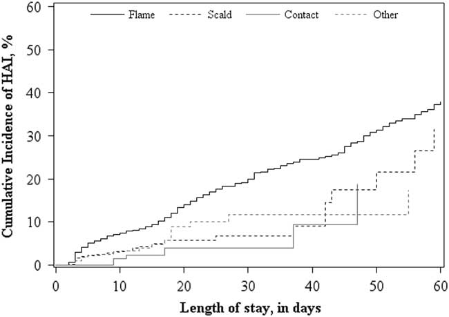
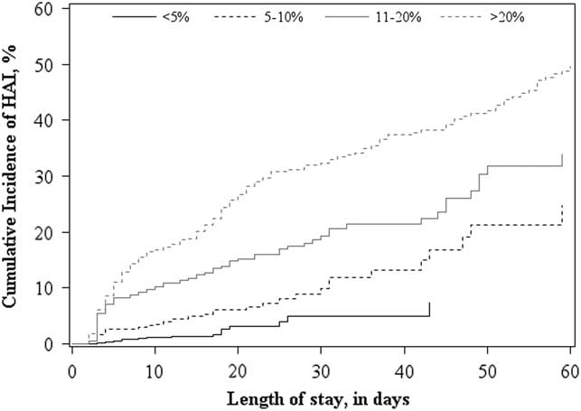
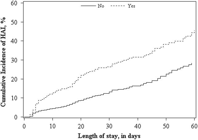
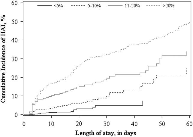
No significant difference in the cumulative incidence of HAI was observed across gender (P=.69), race (P=.62), age (P=.39), diabetes (P=.60), pulmonary disease (P=.24), congestive heart failure (P=.11), prior myocardial infarction (P=.42), pre-existing renal disease (P=.92), peripheral vascular disease (P=.74), or cerebrovascular disease (P=.60) (data not shown). Burn mechanism was significantly associated with HAI incidence (P<.0001), and patients admitted with flame burns had a significantly higher cumulative incidence of infection (Figure 1). Similarly, burn size and inhalational injury were significantly associated with increased cumulative incidence (P<.0001 and P<.0001, respectively) (Figures 2 and 3).
The 60-day cumulative incidence of healthcare-associated infections among adult burn patients, stratified by flame burns (black solid line), contact burns (black dashed line), scald burns (gray solid line), and other burn mechanisms (gray dashed line).

The 60-day cumulative incidence of healthcare-associated infections among adult burn patients, stratified by <5% total burn surface area (TBSA) (black solid line), 5%–10% TBSA (black dashed line), 11%–20% TBSA (gray sold line), and >20% TBSA (gray dashed line).
The 60-day cumulative incidence of healthcare-associated infections among adult burn patients, stratified by patients with (dashed line) and without (solid line) inhalational injury.

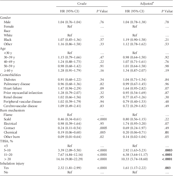
Burn mechanism was significantly associated with time to HAI in the bivariate analysis; however, after adjustment for all other potential risk factors and weighting for censoring, only chemical burns, compared to flame burns, were significantly associated with a lower hazard of infection (HR, 0.20; 95% CI, 0.06–0.71) was observed (Table 3). Both TBSA and inhalational injury significantly changed the hazard of HAI acquisition both before and after adjustment. Compared to patients with <5% TBSA, patients with 5%–10% TBSA were almost 3 times as likely to acquire an HAI (HR, 2.92; 95% CI, 1.63–5.23); patients with 10%–20% TBSA were >6 times as likely to acquire an HAI (HR, 6.38; 95% CI, 3.64–11.17); and patients with >20% TBSA were >10 times as likely to acquire an HAI (HR, 10.33; 95% CI, 5.74–18.60). Patients with inhalational injury were 1.5 times as likely to acquire an HAI (HR, 1.61; 95% CI, 1.17–2.22). Both before and after adjustment, no significant differences in time to first HAI were detected across gender, race, age, and comorbidities.
Unadjusted and Adjusted Effects of Demographics, Comorbidities, and Burn Characteristics on Time to HAI

NOTE. HAI, hospital-associated infection; HR, hazard ratio; CI, confidence interval; Ref, reference; TBSA, total burn surface area. P values <.05 are bold.
a Estimates are adjusted for gender, race, age, comorbidities, burn mechanism, TBSA, and inhalational injury and weighted to account for potentially informative censoring.
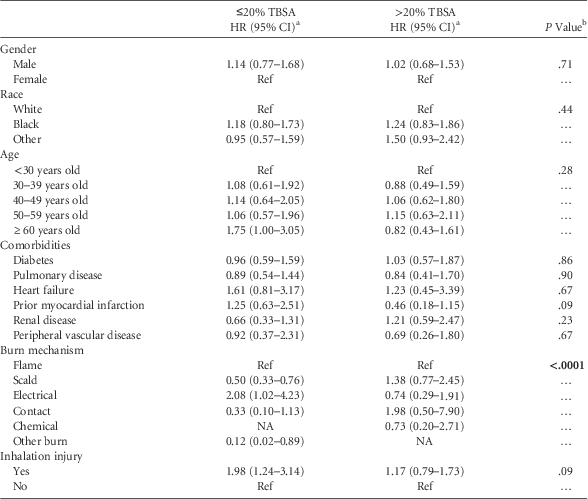
The effect of demographics and most comorbidities were also consistent across smaller (≤20% TBSA) and larger (>20% TBSA) burns (Table 4). However, prior myocardial infarction (P=.09), burn mechanism (P<.0001), and inhalational injury (P=.09) were each found to have differential effects across burn size. Specifically, prior myocardial infarction appeared to increase the incidence of infections among patients with small burns (HR, 1.25; 95% CI, 0.63–2.51) but to decrease the incidence among patients with larger burns (HR, 0.46; 95% CI, 0.18–1.15). Additionally, patients with small burns, scald burns (HR, 0.50; 95% CI, 0.33–0.76), and contact burns (HR, 0.33; 95% CI, 0.10–1.14) appeared to have a lower likelihood of HAI compared to flame burns. However, among patients with large nonflame burns, an increased likelihoods of HAI were observed: scald HR, 1.38 (95% CI, 0.77–2.45) and contact HR, 1.98 (95% CI, 0.50–7.90). Alternatively, the electrical burns increased the likelihood of HAI in small burns (HR, 2.08; 95% CI, 1.02–4.23) and lowered the likelihood of infection among patients with large burns (HR, 0.74; 95% CI, 0.29–1.91). Finally, the effect of inhalational injury appeared to have a smaller effect on time to HAI (HR, 1.17; 95% CI, 0.79–1.73) in patients with large burns compared to those with smaller burns (HR, 1.98; 95% CI, 1.24–3.14).
Assessment of Potential Effect Measure Modification Between Potential Risk Factors and Time to HAI by Burn Size

NOTE. HAI, hospital-associated infection; HR, hazard ratio; CI, confidence interval; Ref, reference; TBSA, total burn surface area; NA, not analyzable. P values <.05 are bold.
a All estimates are adjusted for gender, race, age, comorbidities, burn mechanism, TBSA, and inhalational injury, as well as interaction terms between each potential risk factor and TBSA, and weighted to account for potentially informative censoring.
b Wald tests for significant effect modification of the potential risk factor by burn size.
DISCUSSION
In this analysis of 4,426 burn patients hospitalized for at least 2 days between 2004 and 2013, the 60-day incidence of HAIs among burn patients was 8%. Patients with higher TBSA and inhalational injury were significantly more likely to acquire an HAI during their hospitalization. Moreover, the effect of burn mechanism and inhalational injury was differential across burn size. Neither demographics nor comorbidities were associated with time to HAI, even after allowing for differential effects across burn size. Other studies have also found that burn size, inhalational injury, and cardiac disease were associated with increased risk of infection.Reference Mayhall 3 , Reference Issler-Fisher, McKew, Fisher, Harish, Gottlieb and Maitz 15 – Reference Schultz, Walker and Elligsen 17 However, these previous analyses have been in specific populations (eg, older adults) or only looked at specific infection sites and pathogens. To our knowledge, this is the first study to conduct a comprehensive analysis of risk factors for HAIs, irrespective of site and pathogen, among all adults.
We also found that the effects of burn mechanism were differential across burn size. Among patients with TBSA ≤20%, flame burns and electrical burns had the highest incidence of HAI, and patients with larger scald and contact burns (ie, >20% TBSA) had a higher incidence of HAI. Typically, large scald and contact burns are either secondary to clinically significant neuropathies, where patients are unable to feel the high temperature of the liquid or object they are handling, or are the result of seizing or falling near a hot object and then being unable to move away.Reference Masud, Norton, Smailes, Shelley, Philp and Dziewulski 18 , Reference Rimmer, Bay and Foster 19 While we adjusted for multiple comorbidities, the differential effect in burn mechanism across burn size may be due to the inability to fully control for patient frailty and health status.
Additionally, the effect of inhalational injury was also differential across burn size, with the effect of inhalational injury being more substantial among patients with small burns. There are a few plausible explanations. Possibly, while inhalational injury and burn size are risk factors for infection, when presenting together they have a less than multiplicative relationship (ie, the total effect is less than the sum of the parts). Alternatively, because we used ‘any HAI’ as our outcome, it could be that patients with larger burns were at such an increased risk for skin and soft-tissue infections and death (a competing risk) due to their large burn that inhalational injury was unable to have a meaningful impact using this study design. Future studies should look at the effect of these risk factors on specific infections types (eg, respiratory infections) to determine whether the effects of these risk factors change across infection type and whether the effect of inhalational injury remains differential across burn size.
We have previously described the timeline of HAIs and pathogens among patients with burn injuries, as well as temporal trends in HAIs in our burn unit.Reference van Duin, Strassle and DiBiase 20 – Reference Weber, van Duin and DiBiase 23 Patients tend to start off being colonized with more susceptible Gram-positive organisms early in their hospitalization, whereas after some time in the burn unit, multidrug resistant (MDR) gram-negative bacteria including Pseudomonas aeruginosa tend to predominate.Reference van Duin, Strassle and DiBiase 20 In our hospital, almost half of the pathogens colonizing burn patients were MDR organisms, and the rates of MDR infections increased the longer a patient was hospitalized. Furthermore, the rate of catheter-associated bloodstream infection (CLABSI) has decreased over the past decade, likely due to a bundled intervention introduced to the unit.Reference van Duin, Jones and Dibiase 22 These findings suggest that at least some infections in the burn unit are preventable, and intervention strategies can be used to reduce rates of infection.
This study has several limitations. First, this analysis included patients from a single, large, tertiary-care facility, and these results may not be generalizable to other hospitals, particularly if the patient population, burn characteristics, and antibiotic management strategies differ. Second, we combined all HAIs into a single outcome to increase power and better assess at-risk time for each patient, but outcomes are not all alike and may have different specific risk factors. Future studies should stratify HAIs and account for whether it was a patient’s first, second, etc., HAI for that hospitalization. Changes in HAI management strategies and the CDC definitions for HAIs over time may also impact results. Additionally, because patients could only be followed during their hospitalization, the potential for informative censoring exists; however, inverse probability of censor weighting was used to account for these differences to minimize the potential bias. Similarly, we were unable to assess the fraction of full thickness burn as a potential risk factor because it was not captured in the database. Finally, we have used standardized CDC definitions for HAI as determined in real time by trained infection preventionists. However, these definitions were developed and validated for surveillance purposes, and many more patients are treated for infection by their treating physician in the burn unit than those who meet strict criteria for HAI. Future prospective, multicenter studies are needed to further define infectious risks in the burn population.
In conclusion, larger burns and inhalational injuries are associated with increased incidence of HAIs; moreover, the effects of burn mechanism and inhalational injury on time to HAI are different among patients with larger burns. Clinicians and infection preventionists may use these risk factors to identify potential interventions and increased surveillance systems to reduce the burden of infection among burn patients and to reduce morbidity and mortality.
ACKNOWLEDGMENTS
The authors would like to thank Ali Fokar for his assistance with data management.
Financial support: The project described was supported by the National Center for Advancing Translational Sciences (NCATS), National Institutes of Health (grant no. UL1TR001111). A.M.L. is supported by the NCATS (grant no. KL2TR001109). S.W.J. is supported by NIGMS (grant no. K08GM109106-02). The content is solely the responsibility of the authors and does not necessarily represent the official views of the National Institutes of Health.
Potential conflicts of interest: D.v.D. has served or is serving on the advisory boards of Actavis, Tetraphase, Sanofi-Pasteur, Shionogi, Astellas, Medimmune, and he has received research funding from Steris and Scynexis. A.M.L. has received honoria from Destum Partners and KPB Biosciences and research funding from GlaxoSmithKline. D.J.W. has provided consultation for and has served on the speaker’s bureau (for vaccines) for Merck and Pfizer.