Introduction
The aetiology of sudden sensorineural hearing loss (SNHL) includes various pathophysiological processes. Many investigators have studied the aetiology of SNHL and identified such causal factors as: perilymphatic fistula, viral infections,Reference Fitzgerald and Mark1 autoimmune disorders,Reference Fitzgerald and Mark1 asymptomatic mumps infection,Reference Okamoto, Shitara, Nakayama, Takamiya, Nishiyama and Ono2 inner-ear haemorrhage,Reference Shinohara, Yamamoto, Saiwai, Tsuji, Muneta and Tanabe3 inner-ear anomalyReference Sugiura, Nakashima, Naganawa, Otake, Mukaida and Sone4 and disordered blood flow.Reference Nakashima, Naganawa, Sone, Tominaga, Hayashi and Yamamoto5
Temporal boneReference Sando, Orita and Hirsch6 and computed tomography (CT) studiesReference Krombach, van den Boom, Di Martino, Schmitz-Rode, Westhofen and Prescher7 have demonstrated that the width of the vestibular aqueduct is smaller in Ménière's disease patients than in normal controls. However, the width of the vestibular aqueduct in patients with sudden SNHL has not been reported. This study was conducted to evaluate the width of the vestibular aqueduct in patients with sudden SNHL. We have previously reported that, in patients with sudden SNHL, the frequency of contrast enhancement of the endolymphatic sac was significantly greater than that in control subjects.Reference Naganawa, Koshikawa, Fukatsu, Ishigaki, Nakashima and Ichinose8 In the present study, we attempted to investigate the relationship between the width of the vestibular aqueduct and the presence of endolymphatic sac enhancement, in patients with sudden SNHL, in association with the prognosis for the patient's hearing.
Patients and methods
A total of 19 patients (12 men and seven women; age range, 22–79 years; overall mean age ± standard deviation (SD) = 53.0 ± 14.7 years) and 47 control subjects without SNHL were evaluated prospectively. All the patients attended our university hospital between April 2005 and April 2006. All patients had unilateral sudden SNHL. The criteria for sudden SNHL in this study were: patients being able to describe the day of onset of sudden SNHL for which no cause was known; no hearing loss being observed before the onset of sudden SNHL; and hearing loss occurring in less than three days. We excluded patients with fluctuating hearing loss or progressive hearing loss. All patients were examined using both unenhanced and enhanced magnetic resonance imaging (MRI) and CT. All 47 control subjects suffered from unilateral chronic otitis media and were evaluated with CT.
Audiological findings
Throughout the study, the same audiometer (Model AA-79S, Rion, Tokyo, Japan) was used to evaluate hearing levels in a sound-insulated chamber. Serial audiograms were compared with tympanograms and speech discrimination scores, when available. The average hearing level was expressed as the average score at three frequencies (500, 1000 and 2000 Hz). If the patients did not respond to the maximum sound level produced by the audiometer, we defined the threshold as 5 dB added to the maximum level.
The outcome of sudden SNHL was evaluated using the criteria of the Ministry of Health and Welfare of Japan.Reference Nakashima, Kuno and Yanagita9 By these criteria, the average hearing level is calculated as the average of the hearing levels measured at 250, 500, 1000, 2000 and 4000 Hz. Recovery was ranked as follows:Reference Naganawa, Koshikawa, Fukatsu, Ishigaki, Nakashima and Ichinose8 no change = improvement in hearing of less than 10 dB on average; slight improvement = improvement in hearing of 10 dB or more but less than 30 dB on average; marked improvement = improvement in hearing of 30 dB or more on average; and complete recovery = all five frequencies of the final audiogram were 20 dB or less, or improvement to the same degree of hearing as observed in the contralateral ear. The prognosis score was assigned as follows: zero = no change; one = slight improvement; and two = marked improvement or complete recovery.
Magnetic resonance imaging
From April 2005 to November 2005, MRI scans were performed using a 1.5-Tesla MR system (Visart, Toshiba, Tokyo, Japan) with bilateral, quadrature surface, phased-array coils over both ears. The MRI protocols have been described in detail in previous reports.Reference Naganawa, Koshikawa, Fukatsu, Ishigaki, Nakashima and Ichinose8, Reference Naganawa, Itoh, Fukatsu, Ishigaki, Nakashima and Ichinose10, Reference Naganawa, Itoh, Fukatsu, Ishigaki, Nakashima and Kassai11 The film settings were the same as those previously reported.Reference Naganawa, Koshikawa, Fukatsu, Ishigaki, Nakashima and Ichinose8 From December 2005, MRI scans were performed with a 3-Tesla MR system (Trio, Siemens, Erlangen, Germany) using a receive-only, eight-channel, phased-array coil. The MRI protocols and the film settings have been described in detail in a previous report.Reference Sugiura, Naganawa, Teranishi and Nakashima12
Endolymphatic sac enhancement
Two observers, who were blinded to patients' medical histories, reviewed all images independently with regard to contrast enhancement in the vicinity of the intraosseous or extraosseous endolymphatic sac. Contrast enhancement was judged to be present when comparison of the pre- and post-contrast-enhanced T1-weighted images showed the appearance of a distinct linear or band-like area of increased signal intensity more than 2 mm in length, after administration of the contrast material. The image interpretation has been described in detail in a previous report.Reference Naganawa, Koshikawa, Fukatsu, Ishigaki, Nakashima and Ichinose8 ‘Enhancement’ of the sac does not mean a glittering sac, but the appearance of increased signal intensity after administration of the contrast material. Contrast enhancement in the vicinity of the endolymphatic sac was recorded as present or absent. If there was any disagreement between the observers, a consensus was reached by discussion. The relationship between enhancement and the period from the onset of hearing loss to the MRI examination was also evaluated. The relationship between enhancement and the patient's outcome with regard to hearing was also assessed.
Computed tomography
All CT images were obtained using a CT system with four detector rows (Aquillion, Toshiba) by 0.5 mm collimation, with a 512 × 512 matrix. The film settings have been described in detail in a previous report.Reference Kawase, Naganawa, Sone, Ikeda and Ishigaki13
Vestibular aqueduct measurement
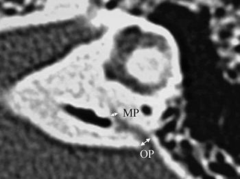
The width of the aqueduct was measured at two points: at the operculum (i.e. a line perpendicular to the posterior surface of the petrous pyramid and extending to the most lateral or postlateral pixel in the medial wall of the operculum) and at the midpoint (i.e. the halfway point between the operculum and the posterior wall of the crus commune or vestibule), according to the method of Madden et al. (Figure 1).Reference Madden, Halsted, Benton, Greinwald and Choo14
Axial computed tomographic scan showing measurement of the vestibular aqueduct width. MP = midpoint; OP = operculum

Two observers, who were blinded to the patients' medical histories, reviewed all images independently with regard to the width of the vestibular aqueduct. The width of the vestibular aqueduct was recorded as the average of the observers' measurements. If there was a large difference between these observations, a consensus was reached by discussion.
Statistical analysis
The width of the vestibular aqueduct at the midpoint and operculum was assessed, comparing: patients with sudden SNHL and control subjects; patients' affected and non-affected ears; and ears affected with otitis media and contralateral ears, in control subjects. The relationship between the width of the vestibular aqueduct and the audiometric threshold (in each of the initial and final audiograms) or the audiogram configuration was evaluated. The relationship between contrast enhancement of the endolymphatic sac and the width of the vestibular aqueduct in patients with SNHL was also evaluated.
The average prognosis scores were compared for patients with and without enhancement. The relationship between the width of the vestibular aqueduct and the prognosis with regard to hearing was assessed.
These statistical analyses were performed using chi-square testing, Mann–Whitney U testing and Pearson correlation coefficients.
Results
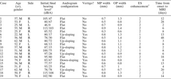
The results for contrast enhancement of the endolymphatic sac and for the width of the vestibular aqueduct are presented in Table I, together with data on hearing outcomes and on the length of time from the onset of hearing loss to the MRI examination.
Clinical and imaging results for patients with sudden sensorineural hearing loss

*Hearing level was expressed as the average score at three frequencies (500, 1000 and 2000 Hz), for the initial and final audiogram. †0 = no enhancement; 1 = ipsilateral enhancement; 2 = bilateral enhancement. Yr = years; MP = vestibular aqueduct at midpoint; OP = vestibular aqueduct at operculum; ES = endolymphatic sac; MRI = magnetic resonance imaging; M = male; F = female; R = right; L = left
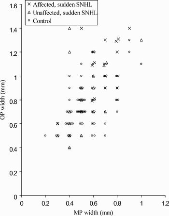
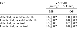
The width of the vestibular aqueduct at the midpoint and operculum in the SNHL-affected ears was significantly greater than that in the contralateral ears (midpoint, p < 0.05; operculum, p < 0.05) and that in the ears of control subjects (midpoint, p < 0.05; operculum, p < 0.0005) (Table II and Figure 2). The width of the vestibular aqueduct at the midpoint and the operculum did not differ significantly, comparing contralateral ears in sudden SNHL and control ears (Table II and Figure 2). In control subjects, the width of the vestibular aqueduct at the midpoint and operculum was not significantly different, comparing the chronic otitis media affected ears and the contralateral ears (Table II).
Scatter plot of vestibular aqueduct widths at the operculum versus those at the midpoint, in affected and unaffected ears of patients with sudden sensorineural hearing loss and in control subjects. MP = midpoint; OP = operculum; SNHL = sensorineural hearing loss

Vestibular aqueduct width at midpoint and operculum

VA = vestibular aqueduct; SD = standard deviation; MP = midpoint; OP = operculum; SNHL = sensorineural hearing loss
The average hearing level (in both initial and final audiograms) showed no correlation with the vestibular aqueduct width. Audiometric configurations in this study were flat in 13 ears (68 per cent) and up-sloping in five ears (26 per cent). A down-sloping configuration was found in one ear (5 per cent). We then analysed the audiogram configuration for each patient and found no correlation with the vestibular aqueduct width. Four out of 19 patients had vertigo. No relationship between the presence of vertigo and the width of the vestibular aqueduct was found.
The MRI scans for nine patients (cases one to nine in Table I) were performed using a 1.5-Tesla MR system, and the MRI scans for 10 patients (cases 10–19) were performed using a 3-Tesla MR system. In patients with sudden SNHL, the frequency of enhancement of the endolymphatic sac did not differ significantly, comparing those patients examined with 1.5-Tesla MRI and those examined with 3-Tesla MRI.
Enhancement of the endolymphatic sac was judged to be present on the affected side in 17 patients (89 per cent). In 11 of these 17 patients, the enhancement was also judged to be present on the contralateral side. However, no enhancement was observed on the contralateral side only.
The frequency of enhancement of the endolymphatic sac was significantly greater in the SNHL-affected ears than in the contralateral ears (p < 0.05). In patients with sudden SNHL, the period between the onset of hearing loss and MRI scanning did not differ significantly between those patients who showed enhancement (average ± SD, 12.6 ± 11.4 days) and those who did not (4.0 ± 2.8 days).
In patients with sudden SNHL, the width of the vestibular aqueduct at the midpoint and operculum in the SNHL-affected ear did not differ significantly, comparing patients with enhancement of the endolymphatic sac (midpoint, 0.6 ± 0.2 mm; operculum, 1.0 ± 0.3 mm) and those without enhancement (midpoint, 0.7 ± 0.2 mm; operculum, 1.1 ± 0.2 mm). Furthermore, no difference in prognosis scores was noted between sudden SNHL patients with and without enhancement.
Discussion
The major finding of this study was that the width of the vestibular aqueduct in the ear affected with sudden SNHL was significantly greater than that in the contralateral ear or in the ears of control subjects. It has been reported that the width of the vestibular aqueduct is smaller in patients with Ménière's disease than in control subjects.Reference Sando, Orita and Hirsch6, Reference Krombach, van den Boom, Di Martino, Schmitz-Rode, Westhofen and Prescher7 Evaluation of the vestibular aqueduct is important to our understanding of the pathophysiological mechanisms involved in sudden SNHL, considering that the vestibular aqueduct size in patients with sudden SNHL differs from that in patients with Ménière's disease.
Enlarged vestibular aqueduct is the most common congenital abnormality in the inner ear on radiological assessment. Enlarged vestibular aqueducts have been associated with a range of congenital disorders, such as the CHARGE association was defined as a non-random association of anomalies (Coloboma, Heart defect, Atresia choanae, Retarded growth and development, Genital hypoplasia, Ear anomalies/deafness). association,Reference Murofushi, Ouvrier, Parker, Graham, da Silva and Halmaqvi15 Alagille syndrome,Reference Okuno, Takahashi, Shibahara, Hashida and Sando16 Pendred syndromeReference Phelps, Coffey, Trembath, Luxon, Grossman and Britton17 and the branchio-oto-renal syndrome.Reference Chen, Francis, Ni, Cremers, Kimberling and Sato18 The criteria to determine an enlarged vestibular aqueduct are vague. However, a vestibular aqueduct diameter larger than 1.5 mm at the midpoint or an opercular measurement of greater than 2 mm are generally considered to be the defining characteristics.Reference Valvassori and Clemis19, Reference Levenson, Parisier, Jacobs and Edelstein20 In the present study, all patients with sudden SNHL showed a vestibular aqueduct diameter of less than 1.5 mm at the midpoint or an opercular measurement of less than 2 mm. Thus, we found that none of the patients in this study showed evidence of an enlarged vestibular aqueduct.
Purcell et al. Reference Purcell, Fischbein and Lalwani21 reported significant differences in the shape of the inner ear in patients with congenital SNHL, even in cases with grossly normal CT scans, when compared in detail with those of patients without SNHL. These authors suggested that hearing loss in SNHL patients with a ‘radiologically normal’ cochlea may be related to dysfunction of both the membranous and bony labyrinths. However, they did not evaluate the vestibular aqueduct quantitatively.
• This study sought to evaluate the vestibular aqueduct in patients with sudden sensorineural hearing loss
• Nineteen patients with sudden SNHL were evaluated using computed tomography and magnetic resonance imaging
• The width of the vestibular aqueduct in SNHL-affected ears at the midpoint and operculum was significantly greater than that in contralateral, unaffected ears or in control ears
• It is possible that sudden SNHL patients with a wider vestibular aqueduct are born with a ‘fragile’ inner ear
• Clarifying the pathophysiological mechanism responsible for endolymphatic sac enhancement may aid the understanding of sudden deafness
Our previous study demonstrated that the endolymphatic sac was enhanced in 75 per cent of sudden SNHL-affected ears, in 53 per cent of contralateral ears in patients with sudden SNHL and in 18 per cent of control ears.Reference Naganawa, Koshikawa, Fukatsu, Ishigaki, Nakashima and Ichinose8 In the present study, the endolymphatic sac was enhanced in 89 per cent of sudden SNHL ears and in 63 per cent of the contralateral ears of patients with sudden SNHL. The frequency of endolymphatic sac enhancement in patients with sudden SNHL did not differ significantly between the previous and the present study (chi-square test). Enhancement of the endolymphatic sac suggests inflammation of the endolymphatic sac tissue or venous enlargement in the region of the sac. Based on the results of this study, we speculate that many patients with sudden SNHL may experience pathophysiological changes in the region of the endolymphatic sac. We have previously reported that the endolymphatic sac was enhanced in 63 per cent of affected ears in patients with acute low-tone SNHL without vertigo.Reference Sugiura, Naganawa, Nakashima, Misawa and Nakamura22 In contrast, the endolymphatic sac was enhanced in 20 per cent of affected ears in patients with Ménière's disease.Reference Naganawa, Koshikawa, Fukatsu, Ishigaki, Nakashima and Ichinose8
It is known that the endolymphatic sac and duct are poorly developed in patients with Ménière's disease. The endolymphatic sac is believed to intervene in the absorption of the endolymphatic fluid, and damage to the endolymphatic sac results in endolymphatic hydrops.Reference Sugiura, Naganawa, Nakashima, Misawa and Nakamura22 It is possible that the narrow vestibular aqueduct in patients with Ménière's disease is associated with an impediment of the absorption of endolymphatic fluid. In contrast, the results of the present study show that the vestibular aqueduct in patients with sudden SNHL was wider than that in controls. A wider vestibular aqueduct might be associated with insufficient maturation of the inner ear, because an increased fluid-filled area of the inner ear may be related to insufficient maturation of the inner ear.Reference Ishida, Sugiura, Naganawa, Teranishi and Nakashima23 It is possible that sudden SNHL patients with a wider vestibular aqueduct are born with a ‘fragile’ inner ear,Reference Sugiura, Nakashima, Naganawa, Otake, Mukaida and Sone4 or are apt to receive abnormal pressure transmission through the vestibular aqueduct.Reference Boston, Halsted, Meinzen-Derr, Bean, Vijayasekaran and Arjmand24
Clarifying the pathophysiological mechanism responsible for endolymphatic sac enhancement in patients with sudden deafness may be a key to understanding the cause of sudden deafness. Precise imaging and evaluation of the inner ear is essential to the investigation of the pathological conditions underlying sudden SNHL.