Introduction
The perception of body and head motion is multisensory, and is subserved primarily by visual and vestibular perception. Visual and vestibular sensory inputs are integrated centrally, and monitored against each other for concurrence. Any persistent discordance between the two can lead either to a central adaptation and recalibration of the abnormal information system, or to its suppression and dominance of the more accurate afferent system (e.g. visual preference following vestibular impairment).Reference Guerraz, Yardley, Bertholon, Pollak, Rudge and Gresty 1 , Reference Devèze, Montava, Lopez, Lacour, Magnan and Borel 2
Reflex eye movements in response to head movement are similarly based on two concurrent reflexes: those based on visual afferents (visual-ocular reflex, pursuit reflex, fixation reflex) and those based on vestibular afferents (vestibulo-ocular reflex). These reflexes are continually monitored in the cerebellum for congruence, and for their accuracy in achieving a stable visual gaze and preventing ‘retinal slide’.Reference Highstein 3 Any persistent inaccuracies are noted, and are corrected for by a continuing and ongoing adaptation and modification of the reflex gain.Reference Highstein 3 – Reference Freedman, Caroll and McElligott 5 The flocculus of the cerebellum is thought to have a major role in this constant monitoring and adaptation.Reference Highstein 3 , Reference Ito 6 Correction for any persistent inadequacies in vestibulo-ocular reflex function may occur in one of two ways: (1) adaptation and recalibration of the vestibulo-ocular reflex,Reference Highstein 3 , Reference Demer 4 , Reference Lisberger 7 – Reference Kramer, Shelhamer and Zee 9 to improve its accuracy; or (2) central suppression of the vestibulo-ocular reflex,Reference Collins and Kornhuber 10 to decrease its effectiveness and enable the other reflexes subserving reflex eye movements to gain primacy.
While the initial evidence for such adaptation was collected by inducing very obvious and gross mismatches of the visual and vestibular inputs,Reference Gonshor and Jones 11 – Reference Miles and Eighmy 13 subsequent studies have demonstrated it to be effective for even minor discrepancies.Reference Collewijn, Martins and Steinman 14 , Reference Cannon, Leigh, Zee and Abel 15 Experiments have been conducted on animals and humans wherein the size of the visual environment has been altered by having the subject wear magnifying or minifying spectacles.Reference Demer, Goldberg, Porter, Jenkins and Schmidt 12 , Reference Miles and Eighmy 13 Alterations in retinal image size lead to changes in the speed with which these images move on the retina, which result in adaptive recalibration of the vestibulo-ocular reflex.
In the real world (outside of experimental laboratories), correction of refractive errors by spectacles or contact lenses also leads to changes in retinal image size. The degree of alteration is related to: the type of correcting lens (concave or convex), the power of the correcting lens and the distance of the correcting lens from the cornea (Figure 1).Reference Abrams and Duke-Elder 16 The estimated change in size with spectacles is approximately 3 per cent per dioptre.Reference Abrams and Duke-Elder 16 In previous years wherein intra-ocular lens implantation following cataract operations was not routine, aphakia corrected with spectacles could lead to the image size being altered by as much as 35 per cent. It is very probable that such alterations in retinal image size would impact on the vestibulo-ocular reflex.
Telescopic effect of placing a corrective concave lens anterior to the myopic eye. (a) Placement of the correcting lens at the ‘anterior nodal point’ (i.e. F1, approximately 15 mms from the cornea) does not alter image size. (b) Placement of the corrective concave lens proximal to the anterior nodal point (F1) leads to divergence of rays (stippled line) from their original path (grey line) and to enlargement of the image size. (c) Placement of the lens distal to the nodal point (F1) leads to diminution of the image size. Most spectacles are worn distal to the nodal point and so lead to a smaller retinal image. Contact lenses are worn on the cornea and lead to a larger retinal image. The opposite effect occurs in hypermetropia corrected with convex lenses.

The present study tested the hypothesis that alterations in retinal image size associated with corrected myopia lead to alterations of the vestibulo-ocular reflex, as measured by the caloric test.
Materials and methods
This prospective, cross-sectional study aimed to generate preliminary hypotheses. Vestibulo-ocular reflex testing, using electronystagmography (ENG) and including caloric testing, was undertaken in healthy volunteers with myopia (more than 2 dioptres) corrected by spectacles or contact lenses (myope group), and the findings were compared with those of volunteers with no refractive error (emmetrope group).
Institutional ethical approval was obtained. All volunteers in both groups were healthy young adults (aged 15–35 years); the participants had no previous vestibular, otological, neurological or significant systemic illness, no previous long-term history of medication or ototoxic medication, and no recent history of medication (3 weeks) or alcohol intake (24 hours). All volunteers were apprised of the study aims and methods, and all gave their informed consent for study inclusion.
Eighteen participants were initially recruited in each group (emmetrope group and myope group). Participant matching was not undertaken, as volunteers in both groups were similarly restricted to the 15–35-year age group and differed only with regard to the myopia in one group. An initial proforma-based evaluation captured baseline demographic data and detailed information regarding: the refractive error correction (spherical, astigmatism, axis), the kind of correction normally used (spectacles or contact lenses), and the proportion of waking hours for which each method of correction (spectacles or contact lenses) was used.
Vestibular evaluation
All volunteers in both groups underwent complete ENG evaluation and recording. The ENG recording was performed on a two-channel recorder with a time constant of 2 seconds. Volunteers in the myope group were asked to remove their spectacles or contact lenses prior to the ENG evaluation. Volunteers were asked to lie on a couch with the head-end elevated by 30 degrees and the tests as listed below were then conducted.
The ENG evaluation included the following: (1) initial calibration, and screening for spontaneous, gaze- and position-induced nystagmus; (2) assessment of voluntary and visually evoked eye movements (i.e. voluntary saccades, pursuit or eye tracking, and optokinetic nystagmus); (3) a bithermal caloric test, undertaken as per the standardised, low volume irrigation technique of our laboratory (which involved irrigating each ear with 20 ml water over 30 seconds at 44 °C and 30 °C;Reference Kirtane 17 all irrigations and recordings were undertaken in the dark); and (4) calculation of the maximum slow phase velocity and the culmination frequency of the recorded nystagmus. The parameters of all four individual caloric responses were averaged for each individual participant to calculate the mean of all four responses in terms of the mean slow phase velocity and mean culmination frequency.
Caloric response analysis
As per standard criteria for low volume irrigations and as per the standardised technique of our laboratory, maximum slow phase velocity values of less than 7 degrees per second, or a culmination frequency of less than 28 beats per 30 seconds, were classified as hypoactive responses.Reference Kirtane 17 , Reference Claussen and von Schlachta 18 Maximum slow phase velocity values of 7–9 degrees per second, or a culmination frequency of 29–33 beats per 30 seconds, were classified as borderline hypoactive. Maximum slow phase velocity values of more than 40 degrees per second, or a culmination frequency of more than 64 beats per 30 seconds, were classified as hyperactive responses.Reference Kirtane 17 – Reference Gonçalves, Felipe and Lima 19
The caloric responses were objectively assigned digital codes on the basis of normative data, and as per the system previously proposed by Claussen and von Schlachta.Reference Claussen and von Schlachta 18 Normal responses were coded 0, hypoactive responses were coded 1 and hyperactive responses were coded 2. As per convention, the caloric responses test was conducted in the sequence of: right ear, warm water; right ear, cold water; left ear, warm water; and left ear, cold water. All four normal responses were therefore reflected by the code 0000 and all four hypoactive responses by the code 1111.
Participants for whom three of four caloric responses were hypoactive and one was borderline hypoactive were classified as bilateral hypoactive. Similarly, three of four normal caloric responses with one borderline hypoactive or hyperactive response was classified as normal. In cases where a clear interpretation based on the above objective criteria was not possible, final interpretation was provided by the reporting physician who was blinded to the status of the participant (myope or emmetrope).
Statistical analysis
Two participants were excluded from analysis on initial screening: one participant (in the myope group) had recently consumed alcohol, and another (in the emmetrope group) demonstrated significant left beating spontaneous nystagmus and a directional preponderance to the same side on ENG. The latter participant was directed towards further evaluation and investigation for this abnormality, and was excluded from further analysis. The final analysis was therefore undertaken on 17 participants in each group.
Statistical comparisons and analyses were conducted using the Epi-Info™ (version 6.04b) software.
Results
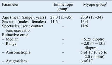
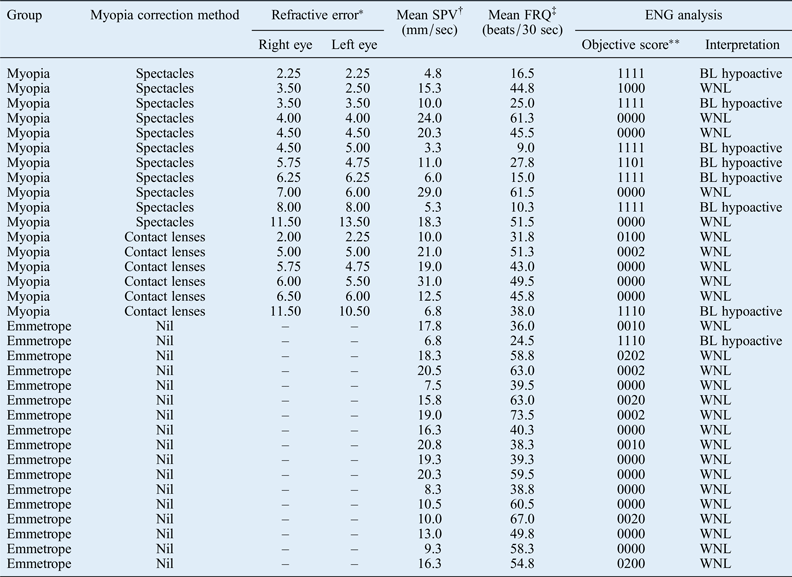
The characteristics of the two group populations are shown in Table I. All spectacle users were wearing their glasses for their entire waking days. Contact lens users tended to use these intermittently, and interspersed contact lens use with spectacle use. The degree of myopia for individual participants and their caloric-induced nystagmus characteristics are illustrated in Table II.
Characteristics of emmetrope and myope groups

*n = 17; † n = 17
Caloric results for myopes and emmetropes

The participants are listed incrementally in order of increasing refractive error. *Cumulative refractive error (i.e. spherical correction plus astigmatism correction), in dioptres. †‘Mean SPV’ refers to the mean maximum slow phase velocity for the four caloric responses. ‡‘Mean FRQ’ refers to the mean of the culmination frequencies for the four caloric responses. **Numeric display of all four caloric responses in the sequence of: right ear, warm water; right ear, cold water; left ear, warm water; and left ear, cold water. Each response is coded as: normal – 0, hypoactive – 1, or hyperactive – 2. Four normal responses are therefore coded as 0000, a hypoactive right ear with cold water response with three other normal responses is coded as 0100, and so on. ENG = electronystagmography; sec = seconds; BL = bilateral; WNL = within normal limits
One of 17 emmetropes and 7 of 17 myopes demonstrated hypoactive nystagmic responses to caloric stimulation (Table II). Among the myopes, hypoactive calorics were particularly frequent in spectacle users (6 out of 11, 55 per cent; p < 0.01 on Fisher's exact two-tailed test). The relative risk of hypoactive caloric responses in spectacle users compared to emmetropes was 9.3 (95 per cent confidence interval = 1.3–66.9).
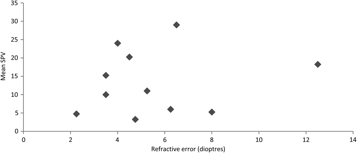
No correlation was observed among the spectacle users with regard to the quantum of refractive error and the slow phase velocity (Figure 2); participants with the least refractive error demonstrated vestibular suppression, and others with the most advanced myopia (refractive error – right eye = 11.50 dioptres and left eye = 13.50 dioptres; and refractive error – right eye = 7.00 dioptres and left eye = 6.00 dioptres) nevertheless had very robust slow phase velocity and frequency characteristics (Figure 2 and Table II).
Scatter plot of spectacle users, demonstrating the mean slow phase velocity (SPV) responses as a function of mean refractive error (mean right and left eye).

One of the six contact lens users had hypoactive responses; this participant also used spectacles for more than 6 hours per day.
Evaluation of slow phase velocity trends in emmetropes (mean, 14.6 degrees per second), contact lens users (mean, 16.7 degrees per second) and spectacle users (mean, 13.4 degrees per second) indicated a trend towards a greater slow phase velocity in the contact lens subgroup (enlarged retinal image) and a diminished slow phase velocity in the spectacle subgroup (minified retinal image). However, this trend was not statistically significant (as determined using the Kruskal–Wallis analysis of variance).
The dataset had few cases of significant astigmatism or anisometropia (Table I), and the sample size was therefore considered too small to assess the impact of these factors.
Discussion
The present study indicates that a significant proportion of myopes using spectacles demonstrate hypoactive vestibular responses when evaluated by caloric testing. The report, from a basic clinical vestibular facility, provides preliminary support for the hypothesis that spectacle use may lead to alteration of the vestibulo-ocular reflex. Further investigation of this hypothesis needs to be undertaken in specialised vestibular research laboratories with facilities that enable a more precise evaluation of the vestibulo-ocular reflex, using rotational tests.
The observed phenomenon may be explained by one of two mutually contradictory hypotheses: either the gain of the vestibulo-ocular reflex is decreased as an adaptive response to the decreased retinal image size, or the vestibulo-ocular reflex is suppressed.
The argument for adaptation is supported by previous experiments reporting recalibration of the vestibulo-ocular reflex in response to experimentally induced gross and sustained changes in retinal image size.Reference Gonshor and Jones 11 , Reference Bello, Paige and Highstein 20 It is thought that with a decrease in retinal image size, as occurs with spectacle use in myopia, the required eye movement to fixate from one point in the environment to another decreases, leading to adaptive recalibration of the vestibulo-ocular reflex.Reference Collewijn, Martins and Steinman 14 Such recalibration would lead to a decrease in the quantum of eye displacement for equivalent stimulation of the semicircular canals.Reference Cannon, Leigh, Zee and Abel 15 The resultant eye movements would be appropriate for the individual subject, and would be the result of an active and appropriately calibrated vestibulo-ocular reflex. Nevertheless, these responses would seem spuriously hypoactive when interpreted in light of the normal limits previously demarcated by observations on normal controls.
Such recalibration of the vestibulo-ocular reflex has previously been demonstrated by rotational testing.Reference Gonshor and Jones 11 – Reference Cannon, Leigh, Zee and Abel 15 , Reference Bello, Paige and Highstein 20 The one available study that assessed vestibulo-ocular reflex gain by rotational chair testing, on seven human participants with assorted refractive errors, reported alterations in vestibulo-ocular reflex gain proportional to calculated changes in image size.Reference Cannon, Leigh, Zee and Abel 15 Data from spectacle users in this present study, however, revealed no consistent relationship between the expected quantum of image diminution (proportional to increasing myopia) and the depression of caloric-induced slow phase velocity (Figure 2).
The correlation between image size and vestibulo-ocular reflex gain has not been found to be consistent in other studies either. Freedman et al. reported that constant wearing of magnifying glasses in cats did not increase vestibulo-ocular reflex gain (as would be expected with adaptation), and gain was instead decreased or suppressed.Reference Freedman, Caroll and McElligott 5
The alternate explanation, in which the observed phenomena are a consequence of vestibulo-ocular reflex suppression, is based on the limitations of visual correction achieved with glasses. One such limitation is that spectacle correction of myopia is optimal only in central gaze. Peripheral vision through spectacles is often impaired by spherical and chromatic aberrations.Reference Freedman, Caroll and McElligott 5 , Reference Abrams and Duke-Elder 16 The inability of the vestibulo-ocular reflex to maintain a clear retinal image in eccentric gaze may therefore lead to its suppression, with central mechanisms facilitating strategies that favour head movements over eye movements.
Another limitation of visual correction with glasses is that the retinal image size in spectacle users is a function of both the power of the correcting lens and the distance of this lens from the cornea.Reference Abrams and Duke-Elder 16 Slipping of spectacles on the nose is not unusual, and would result in frequent changes in retinal image size, leading to frequent demands for adaptation. Previous researchers have demonstrated that the initial response to visual–vestibular mismatch is a suppression of the vestibulo-ocular reflex, with adaptation occurring subsequently over weeks for mismatch that is sustained and unwavering.Reference Gonshor and Jones 11 , Reference Bello, Paige and Highstein 20 Spectacles, however, may not always be worn at the same precise distance from the cornea, and may slip down and be frequently readjusted by the user. The altered visual environment may therefore never be sustained and unwavering so as to enable the adaptive response to move forward from an initial suppression to subsequent recalibration and adaptation.
Contact lens users, with a constant unwavering position of the correcting lens on the cornea, would not be affected by this mechanism. This explanation would also explain why only some, and not all, spectacle users exhibited hypoactive responses.
It is logical to conjecture that a similar suppression would also be encountered with spectacle use for other refractive errors, and that it may be particularly manifested in users of bifocal or multifocal spectacles. As spectacle use is so common, the implications of these findings have significance. The findings suggest that spectacle use may be a possible co-factor in the dysequilibrium symptoms of some individuals, and may be a possible cause of hypoactive caloric responses observed in otherwise asymptomatic patients.
The caloric test, as used in this study, remains the most frequently used clinical test for vestibular evaluation. However, alternative tests such as the rotational test and the head impulse test are more physiologically based and are more sensitive to vestibulo-ocular reflex alterations.Reference Eza-Nuñez, Fariñas-Alvarez and Fernandez 21
-
• The vestibulo-ocular reflex provides a stable visual field by inducing reflex corrective eye movements in response to head movements
-
• Changes in retinal image size can lead to vestibulo-ocular reflex adaptation and recalibration
-
• Concave lenses used for myopia correction lead to retinal image size alteration; spectacle use leads to smaller retinal images and contact lenses to larger images
-
• Spectacle-using myopes have a higher than normal risk of bilateral hypoactive caloric responses, indicative of vestibular suppression
-
• A similar effect may occur in those with other refractory errors (hypermetropia, presbyopia), and may have implications for balance dysfunction in the elderly
-
• Spectacle use may contribute to dysequilibrium symptoms and cause hypoactive caloric responses in otherwise asymptomatic patients
The current study was undertaken as a hypothesis-generating study conducted in a clinical audio-vestibular facility. Further confirmation of these findings with rotational testing in a sophisticated vestibular laboratory, conducted on a larger sample population, is required for confirmation and validation of findings.
Conclusion
A very large proportion of the population has myopia and uses spectacles for correction. Initial evidence is presented to suggest that a proportion of these individuals have hypoactive caloric responses. This observed phenomenon is likely to be secondary to suppression of the vestibulo-ocular reflex, rather than to its adaptation. It is logical to assume that a similar alteration or suppression would also be encountered in individuals using spectacles for other refractive errors, particularly in users of bifocal or multifocal spectacles.
Further testing of this hypothesis is especially important, as it highlights the possibility of a hitherto undetected cause for vestibular dysfunction in the elderly population who are susceptible to falls and its ensuing complications.
Acknowledgement
This study was supported by a research grant from the All India Institute of Medical Sciences, New Delhi, India.