Introduction
The mammalian oocyte is a mysterious cell. Not only can it develop into a new embryo by fertilization with a spermatozoon, but also can become an embryo in the absence of sperm and reprogram somatic nuclei to initiate embryonic development via parthenogenesis (Pangas and Rajkovic, Reference Pangas and Rajkovic2006). However, the molecular mechanisms necessary for the development of the oocyte and embryo are largely unknown. Cells differentiate through the activation and repression of gene transcription. This transcription process might control oocyte- and embryo-specific genes regulating transcriptional cascades (Choi and Rajkovic, Reference Choi and Rajkovic2006; Pangas and Rajkovic, Reference Pangas and Rajkovic2006). Transcriptional control of oocyte and early embryo must include the expression of oocyte-specific genes necessary for oocyte growth and early embryonic development. Oocyte-specific genes are some of the most abundant transcripts in the oolemma, ooplasm and nucleus. Recently, multiple approaches have been undertaken to identify genes preferentially expressed in the oocyte and embryo, and two new key genes in mouse models, including factor in the germline (Figla), newborn ovary homeobox gene (Nobox), were obtained (Liang et al., Reference Liang, Soyal and Dean1997; Rajkovic et al., Reference Rajkovic, Pangas, Ballow, Suzumori and Matzuk2004). Besides, there are some other oocyte- and embryo-specific genes, including growth differentiation factor 9, Gdf9 (McGrath et al., Reference McGrath, Esquela and Lee1995), maternal antigen that embryos require, Mater (Tong et al., Reference Tong, Nelson and Dean2000), zygote arrest 1, Zar1 (Wu et al., Reference Wu, Viveiros, Eppig, Bai, Fitzpatrick and Matzuk2003) and neurotrophins (NTs) (Paredes et al., Reference Paredes, Romero, Dissen, DeChiara, Reichardt, Cornea, Ojeda and Xu2004).
NTs have a wide-ranging role in the development of both the nervous system and the development of non-neuronal systems, including the reproductive, endocrine, immune and respiratory systems (Tessarollo, Reference Tessarollo1998). Brain-derived neurotrophic factor (BDNF) is a member of the NT family, which includes nerve growth factor (NGF), neurotrophin 3 (NT 3), neurotrophin 4/5 (NT 4/5), neurotrophin 6 (NT 6), ciliary neurotrophic factor (CNTF), and glial cell line-derived neurotrophic factor (GDNF). BDNF is known to activate the high-affinity tyrosine kinase B (TrkB) receptor together with the pan-neurotrophin low-affinity co-receptor p75 (Barbacid, Reference Barbacid1994). p75 and TrkB are expressed in the oocytes as well as in the somatic cells of the ovary (Dissen et al., Reference Dissen, Hirshfield, Malamed and Ojeda1995). BDNF and TrkB have been identified in the adult avian and bovine ovary (Jensen and Johnson, Reference Jensen and Johnson2001; Martins da Silva et al., Reference Martins da Silva, Gardner, Taylor, Springbett, Sousa and Anderson2005). A direct effect of BDNF on murine oocyte maturation in vitro has also been reported, with increased first polar body extrusion rate in oocytes stripped off cumulus prior to maturation (Seifer et al., Reference Seifer, Feng, Shelden, Chen and Dreyfus2002). BDNF acts on its receptor, TrkB, in the mouse oocyte to enhance first polar body exclusion and to increase the competence of oocytes for development into early embryos (Kawamura et al., Reference Kawamura, Kawamura, Mulders, Sollewijn Gelpke and Hsueh2005). And BDNF mRNA were expressed in the development of mouse preimplantation embryos (Kawamura et al., Reference Kawamura, Kawamura, Fukuda, Kumagai, Hsueh and Tanaka2007). Supplementation of culture media with BDNF at the concentration of 10 μg/l caused a significant increase in the rates of parthenogenetic blastocyst formation (Martins da Silva et al., Reference Martins da Silva, Gardner, Taylor, Springbett, Sousa and Anderson2005). These data suggest that BDNF may be involved in embryonic development.
There were culture media with BDNF at concentrations of 10 μg/l and 100 μg/l group in SOFaaBSA culture medium (CM) (Martins da Silva et al., Reference Martins da Silva, Gardner, Taylor, Springbett, Sousa and Anderson2005), and we do not know whether supplementing of BDNF at the concentration of 10 μg/l best works in promoting early embryonic development in TCM-199 CM by the co-culture system. In the present study, we have measured the mRNA level of BDNF in bovine oocytes and early embryos and investigated the effects of BDNF on embryo development after in vitro fertilization (IVF), parthenogenetic activation (PA) and nuclear transfer (NT). We provide evidence that cumulus cells and early embryo may have different capacities to respond to NT signaling.
Material and methods
Reagents and media
All of the chemicals used in this study were purchased from Sigma Chemical Company (St Louis, MO, USA), with the exception of recombinant human BDNF (PeproTech EC Ltd, London, UK), TCM-199 powder Gibco BRL (Paisley, Scotland, UK), fetal bovine serum (FBS) and Dulbecco’s Modified Eagle’s Medium (DMEM; Hyclone Company, Logan, UT, USA). The IVM medium was TCM-199, supplemented with 26.2 mM NaHCO3, 5 mM HEPES, 5% estrous cow serum (OCS, self-preparation), 2% bovine follicular fluid (BFF, collected irrespective of the stage of the reproductive cycle) and 0.1 mg/ml FSH. The embryo CM was TCM-199, supplemented with 3% OCS. The basic micromanipulation medium was TCM-199, supplemented with 5 mM NaHCO3, 5 mM HEPES and 5% OCS. The fertilization medium was Tyrode’s medium supplemented with 50 g/ml heparin, 2.5 mM caffeine. All of the media were supplemented with 60 mg/ml penicillin G and 100 mg/ml streptomycin sulfate, then sterilized by passing through a 0.20 mm filter, and stored at 4°C to 8°C for up to 4 weeks (Lu et al., Reference Lu, Shi, Wei, Yang and Wei2005).
Tissues
Bovine ovaries were collected from a local abattoir and kept warm during transportation. In the laboratory, ovaries were washed in Dulbecco’s phosphate-buffered saline at 35°C to 37°C.
Oocyte maturation in vitro
Bovine cumulus–oocytes complexes (COCs) were recovered by aspiration of bovine follicles (diameter, 2 to 6 mm) using a 10 ml disposable syringe with an 18-gauge needle. Only oocytes possessing a compact complete cumulus oophorus and evenly granulated cytoplasm were selected for IVM. Then, COCs were washed twice in the IVM medium and cultured in a 30 mm glass dish containing 1.5 ml IVM medium for 20 to 22 h under a humidified atmosphere of 5% CO2 in air at 38.5°C to 38.8°C.
Nuclear transfer
Firstly, we made a preparation of granulosa cells as donor karyoplasts. The granulosa cells were obtained by removing surrounding cumulus cells after IVM of oocytes. Granulosa cells were pretreated with 0.1 mg/ml aphidicolin for 24 h and then cultured in DMEM + 0.5% FBS for 6 to 9 days before use as donor nuclei.
Bovine donor granulosa cells were placed into a droplet of 100 ml manipulation medium containing 8% polyvinyl pyrrolidone dialyzed and 0.2 mM sucrose. The cytoplasmic membrane of donor cells was ruptured (as indicated by a rough surface) by repeatedly pipetting, using a 10 mm inner diameter injection pipette, before transferring to another manipulation drop containing enucleated oocytes in the injection medium. Thereafter, the donor cell was injected directly into the cytoplasm of a recipient oocyte.
After microinjection, the reconstructed oocytes were incubated in CM under a humidified atmosphere of 5% CO2 in air at 38.5°C to 38.8°C for up to 26 to 28 h after the start of maturation. The activation of reconstructed oocytes was induced by exposure to 5 mM ionomycin in CM for 5 min and subsequent incubation in 2 mM 6-dimethylaminopurine for 3 h at 38.5°C to 38.8°C and 5% CO2 in air. Meanwhile, bovine and buffalo oocytes matured in vitro for 26 to 28 h were activated using the same method described above, and were considered the parthenogenetic control.
Culture of IVF and parthenogenetic activated embryos
After IVM, the oocytes were cultured in fertilization medium. IVF was performed using frozen semen and fertilization medium. The straws of frozen semen were thawed in water at 37.5°C for 0.5 min. The obtained sperm was suspended in the fertilization medium for 30 min. Next, the supernatants were taken to other centrifuge tubes and centrifuged for 5 min (1600 × g). The supernatants were discarded. Sperm were obtained and transferred to the fertilization medium with oocytes under a humidified atmosphere of 5% CO2 in air at 38.5°C to 38.8°C. After 24 to 26 h, the surrounding cumulus cells and sperm were removed by manual pipetting in the CM.
After IVM, surrounding cumulus cells were removed by manual pipetting in the CM, the reconstructed oocytes were incubated in CM under a humidified atmosphere of 5% CO2 in air at 38.5°C to 38.8°C for up to 26 to 28 h after the start of maturation. The activation of reconstructed oocytes was induced by exposure to 5 mM ionomycin in CM for 5 min and subsequent incubation in 2 mM 6-dimethylaminopurine.
In vitro culture of embryos
After fertilization, parthenogenesis or NT activation, oocytes were placed into co-culture with granulosa cell monolayers in a 30 ml droplet of CM overlaid with mineral oil under a humidified atmosphere of 5% CO2 in air at 38.5°C to 38.8°C. The granulosa cell monolayers were established at 48 to 72 h before introduction of embryos. After introduction of embryos, half of the medium was replaced with fresh medium every 24 h. After 2 days of co-culture, cleavage of embryos was verified, and the number of developed blastocysts was recorded within 9 days of co-culture.
Granulosa cell culture
Granulosa cells were obtained by removing surrounding cumulus cells after IVM of oocytes. The rate of live granulosa cells was recorded by trypan blue staining. The number of granulosa cells was adjusted to 2 × 10⁵/ml. Granulosa cells were precipitated in IVM medium. After 4 to 6 h, IVM medium was replaced with DMEM. After 2 days, the granulosa cells were floated by trypsinization. The number of granulosa cells was recorded in cell count slab by trypan blue staining.
Extraction of RNA and synthesis and amplification of cDNA
Total RNA was extracted using an RNA easy Micro kit (Qiagen, Crawley, UK) as previously described. Firststrand cDNA was synthesized from 2 ng of total RNA from the oocytes or embryos. Reverse transcription use was conducted with the PrimeScript™ 1st Strand cDNA Synthesis Kit (Takara, Tokyo, Japan). PCR system use included the TaKaRa Ex Taq (TaKaRa Ex Taq 0.25 μl, 10 × Ex Taq Buffer (Mg2+free) 5 μl, MgCl2 (25 mM) 1.4 μl, dNTP mixture (each 2.5 mM) 4 μl, cDNA 2 μl, forward and reverse primers each 1 μl, ddH2O up to 50 μl) (Takara, Tokyo, Japan). PCR use was conducted with Gene Amp.PCR System 9700 (ABI, MO, USA). Specific primers for each gene are given in Table 1.
PCR primer sequences

BDNF brain-derived neurotrophic factor; SDHA = succinate dehydrogenase flavoprotein subunit A.
aGene-specific primers’ Tm (°C) are 60°C in both BDNF and SDHA.
Quantitative real-time PCR
After total RNA was extracted and firststrand cDNA was synthesized, quantitative real-time PCR was conducted with SYBR Premix Ex Taq (2 × SYBR Premix Ex Taq 12.5 μl, forward and reverse primers each 0.5 μl, cDNA 2 μl, ddH2O up to 20 μl) (Takara, Tokyo, Japan). Q-PCR use was conducted with LightCycler 2.0 (Roche Co., Berlin, DE). The program used for all genes consisted of a denaturing cycle of 30 s at 95°C; 45 to 50 cycles of PCR (95°C for 5 s, 60°C for 15 s, and 72°C for 10 s); a melting cycle consisting of 95°C for 0 s, 65°C for 15 s, 95°C for 0 s, and a step cycle starting at 65°C until 95°C with a 0.2°C/s transition rate; and finally, a cooling cycle of 40°C for 30 s.
Immunohistochemistry
Immunofluorescent detection of BDNF was performed with bovine blastocysts. Blastocysts were fixed with 3.7% paraformaldehyde for 30 min. After three washes in PBS with 100 mM glycine and 0.3% BSA for 5 min, blastocysts were permeabilized in 1% Triton X-100 for 10 min at room temperature. After two washes in T-PBS-BSA (PBS with 0.01% TritonX and 0.3% BSA), blastocysts were blocked by 1% PBS-BSA for 1 h and then incubated with rabbit anti-BDNF polyclonal antibodies in 1% BSA at 1 : 100 dilution for 48 h at 4°C. Blastocysts were incubated for 20 min at 39°C by T-PBS-BSA three additional washes. Blastocysts were incubated with human anti-rabbit immunoglobulin G (IgG) secondary antibodies in 1% PBS-BSA at 1 : 200 dilution for 30 min at room temperature, followed by three additional washes. SABC-FITC was performed for 30 min followed by three additional washes. Counterstaining was performed for 10 min in 10 μg/ml of PI (propidium iodide) dye. After three washes in T-PBS-BSA, blastocysts were analyzed under a confocal laser scanning microscope (TY4653, PerkinElmer, USA). For negative controls, the primary antibody was replaced by non-immune rabbit IgG.
Statistical analysis
Within each experiment, the difference between treatments in rates of oocytes undergoing cleavage and developing to the blastocyst stage was analyzed by a χ 2-test. The BNDF mRNA relative folds were analyzed by one-way ANOVA (GraphPad Software, V2.02).
Results
Evaluation of BDNF mRNA expression
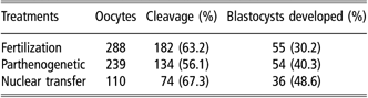
We intended to know whether mRNA for BDNF could be detected in RNA isolated from bovine germinal vesicle (GV) oocytes, metaphase II (MII) oocytes, 4-cell embryos, 8-cell embryos, morulae and blastocysts. Amplification products of the expected size corresponding to BDNF (198 bp) and succinate dehydrogenase flavoprotein subunit A (SDHA) (185 bp) were detected in cDNA sequence, which was reverse transcripted by RNA extracted from bovine oocytes and embryos (Table 2).
In vitro development of embryos
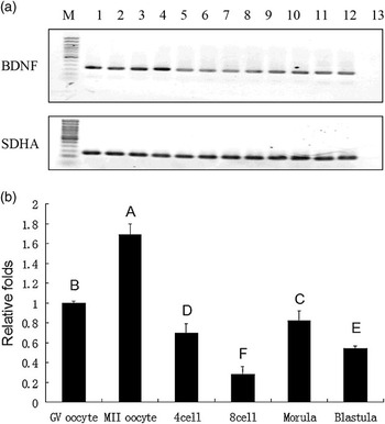
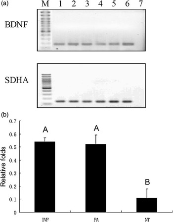
Results from the quantitative analysis of the BDNF gene expression are presented in Figure 1. The mRNA levels for BDNF gene were different in bovine oocytes and IVF embryos at different stages (P < 0.01), with the highest expression in MII oocytes and the lowest expression in 8-cell embryos (i.e. GV oocytes, 1.00 ± 0.18; MII oocytes, 1.69 ± 0.12; 4-cell embryos, 0.70 ± 0.09; 8-cell embryos, 0.28 ± 0.08; morulae, 0.82 ± 0.10; blastocysts, 0.54 ± 0.03; P < 0.01; Figure 1). The BDNF gene was highly expressed in the parthenogenetic and in vitro-fertilized blastocysts compared to the NT blastocysts (i.e. IVF, 0.54 ± 0.03; PA, 0.52 ± 0.07; NT, 0.11 ± 0.08; P < 0.01; Figure 2).
Expression of brain-derived neurotrophic factor (BDNF), succinate dehydrogenase flavoprotein subunit A (SDHA) gene transcripts in cDNA samples derived from oocytes and embryos (a). Stripped single germinal vesicle (GV) oocytes (lanes 1 and 2), stripped single metaphase II (MII) oocytes (lanes 3 and 4), single preimplantation embryos 4-cell (lanes 5 and 6), 8-cell (lanes 7 and 8), morulae (lanes 9 and 10) and blastocyst (lanes 11–12). Lanes 13 represent negative controls omitting the added sample. Quantitative real-time PCR of BDNF mRNA expression in bovine oocytes and in vitro fertilization embryonic at different preimplantation stages (b). Each developmental stage was done in triplicate, and 50 GV or MII oocytes, 40 4-cell embryos, 30 8-cell embryos, 20 morulaes and 10 blastocysts were used for each PCR run. The relative mRNA levels represent the amount in femtograms of the mRNA expression corrected to the ratio of SDHA gene mRNA. Different capital letters indicate high statistical difference (P < 0.01).

Expression of brain-derived neurotrophic factor (BDNF) and succinate dehydrogenase flavoprotein subunit A (SDHA) gene transcripts in cDNA samples derived from 7- to 8-day blastocyst (a). Stripped single blastocysts for parthenogenetic activation (PA) (lanes 1 and 2), stripped single blastocysts for nuclear transfer (NT) (lanes 3 and 4), stripped single blastocysts for in vitro fertilization (IVF) (lanes 5 and 6), and lanes 7 represent negative controls omitting the added sample. Quantitative real-time PCR of BDNF bovine embryonic mRNA expression at different types of blastocysts (b). Each developmental stage was done in triplicate, and 10 IVF, PA or NT blastocysts were used for each PCR run. The relative mRNA levels represent the amount in femtograms of mRNA expression corrected to the ratio of SDHA gene mRNA. Different capital letters indicate high statistical difference (P < 0.01).

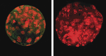
The expression of BDNF in trophectoderm cells was confirmed by using immunofluorescent staining (Figure 3).
Immunohistochemical detection of brain-derived neurotrophic factor (BDNF) proteins in blastocysts. Specific BDNF signals (green) are localized in trophectoderm (TE) cells, but not inner cell mass (red) (ICM) cells (right is negative controls) (scale bars, 20 μm).

Effect of BDNF on IVF embryo development
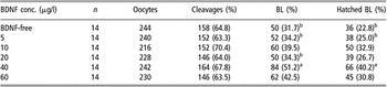
IVF embryo development to cleavage and blastocyst stages was recorded after IVM of oocytes as COCs in the previously defined treatment groups. In this trial, no attempt was made to score the maturation status of oocytes following IVM; thus, most oocytes, except those damaged during removal of cumulus and apoptotic, were subsequently used for IVF. After fertilization, the oocytes were separated into six groups and cultured in media without BDNF or supplemented with 5, 10, 20, 40 or 60 μg/l BDNF, respectively (Table 3).
Effect of brain-derived neurotrophic factor (BDNF) on fertilization embryo development

BL = blastocyst.
Values with different superscripts differ significantly (P < 0.05).
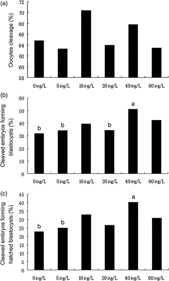
There were no significant differences across treatment groups in the proportion of oocytes undergoing first cleavage after 24 h of fertilization (P > 0.05; Figure 4a). However, blastocyst yields in the 40 μg/l BDNF group was significantly higher than that in the BDNF-free control (BDNF-free, 31.65%; 40 μg/l BDNF, 51.22%; P < 0.05; Figure 4b). Improved hatched blastocyst development following supplementation with the 40 μg/l of BDNF was also apparent (BDNF-free, 22.78%; 40 μg/l BDNF, 40.24%; P < 0.05; Figure 4c).
Development of in vitro fertilization embryos with brain-derived neurotrophic factor (BDNF). Data from replicate trials were pooled to arrive at the average proportion of activated eggs undergoing cleavage (a) or the proportion of cleaved embryos forming blastocysts (b), or hatched blastocysts (c). Significant differences denoted by different letters. Different lower case letters indicate statistical difference (P < 0.05).

Effect of BDNF on parthenogenetic embryo development
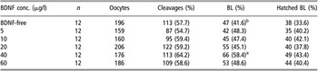
Oocytes, irrespective of maturation status as in the above trial, were parthenogenetic activated after IVM. After PA, oocytes were separated into six groups and cultured in media without BDNF or supplemented with 5, 10, 20, 40 or 60 μg/l BDNF (Table 4).
Effect of brain-derived neurotrophic factor (BDNF) on parthenogenetic embryo development

BL = blastocyst.
Values with different superscripts differ significantly (P < 0.05).
There were no significant differences across treatment groups in the proportion of oocytes undergoing first cleavage after 24 h of activation (P > 0.05; Figure 5a). However, the rates of blastocyst formation in the 40 μg/l BDNF treatment group were significantly higher than that in the BDNF-free control (BDNF-free, 41.59%; 40 μg/l BDNF, 58.41%; P < 0.05; Figure 5b), though there were no significant differences across treatment groups in the proportion of hatched blastocyst development (P > 0.05; Figure 5c).
Development of parthenogenetic embryos with brain-derived neurotrophic factor (BDNF). Data from replicate trials were pooled to arrive at the average proportion of activated eggs undergoing cleavage (a) or the proportion of cleaved embryos forming blastocysts (b), or hatched blastocysts (c). Significant differences denoted by different letters. Different lower case letters indicate statistical difference (P < 0.05).

Effect of BDNF on granulosa cell growth
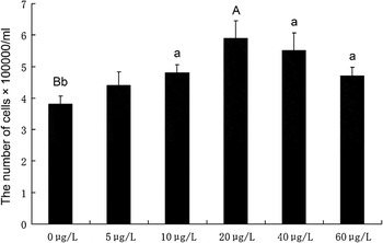
Granulosa cells, obtained by removing surrounding cumulus cells after IVM of oocytes, were separated into six groups and cultured in DMEM without BDNF or supplemented with 5, 10, 20, 40 or 60 μg/l BDNF (Figure 6). The number of granulosa cells was recorded 48 h later. The results showed that the cell number was higher in the 20 μg/l BDNF group than that in the BDNF-free group (BDNF-free, 3.08 × 10⁵/ml; 20 μg/l BDNF, 5.9 × 10⁵/ml; P < 0.01).
Generation of granulose cells with brain-derived neurotrophic factor (BDNF) in Dulbecco’s Modified Eagle’s Medium (DMEM). DMEM without BDNF (3.08 × 10⁵), or in media supplemented with 5 μg/l BDNF (4.4 × 10⁵), 10 μg/l BDNF (4.8 × 10⁵), 20 μg/l BDNF (5.9 × 10⁵), 40 μg/l BDNF (5.7 × 10⁵) and 60 μg/l BDNF (4.7 × 10⁵). Different capital letters indicate high statistical difference (P < 0.01). Different lower case letters indicate statistical difference (P < 0.05).

Discussion
The early development of mammals includes the formation of the zygote, the activation of the embryonic genome and the beginning of cellular differentiation. During this period, histones replace protamines, the methylated haploid parental genomes undergo demethylation following the formation of the diploid zygote and maternal control of development is succeeded by zygotic control. Embryonic genome activation is just after the formation of a chromatin-mediated transcriptionally repressive state and gene expresses efficiently requiring enhancer. The development of this transcriptionally repressive state more likely occurs at the level of the chromation structure, because inducing histone hyperacetylation can relive the requirements for enhancers. Characterization of zygotic mRNA expression patterns during the early period and their relationship to successful development in vitro and in vivo will be essential for defining optimized culture conditions for embryo development.
The cell NT is generally successful, but its overall efficiency is very low. Cloned embryos die at various stages of development, including after birth, due to a variety of developmental defects. Therefore, if methods can be devised for prior selection of NT embryos with the potential to develop into normal offspring, this technology would be of enormous benefit for animal production and conservation practices.
Quantitative real-time PCR is a sensitive and efficient technique to examine gene transcription patterns in oocytes and embryos. The quantification of gene expression in tissue samples requires the use of reference genes to normalize transcript numbers between different samples. Reference gene stability may vary between different tissues, and within the same tissue during different states. The function of SDHA is of an electron transporter in the TCA cycle and respiratory chain. SDHA was used as the reference gene in canine articular connective tissues, bovine polymorphonuclear leukocytes and human primary neuroblastoma (Fischer et al., Reference Fischer, Skowron and Berthold2005; De Ketelaere et al., Reference De Ketelaere, Goossens, Peelman and Burvenich2006; Ayers et al., Reference Ayers, Clements, Salway and Day2007). In order to optimize and validate the reaction to maximize the sensitivity and accuracy in quantitative real-time PCR, we chose SDHA as the reference gene in our research, because SDHA was found to be the most stable gene across the bovine embryonic stages. SDHA is an accurate normalization factor as the reference gene (Goossens et al., Reference Goossens, Van Poucke, Van Soom, Vandesompele, Van Zeveren and Peelman2005).
We have measured BDNF mRNA levels in oocytes and embryos in IVF. The present study demonstrates that mRNA for BDNF could be detected in RNA isolated from bovine GV oocytes, MII oocytes, 4-cell and 8-cell embryos, morulae and blastocysts, respectively, suggesting that this factor could be important for oocytes and embryos (Seifer et al., Reference Seifer, Feng, Shelden, Chen and Dreyfus2002 and Reference Seifer, Lambert-Messerlian and Schneyer2003; Martins da Silva et al., Reference Martins da Silva, Gardner, Taylor, Springbett, Sousa and Anderson2005; Kawamura et al., Reference Kawamura, Kawamura, Fukuda, Kumagai, Hsueh and Tanaka2007). There was the least expression for BDNF in the 8-cell stage. During the 8- to 16-cell period of bovine embryos, the maternal control of development may be succeeded by zygotic control, and the formation of a chromatin-mediated transcriptionally repressive state begins (Heikinheimo and Gibbons, Reference Heikinheimo and Gibbons1998; Young et al., Reference Young, Butterwith and Wilmut1998). There was higher expression in MII oocyte and the morulae stage, suggesting the effect of BDNF on increasing the first polar body extrusion rate in oocytes stripped of cumulus prior to maturation and promoting bovine early embryo development.
The NT embryos developed from adult female granulosa cell nuclear transferred (GNT) oocytes have a lower incidence of perinatal abnormalities than skin fibroblast cell nuclear transferred (FNT) oocytes do, which suggests that reprogramming of GNT may be more complete and stable in gene expression than FNT (Kato et al., Reference Kato, Tani and Tsunoda2000). We thought that gene expression analysis from GNT might offer credible insight into the difference of gene expression in embryos derived from IVF, PA and NT. We found that NT blastocysts contained lower levels of mRNA for BDNF than IVF and parthenogenetic blastocysts did. This finding is novel in the cattle. Incomplete epigenetic regulation is suspected to be the cause of the abnormalities and the low efficiency associated with NT (Suteevun et al., Reference Suteevun, Smith, Muenthaisong, Yang, Parnpai and Tian2006). The aberrant expression pattern in NT embryos was found for transcription factors thought to be involved in stress adaptation, trophoblastic function, DNA methylation and histone acetylation during preimplantation development. These early deviations in gene expression patterns seen in NT-derived embryos warrant further investigations in post-implantation and neonatal development to understand the causative mechanism of the abnormalities and elevated mortality after transfer of NT- and activation-derived embryos (Daniels et al., Reference Daniels, Hall, French, Korfiatis and Trounson2001; Wrenzycki et al., Reference Wrenzycki, Wells, Herrmann, Miller, Oliver, Tervit and Niemann2001; Humpherys et al., Reference Humpherys, Eggan, Akutsu, Friedman, Hochedlinger, Yanagimachi, Lander, Golub and Jaenisch2002). The change of these genes mRNA directly affects the quality of embryos (Rizos et al., Reference Rizos, Lonergan, Boland, Arroyo-García, Pintado, de la Fuente and Gutiérrez-Adán2002; Ruddock et al., Reference Ruddock, Wilson, Cooney, Korfiatis, Tecirlioglu and French2004). By immunohistochemistry, BDNF protein is localized in trophectoderm cells.
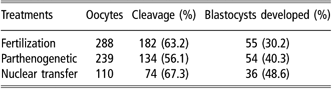
The data in the present study showed that supplementation of culture media with BDNF at the concentration of 40 μg/l caused a significant increase in the rate of parthenogenetic blastocyst formation (31.5% v. 51.2%, P < 0.05) and had a similar effect on IVF blastocyst formation (42.0% v. 58.0%, P < 0.05). These results indicate a role for BDNF in enabling blastocyst formation following IVF and PA. And BDNF in DMEM can accelerate granulosa cells’ proliferation (P < 0.01). We presume that BDNF signals between granulosa cell monolayer and embryos by TrkB and p75 receptor.
The high expression of TrkB in trophectoderm cells of expanded and hatched blastocysts is likely responsible for the observed ability of BDNF to promote early embryonic development, and to prevent blastocyst apoptosis. In the nervous system, BDNF binds TrkB by autocrine and paracrine secretion, which signals cascades of the Ras/Raf/MEK/MAP kinase pathway (Sugimoto et al., Reference Sugimoto, Kuroda, Horii, Moritake, Tanaka and Hattori2001; Ichikawa et al., Reference Ichikawa, Terayama, Yamaai, Yan and Sugimoto2007). However, it has been reported that after the binding of BDNF, TrkB induces a complex signaling cascade, including PI3K pathways, to promote neuronal survival in neurons (Kawamura et al., Reference Kawamura, Kawamura, Fukuda, Kumagai, Hsueh and Tanaka2007). There was evidence demonstrating either the presence or function of the PI3K pathway in mammal preimplantation embryos. The PI3K and Akt pathway is a mediator of growth promoting and cell survival signals. The PI3K and Akt pathway leads to promote blastocyst hatching, which is required for successful implantation of the embryo. PI3K-generated phospholipids regulate Akt activity by directly binding to its pleckstrin homology domain. The consequence of Akt binding to phospholipids is the translocation of Akt from the cytoplasm to the inner surface of the plasma membrane (Currie et al., Reference Currie, Walker, Gray, Deak, Casamayor, Downes, Cohen, Alessi and Lucocq1999). Relocalization of Akt to the plasma membrane brings Akt into the proximity of regulatory kinases that phosphorylate and activate Akt. PI3K and Akt localize to the plasma membrane of the apical trophectoderm. It is this cell layer that directly interacts with the maternal environment and is responsible for transferring external stimuli into the developing embryo (Riley et al., Reference Riley, Carayannopoulos, Wyman, Chi, Ratajczak and Moley2005).
There is evidence that BDNF can promote early embryonic development and growth of granulosa cells. In comparison with the report by Martins da Silva et al. (Reference Martins da Silva, Gardner, Taylor, Springbett, Sousa and Anderson2005), our data show that supplementation of culture media with BDNF at the concentration of 40 μg/l caused a significant increase in the rate of blastocyst formation from IVF. One factor may be that we cultured bovine early embryos by the co-culture system, while Martins da Silva et al. (Reference Martins da Silva, Gardner, Taylor, Springbett, Sousa and Anderson2005) used the SOFaaBSA culture system without granulosa cells. The data in our study suggest that BDNF can promote proliferation of granulosa cells. In the co-culture system, there is a very complicated network of autocrine and paracrine secretion like in vivo. The granulosa cells secrete some growth factors, such as fibroblast growth factor 2 (FGF2), fibroblast growth factor 7 (FGF7) and leukemia inhibitory factor (LIF) (Finch et al., Reference Finch, Rubin, Miki, Ron and Aaronson1989; Van Wezel et al., Reference Van Wezel, Umapathysivam, Tilley and Rodgers1995; Nilsson et al., Reference Nilsson, Kezele and Skinner2002). These growth factors can stimulate the developing embryo. Also, granulosa cells can eliminate factors that hinder the development of embryo, including chelate heavy metal ions and hypoxanthine (Harvey et al., Reference Harvey, Arcellana-Panlilio, Zhang, Schultz and Watson1995). The co-culture system overcomes the 2-cell block in vitro and improves the embryo development. The beneficial effect may be a result of direct cell-to-cell contact between the embryo and helper cells and the removal of deleterious components from medium (Joo et al., Reference Joo, Kim, Na, Moon, Lee and Kim2001).
In conclusion, our study adds BDNF to the list of growth factors that can promote early IVF and parthenogenetic embryonic development by the co-culture system. This effect is likely to be complex and mediated by direct action on the embryo or indirect action through granulosa cells. This study will be of value to the creation of completely defined culture environments for bovine embryos in vitro, which will improve both embryo safety and the efficacy of assisted reproductive technologies applied to cattle and other animals.
Acknowledgment
This work was supported by the Project for Science and Technology Development of Jilin Province, PR China (No. 20070210).