Double-orifice mitral valve is an infrequent cause of mitral valve disease that is usually treated with valve replacement by prosthesis, despite being potentially repairable.
We present the case of a 6-year-old patient with initial dyspnoea and diagnosis of mitral regurgitation. The transthoracic echocardiography showed a double-orifice mitral valve with severe mitral regurgitation and concomitant severe subaortic obstruction.
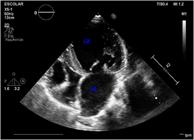
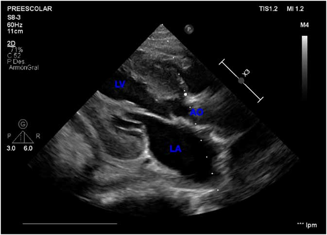
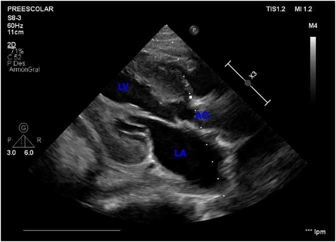
The two-dimensional echocardiography (Fig 1) showed a defect in the commissure of the anterior mitral leaflet (A1), which corresponds to the mitral cleft accessory hole (correlated intraoperatively). In addition, Figure 2 shows a hypertrophic left ventricle and a mitral valve with sub-valvular accessory tissue inserted anomalously from the interventricular septum, which partially occupies the left ventricular outflow, resulting in aortic sub-valvular obstruction.
Two-dimensional echocardiography, apical four chamber. LA=left atrium; LV=left ventricle.

Two-dimensional echocardiography, parasternal long-axis view. AO=aortic valve; LA=left atrium; LV=left ventricle.
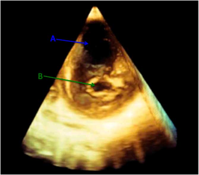
The three-dimensional echocardiography described that the mitral valve main orifice (Fig 3) resembled two apparently coapting cusps, whereas the smaller accessory orifice (Fig 3) located at the theoretical area A1-A2 was responsible for severe regurgitation.
Three-dimensional echocardiography, mitral valve. A=Mitral valve main orifice; B=Mitral accessory orifice.

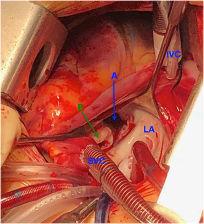
In the operating room, the pre-operative description was confirmed. The “main” orifice (Fig 4) was located at the surgeon’s right-hand side. The accessory one (Fig 4) was at the left and felt very similar to a mitral “cleft”. This secondary orifice had excessive sub-valvular tissue responsible for subaortic obstruction.
Mitral valve, direct vision. A=Mitral valve main orifice; B=Mitral valve accessory orifice; IVC=inferior vena cava; LA=left atrium; SVC=superior vena cava.

The accessory mitral tissue was resected working through the aortic valve, and the “accessory” orifice was closed from the atrium with a continuous monofilament suture. As a result, the “main” orifice previously described remained the only one. The valve competence was verified with intra-operative echocardiography, which confirmed normal valve function.
The post-operative recovery was uneventful. Echocardiography before hospital discharge showed only mild regurgitation and absence of subaortic obstruction.
In conclusion, mitral valve repair in children is always desirable and may be attempted even in complex anatomy such as the double-orifice mitral valve, where it can offer excellent results.
Acknowledgements
Special thanks to the pediatric cardiology team of the Children’s Hospital of Malaga.
Financial support
This study was financially supported by Andalucia Health Service (SAS).
Conflicts of interest
None.