Introduction
Susac syndrome was first described in 1979 by Susac et al. Just over 100 cases have subsequently been described.
The same syndrome has also been termed ‘RED M’ (retinopathy, encephalopathy and deafness-associated microangiopathy), ‘SICRET’ (small infarcts of cochlear, retinal and encephalic tissues) and retinocochleocerebral vasculopathy.
The classical triad of Susac syndrome may not be present initially, and may take from several weeks to two years to become clinically apparent.Reference Do, Fisch and Francois1 Audiological symptoms are only seen in 22 per cent of patients on first presentation, but bilateral hearing loss will usually develop eventually.Reference Aubart-Cohen, Klein, Alexandra, Bodaghi, Doan and Fardeau2 Despite a body of evidence in favour of a cochlear cause of hearing loss, there have also been reports suggesting retrocochlear pathology.
Our patient presented with fluctuating hearing loss alongside neurological symptoms, and eventually developed a severe hearing loss in the right ear and profound loss in the left. She underwent successful left implantation of an Advanced Bionics (California, USA) cochlear implant.
Eighteen months after the first onset of symptoms, the patient resumed her daily activities, with rehabilitation of hearing loss and no severe impairment otherwise.
Case report
A 36-year-old woman was admitted with a one-week history of increasing headache, agitation, confusion and amnesia.
Following admission, she developed hearing loss and tinnitus. Pure tone audiometry showed a 60 dB hearing threshold in the right ear and normal thresholds in the left.
A magnetic resonance imaging (MRI) scan showed subtle hyperintensity in the midbrain and left caudate regions, suggestive of inflammatory encephalitis.
The patient was readmitted after three weeks with vestibular symptoms and progressive hearing loss. Pure tone audiometry revealed an improvement in thresholds on the right but a 60 dB threshold in the left ear. Clinical examination revealed jerky pursuit, hypometric saccades, a positive Fukuda's head thrust test bilaterally and a reduction of dynamic visual acuity.
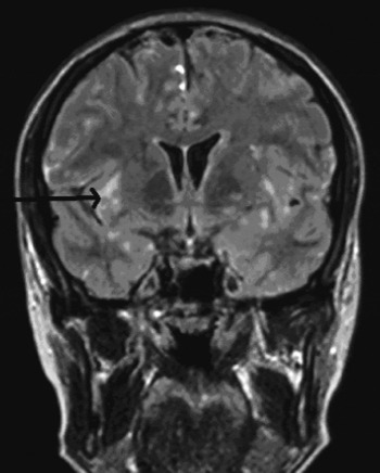
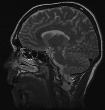
At this stage, MRI scanning showed multiple disseminated hyperintense lesions in the subcortical white matter, cerebral peduncle and corpus callosum (Figures 1 and 2), together with leptomeningeal enhancement, raising the possibility of Susac syndrome.
Coronal magnetic resonance imaging scan, Fluid attenuated inversion recovery (FLAIR) sequence, showing hyperintense subcortical and basal ganglia lesions (arrow).

Sagittal, T2-weighted magnetic resonance imaging scan showing hyperintense lesions in the basal ganglia and cerebral peduncle (arrow).

One month after initial presentation, the patient developed blurred vision and reported seeing ‘flashes of light’. Retinal examination revealed right arteriolar occlusion, while fluorescein angiography showed multiple peripheral retinal arterial occlusions. At this stage, the diagnosis of Susac syndrome was confirmed.
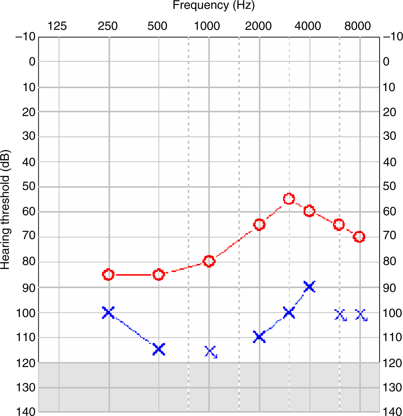
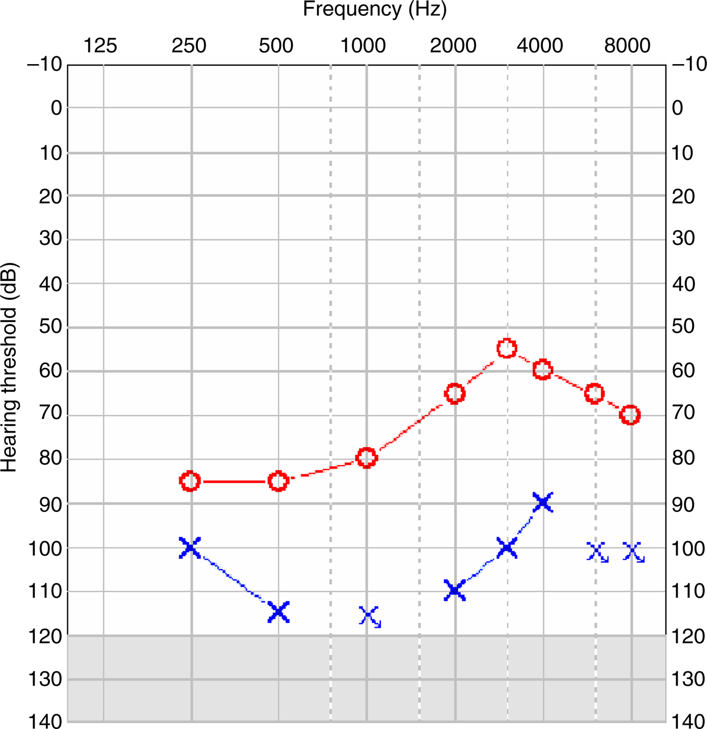
The patient's treatment included steroids, immunoglobulins, cyclophosphamide, rituximab and mycophenolate. Her hearing continued to fluctuate over the subsequent months, leaving her with profound hearing loss on the left side and severe loss on the right (Figure 3). The patient's average aided thresholds were 30 dB; however, her speech discrimination was poor, with a Bamford–Koval–Bench score of 4 per cent with listening and lip-reading, and her hearing handicap score was 100 per cent.Reference Green, Bhatt, Mawman, O'Driscoll, Saeed and Ramsden3
Pre-operative pure tone audiogram.
Owing to negligible functional gain from hearing aids, the patient underwent implantation of a left Advanced Bionics cochlear implant, with full insertion of electrodes into the cochlea. Neural response telemetry showed excellent responses in electrodes 1 to 14, with normal impedances.
Six months post-operatively, the patient's Bamford–Koval–Bench scores were 84 per cent with listening and lip-reading and 68 per cent with listening alone. Free-field audiogram thresholds averaged 20 dB across all frequencies. The patient continued to show signs of vestibular failure.
Discussion
Susac syndrome was first described in 1979 by Susac et al. Reference Susac4 It has a clear predilection for females, with only approximately 17 per cent of cases occurring in males. Age at diagnosis ranges between 18 and 69 years, with a mean age of 30 years.Reference Roeser, Driscoll, Shallop and Gifford5–Reference Papo, Biousse, Lehoang, Fardeau, N'Guyen and Huong7 At presentation, the full triad of clinical features is seen in just 11 per cent of patients, and cochlear symptoms in 22 per cent.
Inner ear involvement is manifested by hearing loss, tinnitus and vertigo. Low and mid-range frequencies are most commonly affected, although all frequencies may eventually become involved.Reference Bateman, Johnson and Gibbin8 Hearing loss may be fluctuant in 45 per cent of patients; although initially unilateral in 52 per cent, hearing loss will eventually involve both ears, given time.Reference Aubart-Cohen, Klein, Alexandra, Bodaghi, Doan and Fardeau2 The majority of patients will have persistent mild to moderate hearing loss as one of the sequelae of the disease, even after stabilisation by medical therapy.
The pathophysiological effects of Susac syndrome consist of microangiopathy affecting the brain, cochlea and retina; microvascular changes have also been demonstrated in muscle biopsies from patients with constitutional symptoms. The exact aetiopathogenesis is not well defined, although one author has proposed a theory of an immune-based mechanism triggered by previous infection.Reference Do, Fisch and Francois1
The diagnosis of Susac syndrome is based upon T2-weighted MRI evidence of multifocal, hyperintense lesions in characteristic locations: the periventricular area, corpus callosum, basal ganglia and cerebellum. Encephalopathy can be acute or subacute, and is frequently seen with impaired cognition and memory, ataxia, dysarthria and corticospinal tract dysfunction.
The other important aspect of diagnosis is the presence of retinal arteriolar branch occlusions and arterial wall hyperfluorescence on fluorescein angiography or fundoscopy. The clinical manifestation of retinal involvement varies with the extent and position of arteriolar occlusion. Interestingly, however, signal intensity or enhancement abnormalities have never been reported in the cochlea.
The diagnosis of Susac syndrome can be difficult owing to the variable time of symptom onset, compounded by encephalopathic changes in mentation which may obscure initial visual or auditory symptoms.Reference Do, Fisch and Francois1 At presentation, vestibulocochlear symptoms can mimic Ménière's disease. Multiple sclerosis is the other demyelinating disorder which presents similarly with associated central nervous system symptoms; however, it can be distinguished on MRI. Hence, unexplained encephalopathy in a young patient, especially if associated with hearing loss, should prompt repeated fundoscopy to detect vital retinal signs and aid the diagnosis.
• Susac syndrome is a rare disorder with a potential for misdiagnosis
• The key to diagnosis is recognition of T2-weighted magnetic resonance imaging signal abnormalities in characteristic locations in the brain, together with branch retinal artery occlusion and associated hearing loss
• Hearing may fluctuate initially but will eventually stabilise; cochlear implantation should be considered as a viable option in patients meeting the criteria
• Cochlear implant outcomes in such patients are encouraging, despite the possible involvement of retrocochlear pathways
Sensorineural hearing loss is an important feature of Susac syndrome. Previous studies have suggested a cochlear cause, based upon poor word recognition scores, absent otoacoustic emissions and normal acoustic reflexes, and supported by microangiopathy (seen in biopsy specimens) indicating damage to end-arterioles supplying the apical cochlea (explaining the observed pattern of low frequency hearing loss).Reference Roeser, Driscoll, Shallop and Gifford5 Bateman et al. have proposed the presence of retrocochlear pathology, based upon the presence of prolonged wave I–V latency and the absence of wave I during auditory brainstem response (ABR) testing.Reference Bateman, Johnson and Gibbin8 However, our patient's experience, and that of a previous Susac syndrome patient undergoing bilateral cochlear implantation with a successful outcome, do not lend support to this theory. We would suggest that surgery is indicated in patients with thresholds severe enough to merit a cochlear implant.