Introduction
Ossicular pathologies can be classified as congenital malformations or as acquired ossicular discontinuity or fixation. The incus is the most commonly affected ossicle within the middle ear.Reference Nomura, Nagao and Fukaya1 The lenticular and distal long processes of the incus are most vulnerable, and the malleus and footplate are most resistant.Reference Tuz, Dogru, Yasan, Doner and Yariktas2 Chronic otitis media is the commonest cause of ossicular defects, and the most frequent of these is necrosis of the long process of the incus.Reference Watson3 Other less common causes include trauma, ‘local pressure from the chorda tympani nerve’,Reference Watson3 and, rarely, systemic diseases, including diabetes mellitusReference Tuz, Dogru, Yasan, Doner and Yariktas2 and sarcoidosis.Reference Banerjee and Gleeson4
To our knowledge, our patient constitutes the first reported case of an absent proximal long process of the incus, with preservation of the lenticular process and incudo-stapedial joint, in the absence of any previous history of chronic serous otitis media.
Case report
A 53-year-old man presented with a two-year history of progressive hearing loss and tinnitus in his right ear. He himself felt that he had previously had subjectively normal hearing. He denied any history of previous ear infections, trauma or family history of deafness.
Ear examination confirmed normal ear canals and tympanic membranes bilaterally.
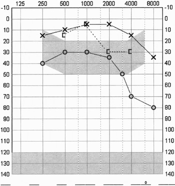
Pure tone audiometry (Figure 1) revealed a 25 dB conductive hearing loss at the mid-frequencies in the right ear, with a Carhart's notch at 2 kHz, and also marked hearing loss at 4 kHz, which was also mainly conductive. The patient had normal hearing in his left ear.
Pre-operative pure tone audiogram. ×= Air conduction, left ear; O = Air conduction, right ear; [ = Bone conduction, right ear

Tympanometry showed a normal type A graph for both ears.
A clinical diagnosis of right-sided otosclerosis was made, and all management options were discussed with the patient. He was keen to pursue surgery.
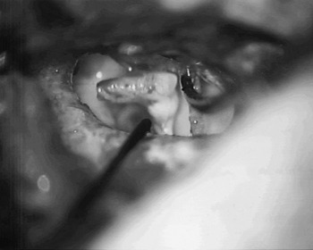
The patient thus underwent an exploratory tympanotomy, which revealed unsuspected pathology; there was erosion of the proximal long process of the incus adjacent to its body, with a normal intact long process and incudo-stapedial joint. The rest of the middle-ear examination was normal, with a mobile stapes footplate and no evidence of any granulation tissue, or any other signs of middle-ear disease. The redundant long process of the incus was excised and a partial ossicular replacement prosthesis was inserted (Figure 2).
Intra-operative finding of necrosis of the proximal long process of the incus.

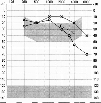
The patient subsequently made an uneventful recovery, and his post-operative pure tone audiometry results confirmed improved hearing, with complete closure of the air–bone gap (Figure 3).
Post-operative pure tone audiogram. ×= Air conduction, left ear; O = Air conduction, right ear; [ = Bone conduction, right ear

Discussion
There have been a number of theories attempting to explain why necrosis of the long process of the incus is the most commonly encountered abnormality. This is mostly attributed to its blood supply, which is said to be particularly precarious.Reference Lannigan, O'Higgins and McPhie5, Reference Alberti6 However, alternative explanations include an osteoclastic theory.Reference Lannigan, O'Higgins and McPhie7 More recently, there have also been cases of incus necrosis described in association with some systemic diseases.
Alberti was the first to describe the arterial supply of the lenticular and long processes of the incus.Reference Alberti6 The predilection of these parts of the incus to undergo resorption is attributable to their tenuous blood supply. The long process of the incus receives its blood supply from three groups of vessels, which all freely anastomose.Reference Alberti6 The main blood supply is from a number of small vessels passing from the medial wall of the tympanum along the crura of the stapes. These include the stylomastoid artery, superficial petrosal artery, carotico-tympanic artery, and superior and inferior tympanic arteries. In addition, there is also a contribution from vessels passing along the stapedius tendon, which are usually branches of the stylomastoid artery, and from the vessels of the posterior crus, a branch of the superficial petrosal artery. The final contribution comes from vessels descending the long process of the incus from above.
When parts of this vascular supply are impaired and there is discontinuity in the free communication of the anastomotic plexus, the distal tip of the incus is susceptible to erosion. Pressure from a severely retracted eardrum, for example in chronic adhesive otitis media, is thought to cause interference with the blood supply of the long process of the incus, resulting in pressure necrosis. The same effect has also been described as a result of pressure from the chorda tympani intervening between the incus and the eardrum.Reference Watson3
However, such theories of a tenuous vascular supply to the long process of the incus have been challenged. Lannigan et al. have shown that the vascular anatomy of the long process is similar to that of the short process and the body of the incus.Reference Lannigan, O'Higgins and McPhie5 Using electron microscopy studies of the vascular foramina on the cortical bone surfaces of incudes, these authors have demonstrated that there is no statistical difference between the numbers of vascular foramina on the lenticular and long processes, as opposed to the body and the short process.Reference Lannigan, O'Higgins and McPhie5
• Ossicular pathology most commonly affects the incus
• The lenticular and distal long processes of the incus are most vulnerable
• Chronic otitis media is the most frequent cause of acquired ossicular defects
• This paper describes an unusual case of an absent proximal long process of the incus, with preservation of the lenticular process and incudo-stapedial joint, in the absence of any previous history of chronic serous otitis media
The main alternative theory of incus necrosis involves failure of bone remodelling.Reference Lannigan, O'Higgins and McPhie7 The normal incus undergoes constant remodelling through resorption and rebuilding throughout life, via osteoblast and osteoclast activity. Scanning electron microscopy studies have been used to assess the pattern and degree of bony remodelling at different sites of the incus.Reference Lannigan, O'Higgins and McPhie7 These have demonstrated that the body and short process appear stable throughout life. However, by contrast, the lenticular and long processes show marked osteoclastic activity, with a tendency to undergo progressive erosion with advancing age.Reference Lannigan, O'Higgins and McPhie7 The site of abnormal osteoclastic activity has also been suggested to be developmental in origin, relating to the line of fusion of branchial components of the ossicular chain; alternatively, the distribution may relate to normal stresses within the ossicular chain.Reference Lannigan, O'Higgins and McPhie7
More recently, there has been a suggested association of incus necrosis with some systemic conditions. These include diabetes mellitus.Reference Tuz, Dogru, Yasan, Doner and Yariktas2 In this condition, incus necrosis has been attributed either to increased levels of circulating prostaglandins, which may result in bone erosion by inducing osteoclastic activity,Reference Nasrullah, Landry, Singh, Sklepowicz and Herbert8 or alternatively to basal membrane thickening in the arterial supply, which may result in insufficient local blood flow.Reference Masharani, Karam, German, Greenspan and Gardener9 There has also been a reported case of mesotympanic sarcoidosis which resulted in necrosis of the long process of the incus; this was thought to be a direct consequence of the granulomatous process.Reference Banerjee and Gleeson4
Conclusion
It would seem that there is still no consensus on the pathophysiology of ossicular discontinuity. Previously, this condition has always been described in association with some local or systemic disease. To our knowledge, there have been no previous reports of ossicular necrosis in the absence of chronic middle-ear pathology, particularly necrosis of the proximal segment of the long process of the incus.