Introduction
Several procedures for reconstruction of the middle-ear mechanism have been attempted. The purpose of each procedure has been to achieve stable, long-term hearing improvement following tympanoplasty.Reference Yung1, Reference Merchant, McKenna and Rosowski2 Jansen first reported the use of auricular and nasal cartilage to reconstruct the ossicular chain in ears without a stapes suprastructure in 1961. In 1963, both SalenReference Salen3 and JansenReference Jansen4 reported the use of a cartilage-perichondrial composite graft. In contrast, Heerman was a great advocator of cartilage tympanoplasty, and had many German articles published on the topic (as cited in YungReference Yung1). Sheehy,Reference Sheehy5 Tjellstrom, Albrektsson and colleagues,Reference Tjellstrom, Lindström, Albrektsson, Brånemark and Hallén6–Reference Tjellstrom and Albrektsson8 Kapur and Jayaramachandran,Reference Kapur and Jayaramachandran9 and several others have recommended the use of autologous bone grafts, including autologous ossicles, for reconstruction of the middle ear. In 1997, Lacosta et al.Reference Lacosta, Infante and Pisón10 reported comparable results using cortical and incus autografts and total ossicular replacement prostheses (TORPs). In 2001, Malard et al.Reference Malard, Daculsi, Toquet, Beauvillain De Montreuil, Legent and Bordure11 conducted a comparative study of functional outcome in 100 cases using ossicular autografts or calcium phosphate biomaterials for ossiculoplasty, and reported that 60 per cent of patients in both groups showed a post-operative hearing gain.
Titanium was introduced by a small number of US otolaryngologists in the late 1990s. European otologists were the first to use titanium middle-ear implants in a significant number of patients, in the mid-1990s.Reference Zenner, Stegmaier, Lehner, Baumann and Zimmermann12 For the last decade, titanium has become a popular material in ossicular implants. The advantages cited include improved visibility via an open head, possible improved signal transfer at 2 kHz, improved handling to adjust to individual anatomy, and tissue and magnetic resonance imaging (MRI) compatibility.Reference Zenner, Stegmaier, Lehner, Baumann and Zimmermann12–Reference Martin and Harner14
This study describes and tests a type of autologous TORP using cortical bone and cartilage from the pinna. The purpose is to introduce a TORP that is autologous, stable, integrates with the stapes, is biocompatible, has adequate rigidity for sound transmission, has no chance of extrusion, is MRI compatible and provides sustainable hearing improvement. Data have been presented in accordance with the American Academy of Otolaryngology–Head and Neck Surgery (AAO-HNS) ‘Committee on Hearing and Equilibrium guidelines for the evaluation of results of treatment of conductive hearing loss’.Reference Monsell, Balkany, Gates, Goldenberg, Meyerhoff and House15
Materials and methods
The ears of 40 patients, who had chronic otitis media with absent suprastructure of the stapes and long process of the incus, were repaired using the total ossicular replacement technique at Lala Lajpat Rai Memorial Medical College, Meerut, India. Patients with extensive cholesteatoma and with average bone conduction findings (at frequencies of 0.5, 1, 2 and 3 kHz on pure tone audiometry) above 25 dB sensorineural hearing loss were excluded from the cohort to maintain homogeneity of the results.
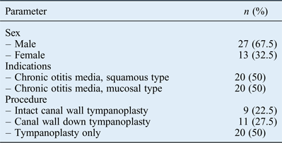
The study comprised 27 males and 13 females aged between 15 and 50 years (median age of 24 years) (Table I). All patients underwent a routine ENT examination and the necessary pre-operative investigations including pure tone audiometry. Fifty per cent of the patients had the squamous variety of chronic otitis media and the other 50 per cent had the mucosal variety. Twenty patients (50 per cent) underwent tympanoplasty only, 9 (22.5 per cent) underwent intact canal wall tympanoplasty, and 11 (27.5) underwent canal wall down mastoidectomy and tympanoplasty (Table I).
Patient and surgical data

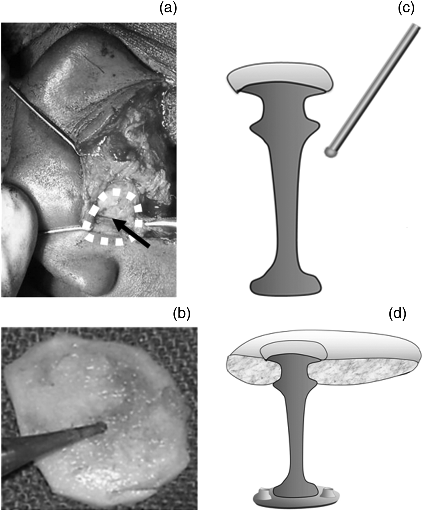
The vault of the ‘umbrella’ graft was constructed using concavo-convex portions of conchal cartilage (cut at the place where it is thinnest, in a circular manner) and its perichondrium. Using a 0.5 mm cutting burr, a hole was drilled in the vault to fit the stalk or handle of the TORP, keeping in mind the distance between the centre of the stapes and the posterior wall of the middle ear. A block or chip of bone was taken from the mastoid with the help of a chisel, and carved out in the desired shape (like an umbrella handle or stalk) using a 0.5 mm cutting burr, as shown in Figure 1. The lower end of the stalk was shaped to be slightly broader (diameter, 2–2.5 mm), and was flat on its base to increase its contact area with the stapes footplate. A very tightly fitting vault would impede movements; therefore, the diameter was kept to 1 mm less than the annulus. The malleus was intact in 35 cases (87.5 per cent). The mean height of the TORP was 5 ± 0.9 mm (range, 3.5–7 mm). After the graft was placed over the footplate of the stapes, it was given a support of gel foam in situ and, finally, was covered with a temporalis fascia graft. In cases where the middle-ear mucosa was damaged or a raw area was seen over the footplate and the edges of the oval window, it was covered with thin temporalis fascia so that the cortical bone graft did not adhere to the edges.
The autologous total ossicular replacement prosthesis (TORP) surgical procedure entails: (a) exposure of conchal cartilage (arrow) via post-aural incision, (b) drilling of a hole in conchal cartilage and (c) reshaping of the bone with a drill. The image in part (d) shows a vertical section of the TORP resting on the stapes footplate.
Audiograms were taken first at 6 months and then at 12 months post-operatively, and compared with pre-operative audiograms. The average pre- and post-operative air–bone gaps (ABGs) were calculated using air and bone conduction thresholds at frequencies of 0.5, 1, 2 and 3 kHz. The AAO-HNS guidelines define a mean post-operative ABG of 20 dB or less as a successful hearing result.Reference Monsell, Balkany, Gates, Goldenberg, Meyerhoff and House15 Hearing gain or loss was calculated in terms of the difference between pre- and post-operative mean ABGs.
All data were analysed using IBM SPSS® statistics software, version 20. Continuous data were initially assessed for normality. Continuous data were expressed as mean ± standard deviation, and categorical data were expressed as counts and proportions. The pre- and post-operative comparison of continuous variables was conducted using a paired t-test. Data from two separate and unequal groups were assessed for significant differences using an unpaired t-test. A two-sided p value of 0.05 was considered statistically significant.
Results
Overall hearing results
Table II shows an improvement in pure tone average (PTA) and average ABG for all patients (n = 40). The PTA improved from a pre-operative average of 55.7 ± 4.7 dB to a post-operative average of 31.8 ± 9.9 dB, representing an improvement of 23.9 ± 8.5 dB. This improvement was statistically significant (p < 0.001). The ABG improved from a pre-operative average of 39.3 ± 4.9 dB to a post-operative average of 15.3 ± 6.4 dB, an improvement of 23.9 ± 8.5 dB. The improvement was again statistically significant (p < 0.001).
Overall ABG and PTA pre- and post-operative, and improvement data
Data represent means ± standard deviations. *n = 40. †For 0.5, 1, 2 and 3 kHz. ABG = air–bone gap; PTA = pure tone average (air conduction); pre-op = pre-operative; post-op = post-operative
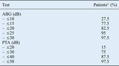
Table III shows the post-procedure ABG values for all patients; the ABG was less than or equal to 10 dB in 27.5 per cent of patients, less than or equal to 15 dB in 77.5 per cent of patients, less than or equal to 20 dB in 82.5 per cent of patients, less than or equal to 25 dB in 95 per cent of patients and less than or equal to 30 dB in 97.5 per cent of patients. Figure 2 shows the ABG data in bins of 0–10 dB, 11–20 dB, 21–30 dB and over 30 dB. Only 5 per cent of patients had an ABG greater than 25 dB post-operatively.
Overall post-operative air–bone gap findings.
Post-operative ABG and PTA data
*n = 40. ABG = air–bone gap; PTA = pure tone average (air conduction)
Successful rehabilitation of the PTA to 30 dB or less was achieved in 75 per cent of patients, and ABG to 20 dB or less was attained in 82.5 per cent of the patients (Table III).
Hearing results by procedure type
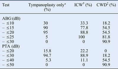
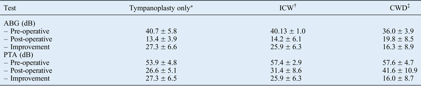
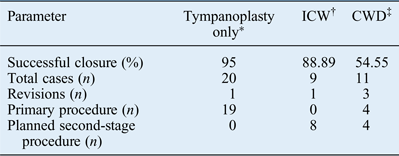
The hearing results for patients who received an autologous TORP but underwent different types of procedure were analysed separately and compared with each other. An ABG of 20 dB or less was achieved by 95 per cent of patients who underwent tympanoplasty only (n = 20), by 88.8 per cent of patients who underwent intact canal wall tympanoplasty (n = 9) and by 54.5 per cent of patients who underwent a canal wall down tympanoplasty procedure (n = 11) (Table IV). A PTA of 30 dB or less was achieved by 94.7 per cent of patients who underwent tympanoplasty only (n = 20), by 88.9 per cent of patients who underwent intact canal wall tympanoplasty (n = 9) and by 18.2 per cent of patients who underwent a canal wall down tympanoplasty procedure (n = 11) (Table IV).
Post-operative ABG and PTA data by procedure type
*n = 20; †n = 9; ‡n = 11. ABG = air–bone gap; PTA = pure tone average (air conduction); ICW = intact canal wall tympanoplasty; CWD = canal wall down tympanoplasty
The improvements in mean ABG for the three groups are shown in Table V. The mean improvement in ABG was 27.3 ± 6.6 dB in the tympanoplasty only group, 25.9 ± 6.3 dB in the intact canal wall tympanoplasty group and 16.3 ± 8.9 dB in the group who underwent a canal wall down tympanoplasty procedure. Hearing gain in the form of mean ABG was found to be statistically significant when all three groups were analysed separately (p < 0.001). No statistically significant difference in mean improvement was found between the tympanoplasty only and intact canal wall tympanoplasty groups (t-test, p > 0.05). However, the mean improvement in the tympanoplasty only group was significantly greater than in the canal wall down tympanoplasty group (t-test, p < 0.05). In addition, the mean improvement in the intact canal wall tympanoplasty group was significantly greater than in the canal wall down tympanoplasty group (t-test, p < 0.05).
ABG and PTA hearing gains by procedure type
Data represent means ± standard deviations. *n = 20; †n = 9; ‡n = 11. ABG = air–bone gap; PTA = pure tone average (air conduction); ICW = intact canal wall tympanoplasty; CWD = canal wall down tympanoplasty
Table VI summarises the results of the different procedures carried out with the autologous TORP. In the tympanoplasty only group, repair was performed as a primary procedure in 19 patients and as revision surgery in 1 patient. These were all non-cholesteatoma patients. In the nine cholesteatoma patients who underwent intact canal wall tympanoplasty, apart from one revision case all other operations were conducted as a planned second-stage procedure, three to six months after the first procedure. This was done to rule out the possibility of residual disease or recurrence of disease. In patients who underwent canal wall down surgery, four operations were conducted as a primary stage procedure and four as a planned second-stage procedure; three were revision cases.
Summary of hearing results
*n = 20; †n = 9; ‡n = 11. ICW = intact canal wall tympanoplasty; CWD = canal wall down tympanoplasty
Discussion
The search for the ideal prosthesis to reconstruct middle-ear anatomy has a long history that continues to evolve. The ideal prosthesis should be lightweight, rigid, biocompatible, have a low extrusion rate and produce acceptable hearing results. For clinical purposes, prostheses made of ossicles, cortical bone and some synthetic materials generally meet this requirement.Reference Roth, Pandit, Soma and Kertesz16, Reference Merchant, McKenna and Rosowski17 Autologous cortical bone grafts do not stimulate formation of bone nor do they undergo resorption. Like autologous ossicles, they undergo slow replacement of non-viable bone by bone formation through a process of ‘creeping substitution’, and can be used for ossicular chain reconstruction.Reference Merchant, McKenna and Rosowski17–Reference Schuknecht and Shi20 In 1995, Mills and CreeReference Mills and Cree21 reported a histological study of cortical bone grafts that were removed between 6 and 232 months after implantation in ossicular reconstructions. They concluded that the cortical bone grafts appeared to survive in the middle ear.
Several reports on the successful use of cortical bone grafts have been published. In 1983, Ojala et al.Reference Ojala, Sorri, Vainio-Mattila and Sipilä22 concluded that an autologous cortical bone columella can be recommended in cases where the patient's own ossicles are affected by disease. In 1993, MillsReference Mills23 compared the hearing results of patients with ossicular or cortical bone grafts, and showed that there was no significant difference between the two. He suggested that cortical bone is a satisfactory material for ossiculoplasty. In 2009, Ceccato et al.Reference Ceccato, Portmann and Davis24 described a technique of sculpting and shaping the ossicles while still attached to the cortical bone. Cartilage grafts also have a long history of being used for tympanoplasty.Reference Yung1 These grafts, which are not very rigid, have been used adequately for reconstruction or reinforcement of the tympanic membrane, and as a buffer between a prosthesis and the tympanic membrane.Reference Merchant, McKenna and Rosowski17 Most surgeons who use titanium prostheses use a cartilage buffer between the prosthesis and the tympanic membrane to prevent its extrusion.Reference Zenner, Stegmaier, Lehner, Baumann and Zimmermann12–Reference Martin and Harner14, Reference Roth, Pandit, Soma and Kertesz16, Reference Gardner, Jackson and Kaylie25, Reference Fisch, May, Linder and Naumann26 ElwanyReference Elwany27 has reported that preservation of perichondrium on both sides of a piece of cartilage increases the chances of chondrocyte survival.
Recently, titanium has been a popular synthetic material in ossicular reconstruction. One reported difficulty with the titanium prosthesis is that it cannot be naturally secured to the footplate, and extrusion of the titanium prosthesis, however small the risk, has been noted in most studies published so far.Reference Zenner, Stegmaier, Lehner, Baumann and Zimmermann12–Reference Martin and Harner14 Moreover, a titanium prosthesis needs a cartilage cap to prevent extrusion.Reference Zenner, Stegmaier, Lehner, Baumann and Zimmermann12–Reference Martin and Harner14, Reference Roth, Pandit, Soma and Kertesz16, Reference Gardner, Jackson and Kaylie25, Reference Fisch, May, Linder and Naumann26 Another disadvantage of titanium that has not been discussed so far in the literature is its cost, which is important in developing countries where chronic otitis is more prevalent. This factor becomes even more important if the cost of revision surgery is also considered.
An autologous TORP has already been described by an author of this paper as a cost-effective alternative to titanium and other synthetic TORPs.Reference Malhotra28 The procedure entails constructing an umbrella-shaped TORP using a vault of conchal cartilage; a stalk resembling the handle of an umbrella is carved out (drilled) from the malleus and fitted into a hole made in the vault. However, the removal of the malleus in this technique is questionable. The literature clearly shows that preservation of the malleus, wherever possible, is beneficial acoustically in ossiculoplasty.Reference Dornhoffer and Gardner29–Reference Fisch33 Moreover, if the malleus is used as the autologous TORP handle, it would never be straight, and its terminal end (which is kept pointed) would not be stable on the stapes footplate.
• Autologous cortical bone and cartilage from the pinna can be used to construct a stable total ossicular replacement prosthesis (TORP)
• In this study, the autologous TORP resulted in successful rehabilitation of conductive hearing loss in 82.5 per cent of patients
• Results were better for tympanoplasty only or canal wall up mastoidectomy cases than canal wall down mastoidectomy cases
• The autologous TORP described is biocompatible, stable, magnetic resonance imaging compatible and cost-effective
The autologous TORP described in this study incorporates the advantages of titanium and umbrella grafts described previously, and it overcomes their disadvantages. The cartilage vault provides strength to the tympanic membrane, prevents its retraction, helps in the coupling of sound energy to the stalk and keeps the graft stable as it is supported from the lateral side by the tympanic membrane. The TORP is biocompatible. In addition, the cortical bone used for the stalk provides adequate rigidity for sound transmission, and the wide base of the stalk gives sufficient stability on the stapes footplate. It has a better chance of integration with the stapes footplate than other grafts. Furthermore, the hourglass shape, drilled into the handle, which fits into the hole made in the vault, gives stability to the assembly without the use of any tissue glue. The TORP is MRI compatible and, above all, gives sustainable hearing results. Although revision surgery is required, there is no additional implant cost involved.
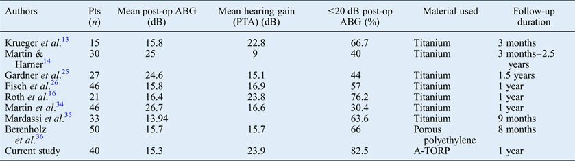
The results of various studiesReference Krueger, Feghali, Shelton, Green, Beatty and Wilson13, Reference Martin and Harner14, Reference Roth, Pandit, Soma and Kertesz16, Reference Gardner, Jackson and Kaylie25, Reference Fisch, May, Linder and Naumann26, Reference Martin, Silva, Certal, Amorim and Carvalho34–Reference Berenholz, Burkey and Lippy36 are compared with those of the present study in Table VII. It is evident that the hearing results with the autologous TORP are comparable with those of the titanium prosthesis. Another reason for the improved hearing results in the current study may relate to the authors' preference for performing intact canal wall mastoidectomy in two stages for cholesteatoma, whenever possible. Canal wall down mastoidectomy (which showed significantly inferior results) was limited to patients with sclerotic mastoids with large volumes of disease, in which access to all parts of the matrix was difficult.
comparison of ABG and PTA results with previous literature on other prostheses
ABG = air-bone gap; PTA = pure tone average (air conduction); pts = patients; post-op = post-operative; A-TORP = autologous total ossicular replacement prosthesis
In conclusion, our study indicates that the autologous TORP gives sustainable hearing results. It is a good alternative to titanium TORPs, both in terms of hearing improvement and its autologous nature. This may renew our interest in autologous grafts and prompt more research on middle-ear reconstruction, as more experimentation may be required to improve the design further and to study the long-term results of the autologous TORP.