1. Introduction
Over several decades, several researchers have investigated the effectiveness of implants in stabilizing the craniovertebral junction (CVJ), all of whom have conducted their investigations on decapitated specimens. It is reported in the article [Reference Schulze, Trautwein, Vordemvenne, Raschke and Heuer1] that a KUKA KR125 manipulator is used and that X-ray images are taken with the manipulator for implant study on a harvested specimen from the Cadaver using the X-ray images manipulator. When the Cadaver was installed, it was potted to the frame of the ground system, which a KUKA-KR125 powers. Because of this, it was possible to measure the range of motion. The study [Reference Puttlitz, Melcher, Kleinstueck, Harms, Bradford and Lotz2] used a pulley, nylon thread, and weight system to apply pure couples on the Cadaver CVJ. According to the article [Reference Helgeson, Lehman, Sasso, Dmitriev, Mack and Riew3], the paper uses the MTS 858 Bionix testing system outfitted with a custom-built 6-DOF spine simulator. Data for 10 specimens harvested from the Cadaver were occipitocervical specimens used to determine the stability of the implant apparatus. The spinal column’s CVJ region’s range of motion was calculated using the CA 6000 spinal motion analyzer [Reference Dvorak, Antinnes, Punjabi, Oustalot and Bonomo4]. Serial manipulators have been used for manipulating the CVJ in the clinical literature. In the paper [Reference Helgeson, Lehman, Sasso, Dmitriev, Mack and Riew3], significant kinematic workspaces are reported [Reference Tsai5].
In ref. [Reference Garrido-Jurado, Muñoz-Salinas, Madrid-Cuevas and Marín-Jiménez6], the authors presented a single-camera approach for posture estimation using fiducial markers. The marker image distortion must be compared to the library’s distortion to calculate the pose at the target position. The marker pattern improves accuracy [Reference Garrido-Jurado, Muñoz-Sainas, Madrid-Cuevas and Medina-Carnicer7–Reference Kansal12]. The authors describe the normal CVJ anatomy and numerous CVJ disorders [Reference Zubair, Kansal and Mukherjee10]. The CVJ is anatomically and biomechanically distinct from the sub-axial spine [Reference Reddy, Kavitha, Gafoor, Suresh and Harinath13, Reference Hong, Kim, Lee, Park, Hur, Lee, Lee and Lee14]. These specializations allow for significant mobility while maintaining stability, though fixation is complex. Specifically, this study has explained how to determine the pose estimation of the Cadaver’s CVJ region. The proposed technique has been detailed in Section 2. Section 3 will discuss Cadaver’s posture estimate and the vision-based CVJ studies. The conclusion has been discussed in detail in Section 4.
2. Methodology
2.1. Cadaver CVJ pose estimation using vision sensor
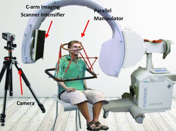
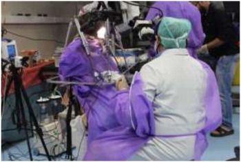
Figure 1 depicts a method for determining intervertebral mobility in a Cadaver’s CVJ. The first three vertebrae of the spinal column delineate the CVJ area. It helps reduce the majority of neck mobility. Intervention in this location necessitates surgical placement of an implant to stabilize the entire system [Reference Zubair, Kansal and Mukherjee10]. The experimental setup activates the neck motion in a Cadaver close to the human subject. This paper uses computer vision techniques to analyze the Cadaver’s CVJ, as shown in Fig. 2. Initially, the experiment will be conducted in the prone position. Still, due to the placement of the experimental setup, mounting was a challenging task. Inherently, we have switched to experimenting with a sitting posture. In the sitting posture, multiple regions were quickly exposed to the CVJ exploration compared to the prone position.
Experimental setup [Reference Zubair, Kansal and Mukherjee10].

Odontoidectomy procedure in Cadaver.

A predetermined library of ArUco markers [Reference Garrido-Jurado, Muñoz-Salinas, Madrid-Cuevas and Marín-Jiménez6, Reference Garrido-Jurado, Muñoz-Sainas, Madrid-Cuevas and Medina-Carnicer7, Reference Garrido-Jurado, Munoz-Salinas, Madrid-Cuevas and Marin-Jimenez15] was used to estimate the pose of an object. Using monocular vision and markers to achieve a Cadaver CVJ pose [Reference Kansal, Kumar and Mukherjee8, Reference Kansal and Mukherjee9, Reference Kansal and Mukherjee16] is possible. The legs are connected to the base platform by universal (U) joints and the moving platform by spherical (S) joints. These legs are manually actuated using screw (Sc) action. The 6-DOF parallel manipulator is named a 6-UScS manipulator. The 6-UScS manipulator moves the relocating platform by adjusting the leg length via screw action. This distinguishes it from the typical Stewart platform. The 6-UScS manipulator’s movable platform will be called the top platform. The upper platform holds the cadaver’s cranium using bespoke fasteners, while the bottom platform is secured to the chair. The motion of the 6-UScS manipulator is controlled by modifying the length of its legs by turning the screw (variable length).
3. Cadaveric experiment
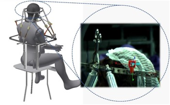
A parallel manipulator-based experimental setup for measuring the CVJ load after implant instrumentation has been created. Prior studies conducted experiments on the decapitated specimen to quantify the reaction of the CVJ implant. An earlier experiment was conducted using decapitated specimens in which the soft tissues were severed at the spine. To account for the effects of passive loading on the soft tissues, an approach closer to the natural anatomical function of the CVJ is preferred. This will require developing a novel method for measuring forces during CVJ manipulation without decapitation. Thus, among other benefits, the capacity to undertake an in situ evaluation of the implant in the CVJ without decapitation has been pursued. Therefore, the experimental setup shown in Fig. 3 for the in situ implant study in the Cadaver’s CVJ region is unique and has not been attempted before to the author’s knowledge. The experimental setup shown in Fig. 3 consists of two central units: a customized chair to support a Cadaver in a sitting position and a manually actuated 7-DOF parallel manipulator (MA7DPM). The MA7DPM is an integration of two parallel manipulators which have a common base. The moving platform of the 1-DOF parallel manipulator can be fixated to any cervical vertebra (i.e., C1–C7). The moving platform of the 6-DOF parallel manipulator is designed to be fixated on the cranium.
Experimental setup for Cadaver’s pose estimation.

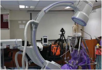
The layout of the experimental setup.

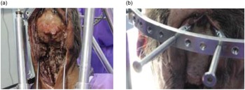
A series of experiments are conducted on a single Cadaver by changing the implant to evaluate its performance. As Fig. 5(a) depicts a typical experimental layout that was set up at the Cadaver Training and Research Facility of the Jai Prakash Narayan Apex Trauma Center (JPNATC) at the All India Institute of Medical Sciences (AIIMS) in New Delhi under the supervision of clinical staff after receiving appropriate ethical clearance. Figure 5(b) depicts a typical experimental setup at the Cadaver Training and Research Facility of the Jai Prakash Narayan Apex Trauma. Getting the Cadaver into the chair and putting his torso between two vertical pillars will require some maneuvering. The Cadaver is strapped to the table to prevent movement in the thoracic region.
(a) Exposure of cranium and cervical vertebrae to fixated with the test setup. (b) The customised polyaxial screw fixated with the test setup.

On the other hand, movement in the cervical spine is permitted because it is the area of concern. The pillar is equipped with a T-slot, into which the sliding L-clamp is screwed to hold the pillar in place. A set of screws contains the MA7DPM to the L-clamps in place. Because of these sliding clamps, the elevation of the MA7DPM was altered, resulting in the top platform being placed at the same level as the Cadaver’s occiput. Load is essential to measure the behavior of the CVJ junction load-to-deformation to the applied motion. This will give us an in-depth understanding of the CVJ loading conditions per the actuation provided.
3.1. Experiment layout
The monocular vision sensor estimates the cadaver CVJ pose estimation via a 6-UScS manipulator using ArUco-based markers and then transmitted to the manipulator. A set of images were taken from a calibrated vision sensor after the manual actuation of the 6-UScS manipulator was completed. The processed images were used to determine the pose of the Cadaver CVJ after the desired neck trajectory, including lateral bending, flexion/extension, and axial rotation. In radiology, a C-arm is a mobile X-ray machine that can be moved around and adjusted manually to accommodate the desired location for taking X-ray images. The height and orientation of the X-ray camera can be adjusted manually. It was placed around the Cadaver to obtain X-ray images, ensuring the polyaxial screw was adequately secured to the bone.
3.2. Experiment preparation
Initially, the Cadaver was assessed for the absence of rigor mortis. The neck and the head region were exposed without damaging the soft tissues, as shown in Fig. 5(a) following the clinical procedure. The occiput to the (C5) vertebra was evident for fastening with the experimental setup. The thoracic portion of the Cadaver was positioned on the customized chair in a sitting posture and immobilized by strapping between the two pillars. Following this, the base platform of the MA7DPM, common to the Sarrus mechanism and 6-UScS, was attached to the chair. Coupling and extender screws were assembled with the polyaxial screws to connect the cranium with the manipulator’s top platform, as shown in Fig. 5(b). X-ray images were taken to confirm the proper insertion of polyaxial screws. Finally, the calibrated vision sensor [Reference Basler17] determines the Cadaver’s pose estimation with respect to the base platform.
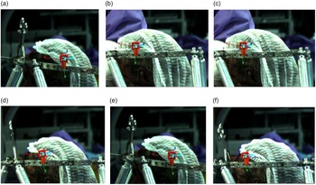
The ArUco markers are employed to estimate the pose of a Cadaver. A vision sensor and marker(s) provide the pose estimation concerning the stationary base platform, as seen in Fig. 6(a–f).
(a–f) Vision-based pose estimation of the top platform by inducing the manual actuation.

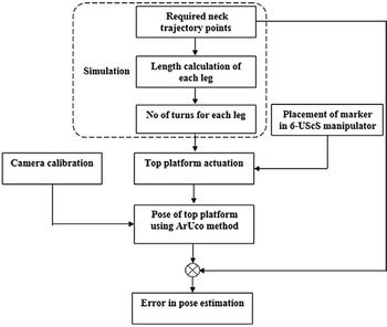
Flow diagram for estimating the error in the pose of the experimental setup [Reference Zubair, Kansal and Mukherjee10].

The pose of the marker is estimated with reference to the base fixed frame (B ff ) as:
 \begin{equation} _{\text{Marker}}\mathbf{T}^{Bff}={\left(_{\text{Table}}\mathbf{T}^{C}\right)^{-1}}_{\text{Marker}}\mathbf{T}^{C} \end{equation}
where Marker TBff is the transformation between the cube coordinate system and the base fixed frame, Table T C is the transformation in the coordinate frame fixed to the table and the frame fixed to the camera, and Marker T C is the transformation in the coordinate marker frame and the frame fixed to the camera. Since the table and camera have no relative movement, Table T C is fixed throughout the experiment. After executing the table calibration and estimating the marker pose with reference to the base fixed frame [Reference Babinec, Jurišica, Hubinský and Duchoň18], the pose of the marker is transformed from the camera coordinate frame to the base fixed frame (B ff ) and then to the platform coordinate frame ( P TMarker). The inverse kinematics modules operate with the pose of the marker defined with respect to the plane fixed to the base of the platform coordinate frame. The platform motion is controlled manually by rotating the actuators, and the vision modules run in Visual C++. The kinematic transformation has been established to measure the head motion with respect to the marker position as shown in Fig. 7.
3.3. Experiment and results
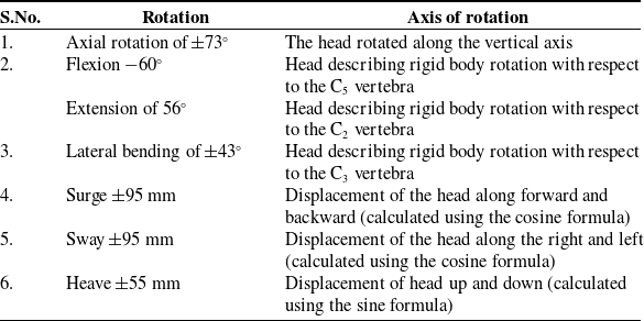
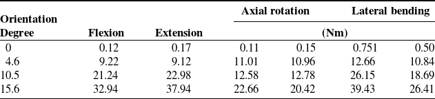
Using the screw joints on the legs of the MA7DPM in conjunction with a 6-UScS manipulator allows the user to move in all three directions: lateral bending, axial rotation, and flexion/extension, by activating the screw joints on the legs. This table shows the precise trajectory of the skull used in this experiment and additional information about it. Table I shows the data. In the flexion/extension and side-bending motions performed at their maximum values in a given direction, these modes represent the maximum translation values encountered.
The Cadaver’s range of motion.

Once the Cadaver was placed in the experimental setup, a surgical procedure (Odontoidectomy) was performed to remove the odontoid from the C2 vertebra. Transporal exposure using retainers was used to expose the odontoid for surgery by the surgical team of AIIMS. In clinical practice, the procedure, in general, is performed in a prone position where the Cadaver is placed on the stretcher. It has been mentioned earlier that the experiment requires a sitting posture of the Cadaver, and allowing access was a significant design challenge. Table II shows the load response measured using the experimental setup for the C1–C2 rod and screw instrumented in the CVJ region.
Load response was measured using the experimental setup for the C1–C2 rod and screw implant instrumented in the CVJ region.

3.4. Discussion
The ArUco approach and the ArUco method were used to analyze the process of Cadaver pose estimation using a vision sensor, and the ArUco method was used to accomplish this. This study aimed to determine how well the camera-based measurement performed when estimating the pose faults caused by fixed leg movement (incremental change). This experiment set had its load response measured and analyzed. After conducting two distinct series of tests, it was possible to determine how much load was being measured and, as a result, how much error was being introduced into the load sensor reading.
4. Conclusion
As a result, the posture estimation of the Cadaver using ArUco markers has proven to be a viable method in the experimental setup that has been constructed [Reference Zubair, Kansal and Mukherjee10]. The number of markers on the top platform can be increased to obtain a more stable pose estimation. In some cases, performing CVJ on a decapitated specimen does not provide the best results in removing soft tissue effects from the decapitated specimen. No decapitation tool was used on the specimen during the experiment, and the results were recorded. A future research extension could include establishing an experimental setup in the CVJ region with different Cadavers, such as those of various ages and genders, who could then be instrumented and investigated.
Financial support
We acknowledge Mechatronics Lab, Mechanical Engineering Department, IIT Delhi, New Delhi, India, and (JPNATC) Jai Prakash Narayan Apex Trauma Centre, AIIMS, New Delhi, India, to provide the environment for carrying out the research activity.
Author contributions
MZ and SM conceived and designed the study. MZ and SK conducted data gathering. MZ and SM performed statistical analyses. MZ and SK and wrote the article.
Competing interests
The authors do not have any type of conflict of interest.
Ethical standards
Not applicable.