Intrauterine growth restriction is recognised as the failure of an infant to achieve his or her genetic growth potential in utero.Reference Comas, Crispi, Cruz-Martinez, Martinez, Figueras and Gratacós 1 The causes of intrauterine growth restriction are broadly described under three main categories: maternal, foetal, and placental. Several maternal demographic factors have been associated with intrauterine growth restriction. Foetal factors can vary from genetic causes, congenital malformations, foetal infection, or other causes, including multiple pregnancies. Placental insufficiency accounts for many cases of intrauterine growth restriction and can affect up to 3% or more of all pregnancies. The pathogenesis of intrauterine growth restriction is not well defined; defects in the placental circulation and transport can affect the nutrient transport to the foetus, resulting in intrauterine growth restriction.Reference Suhag and Berghella 2 Intrauterine growth restriction can either be symmetric or asymmetric. Symmetric intrauterine growth restriction is characterised by a similar and proportionate reduction in parameters such as weight and length, as well as cranial and abdominal circumference. Asymmetric intrauterine growth restriction is characterised by a greater reduction in body weight, when compared with length.Reference Puccio, Giuffré, Piccione, Piro, Rinaudo and Corsello 3 Whereas symmetric intrauterine growth restriction is often due to congenital infections and foetal congenital anomalies, asymmetric growth retardation is generally caused by lack of substances that are required for foetal metabolism in late pregnancy.Reference Wollmann 4
Foetuses with intrauterine growth restriction are exposed to hypoxia.Reference Regnault, de Vrijer, Galan, Wilkening, Battaglia and Meschia 5 Owing to adaptation mechanisms against placental failure and hypoxia, various alterations occur in the cardiovascular system. Barker et alReference Barker 6 suggested that certain alterations are observed in cell and tissue structures as an adaptation mechanism against alterations in intrauterine environment, and that these changes become permanent in due course, exerting a negative impact on cardiovascular structure, function, and integrity. Cardiac dysfunction with maintained cardiac output has consistently been reported to be present in intrauterine growth restriction. Although earlier studies suggested that cardiac parameters became abnormal only in severely affected foetuses, more recent research strongly suggests that subclinical cardiac dysfunction could be present from early stages of foetal deterioration. Comas et alReference Comas, Crispi, Cruz-Martinez, Martinez, Figueras and Gratacós 1 demonstrated the presence of both left ventricular systolic and diastolic dysfunction in intrauterine growth restriction foetuses. There are no available data that evaluated left ventricular dimensions, and mass in neonates with symmetric and asymmetric intrauterine growth restriction. Therefore, we aimed to evaluate left ventricular dimensions, systolic functions, and mass in neonates with symmetric and asymmetric intrauterine growth restriction. We also assessed associated maternal risk factors and compared results with healthy appropriate for gestational age neonates.
Materials and methods
Study population
This cross-sectional study enrolled all term neonates who were born in Konya Training and Research Hospital and had a diagnosis of intrauterine growth restriction between June, 2011 and April, 2012. Gestational age was determined by the mothers’ last menstrual period and second trimester ultrasound date if the last menstrual period was unknown. Term neonates were defined as neonates who were born between 37 and 42 completed gestational weeks. Intrauterine growth restriction was defined as birth weight <10th percentile, whereas appropriate for gestational age was defined as birth weight between the 10th and 90th percentiles based on the intrauterine growth curves.Reference Alexander, Himes, Kaufman, Mor and Kogan 7 Ponderal index was calculated using the following formula: weight (g)×100/length3 (cm). Asymmetric intrauterine growth restriction was accepted if the Ponderal index value was smaller than 2.25. Weight was measured at birth by using a digital scale. Height and head circumference were measured using a standard board and tape. Body surface area was calculated using the Du Bois formula: 0.007184×weight0.425×height0.725.Reference Du Bois and Du Bois 8 Intrauterine growth restriction neonates were classified into two groups based on the Ponderal index. (1) Asymmetric intrauterine growth restriction neonates (n=62, 42 girls and 20 boys, mean Ponderal index=2.045±0.19) and (2) Symmetric intrauterine growth restriction neonates (n=39, 30 girls and nine boys, mean Ponderal index=2.68±0.25). The appropriate for gestational age group (n=50, 20 girls and 30 boys) consisted of age- and sex-matched full-term healthy neonates without evidence of congenital defects and congenital heart disease. Neonates with asphyxia, prematurity, respiratory distress, dysmorphic features, a stressful birth history, multiple birth history, and intrauterine infection were not included in the study. Maternal risk factors known to be associated with intrauterine growth restriction included maternal socio-economic status, maternal age, gestational weight gain, pre-pregnancy body mass index, parity, hypertension, infections such as cytomegalovirus or rubella, smoking and use of drugs. Maternal socio-economic status was evaluated as a combination of factors including level of education, occupation, and income based on data in Turkey.Reference Torun 9
Number of pregnancies, live births, and stillbirths were also evaluated. The study was approved by the local ethics committee of our institution, and parents provided written informed consent.
Transthoracic echocardiographic examination
All echocardiographic assessments were performed in the first 48–72 hours of life by the same expert paediatric cardiologist, who was blinded for the clinical features of the study group. Analyses were made in a quiet setting and during a time interval when the neonates were in a calm mood; no medication was administered for sedation. ProSound Alpha 7 echocardiography equipment (Aloka, Hitachi-Aloka Medical, Tokyo, Japan) with a range from 7 to 10 MHz phased-array transducer was used according to each study subject’s weight and obtained the best image from subjects. Conventional echocardiographic evaluation from the parasternal long-axis view included left ventricular end-diastolic and end-systolic diameters, septum, and left ventricular posterior wall thicknesses in diastole and systole, left ventricular ejection fraction, and fractional shortening. Teichholz’s M-mode Formula was used to calculate left ventricular ejection fraction and fractional shortening. All data were calculated by taking the mean value of three measurements. All data were obtained according to the recommendations of the American Society of Echocardiography.Reference Sahn, De Maria, Kisslo and Weyman 10 Relative posterior wall thickness was calculated by the following formula: (Interventricular septum diameter in diastole+Left ventricular posterior wall diameter in diastole)/Left ventricular end-diastolic diameter.Reference Schuster, Karpoff and Perez-Martin 11 Left ventricular mass was calculated using the following formula: 0.8{1.04[(Left ventricular end-diastolic diameter+Left ventricular posterior wall thickness+interventricular septal thickness)Reference Puccio, Giuffré, Piccione, Piro, Rinaudo and Corsello 3 −Left ventricular end-diastolic diameter3]}+0.6 and indexed for body surface area.Reference de Simone, Daniels and Devereux 12
Statistical analysis
Data were reported as mean±standard deviation, minimum–maximum (range) or per cent. Normality assumptions were assessed before conducting parametric tests. Differences in the means of variables were evaluated using both parametric and non-parametric tests depending on the distribution of the variables. χ 2 test was used to compare categorical variables. When all groups were compared for parameters, analyses of variance were used; post hoc analysis was performed using Tukey’s honestly significantly different test. Results were considered significant if p<0.05. Statistical analyses were performed with the Statistical Package for Social Science program (SPSS version 15.0 for Windows; Chicago, Illinois, United States of America).
Results
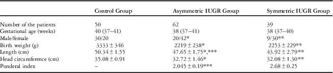
The clinical features of the neonates with intrauterine growth restriction and healthy control groups were shown in Table 1. In all, four intrauterine growth restriction neonates with incomplete records and one intrauterine growth restriction neonate with Noonan syndrome were excluded from the study. The remaining 101 patients comprised 29 (28.7%) boys and 72 (71.3%) girls. Girls were found to be more prone to developing intrauterine growth restriction. Intrauterine growth restriction neonates were categorised into two groups based on the Ponderal index. The asymmetric intrauterine growth restriction group included 62 neonates (61.4%) and the symmetric intrauterine growth restriction group included 39 neonates (38.6%). The intrauterine growth restriction groups had significantly lower birth weight (2253±229 g versus 2219±238 g versus 3333±346 g), height (43.92±2.79 cm versus 47.65±1.75 cm versus 50.34±1.55 cm), and head circumferences (32.08±1.30 versus 32.72±1.46 versus 35.08±0.91) than the control group.
Clinical features of the intrauterine growth restriction neonates and healthy controls.

Data are shown as mean±standard deviation, median (range)
* Asymmetric intrauterine growth restriction versus Control, p<0.05
** Symmetric intrauterine growth restriction versus Control, p<0.001
*** Asymmetric intrauterine growth restriction versus symmetric intrauterine growth restriction, p<0.05
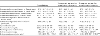
Left ventricular systolic functions, dimensions, and wall thicknesses of the neonates with intrauterine growth restriction and healthy control groups were presented in Table 2. The asymmetric intrauterine growth restriction group had significantly lower left ventricular end-diastolic and end-systolic diameters and posterior wall thickness in systole and diastole than the control group. The symmetric intrauterine growth restriction group had significantly lower left ventricular end-diastolic diameter (15.49±3.17 mm versus 16.83±1.75 mm) (p<0.001) than the control group. By conventional echocardiography, left ventricular ejection and shortening fractions were similar in both intrauterine growth restriction groups and the control group (p>0.05). All left ventricular dimensions were lower in the asymmetric intrauterine growth restriction neonates compared with the symmetric intrauterine growth restriction neonates (p>0.05), but this finding was not statistically significant except for left ventricular posterior wall thickness in diastole (3.08±0.83 mm versus 3.54±0.72 mm) (p<0.05). Both symmetric and asymmetric intrauterine growth restriction groups had significantly lower relative posterior wall thickness (0.54±0.19 versus 0.48±0.13 versus 0.8±0.12), left ventricular mass (9.8±4.3 g versus 8.9±3.4 g versus 22.2±5.7 g), and mass index (63.6±29.1 g/m2 versus 54.5±24.4 g/m2 versus 109±28.8 g/m2) when compared with the control group.
Left ventricular dimensions and systolic functions of the intrauterine growth restriction neonates and healthy controls.

Data are shown as mean±standard deviation, median (range)
* Asymmetric intrauterine growth restriction versus Control, p<0.05
** Symmetric intrauterine growth restriction versus Control, p<0.001
*** Asymmetric intrauterine growth restriction versus symmetric intrauterine growth restriction, p<0.05
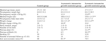
Maternal features of the neonates with intrauterine growth restriction and healthy controls are shown in Table 3. The percentage of the mothers who had a pre-pregnancy body weight <50 kg was higher in the symmetric intrauterine growth restriction group when compared with the control group (25.6% versus 8%) (p<0.05). The mean height of the mothers in the symmetric intrauterine growth restriction group was lower than the control group (159.1±4.5 cm versus 161.9±6.06 cm) (p<0.05). The history of previous abortion was higher (33.3% versus 16.1%) (p<0.05) in the symmetric intrauterine growth restriction group when compared with the asymmetric intrauterine growth restriction group. The percentages of the mothers with low socio-economic status were significantly higher in the asymmetric intrauterine growth restriction group when compared with the control group. (85.5% versus 68%) (p<0.05).
Maternal features of the intrauterine growth restriction neonates and healthy controls.
Data are shown as mean±standard deviation, median (range), or per cent
* Asymmetric intrauterine growth restriction versus Control, p<0.05
** Symmetric intrauterine growth restriction versus Control, p<0.05
*** Asymmetric intrauterine growth restriction versus symmetric intrauterine growth restriction, p<0.05
Discussion
To our knowledge, our study is the first to evaluate left ventricular dimensions, wall thicknesses, mass, and systolic functions in neonates with intrauterine growth restriction and compare results with respect to asymmetric or symmetric subgroups. This cross-sectional study provides evidence that intrauterine growth restriction neonates have lower left ventricular dimensions, wall thicknesses, mass, and mass index when compared with healthy term appropriate for gestational age neonates. Left ventricular ejection fraction and shortening fraction were found to be preserved in neonates with intrauterine growth restriction. Our study also demonstrated that both symmetric and asymmetric intrauterine growth restriction groups had similar relative posterior wall thickness, left ventricular mass, and mass index.
Various maternal and foetal risk factors may lead to intrauterine growth restriction. The confirmed maternal risk factors include short stature, low weight, ethnicity, nulliparity, mother born intrauterine growth restriction, cigarette smoking, and cocaine use. Maternal medical history of chronic hypertension and renal disease are also associated risk factors with increased intrauterine growth restriction birth. On the other hand, risk factors that develop during pregnancy include placental abruption, pre-eclampsia, and gestational hypertension.Reference Meas, Deghmoun and Alberti 13 In our study, low socio-economic level, short maternal stature, and low maternal weight were found as risk factors to develop intrauterine growth restriction.
In order to evaluate the effect of foetal sex on pregnancy outcome, Melamed et alReference Melamed, Yogev and Glezerman 14 conducted a retrospective study on 66.387 (34.367 to 51.8% male foetuses and 32.020 to 48.2% female foetuses) singleton pregnancies over a period of 11 years. They concluded that although the incidence of pre-term delivery and caesarean section was higher for male foetuses, female foetuses were more likely to develop intrauterine growth restriction. However, other studies have shown that male sex is an independent risk factor for poor pregnancy outcome, although ultrasound diagnostic rate of intrauterine growth restriction was higher in female foetuses.Reference Radulescu, Ferechide and Popa 15 In our study, we found that the number of girls with intrauterine growth restriction was 2.5 times more than boys.
Recent studies have shown structural cardiac changes and cardiac dysfunction in intrauterine growth restriction patients. Crisspi et alReference Crispi, Bijnens and Figueras 16 showed morphological cardiac changes and subclinical cardiac dysfunction in 5-year-old children born with intrauterine growth restriction. Morphological alterations were directly correlated with the severity of intrauterine growth restriction. Intrauterine state of chronic hypoxia and undernutrition, together with an increased placental vascular resistance, results in a combined pressure and possible volume overload on foetal heart, which induces abnormal cardiac function. The ensuing increased wall stress on the developing myocardial fibres should trigger a cardiac remodelling response to compensate for local stress.Reference Verburg, Jaddoe, Wladimiroff, Hofman, Witteman and Steegers 17 , Reference Gardiner, Brodszki and Marsal 18 Under normal situations, an acquired mild pressure overload leads to hypertrophy in the region of highest stress;Reference Baltabaeva, Marciniak and Bijnens 19 however, in the developing heart under conditions of sustained hypoxia and undernutrition, the myocardium may be unable to develop hypertrophic changes. Consequently, increased wall stress can only be compensated for by an increase in the local radius of curvature resulting in dilated changes and a more spherical cavity.Reference Ream, Ray, Chandra and Chikaraishi 20
In embryonic animal studies, where experimental intrauterine growth restriction was developed by exposure to hypoxia, various alterations were observed such as decrease in the amount of myosin heavy chain in cardiac sarcomeric proteins, loss of cardiac myocytes and tissue, decrease in left ventricular wall thickness, and increase in apoptosis and glycogen.Reference Morsing, Liuba, Fellman, Marsál and Brodszki 21 In a study by Crispi et al,Reference Crispi, Bijnens and Figueras 16 in addition to normal interventricular septum and left ventricular posterior wall diameters in diastole values, short left ventricular longitudinal diameter, long transverse diameter, and low sphericity index were found in intrauterine growth restriction cases. The authors suggested that in foetuses with intrauterine growth restriction, cardiac remodelling and disorders of longitudinal functions persisted during childhood.
Morsing et alReference Morsing, Liuba, Fellman, Marsál and Brodszki 21 evaluated the effect of intrauterine growth restriction on cardiovascular outcome with abnormal foetal blood flow in children born pre-term. In this study, 7-year-old children born with pre-term and intrauterine growth restriction were assessed and it was concluded that there is no effect of intrauterine growth restriction on cardiac size and function. In our study, we found significantly lower left ventricular end-diastolic and end-systolic diameters and posterior wall thickness in systole and diastole in asymmetric intrauterine growth restriction neonates when compared with controls. Left ventricular dimensions were lower in the asymmetric intrauterine growth restriction neonates compared with the symmetric intrauterine growth restriction neonates, but this finding was not statistically significant except for posterior wall thickness in diastole.
In recent studies, echocardiographic examination of foetuses with intrauterine growth restriction has revealed abnormalities in both systolic and diastolic functions and significant elevations in B-type natriuretic peptide levels in the umbilical cord blood.Reference Crispi, Hernandez-Andrade and Pelsers 22 , Reference Robel-Tilling, Knüpfer and Voqtmann 23 Left ventricular ejection fraction and fractional shortening were reported to be maintained in cases with intrauterine growth restriction, unless growth retardation is severe.Reference Ream, Ray, Chandra and Chikaraishi 20 , Reference Mikkola, Leipala, Boldt and Fellman 24 In a recent study by Altin et al,Reference Altın, Karaarslan, Karataş, Alp, Şap and Baysal 25 intrauterine growth restriction neonates had lower interventricular septum and posterior wall thickness in diastole than the control group. However, in this study both intrauterine growth restriction and control groups have similar left ventricular ejection fraction and fractional shortening. In our study, left ventricular ejection fraction and fractional shortening were normal in both symmetric and asymmetric intrauterine growth restriction groups, which is similar to earlier reports.Reference Meas, Deghmoun and Alberti 13 , Reference Altın, Karaarslan, Karataş, Alp, Şap and Baysal 25 , Reference Aburawi, Malcus, Thuring, Fellman and Pesonen 26
It has been known that individuals with intrauterine growth restriction birth are prone to high blood pressure values in advanced adulthood.Reference Barker 6 , Reference Levy-Marchal and Czernichow 27 There are two new studies that show that during childhood and young adulthood high systolic blood pressure values were determined in patients with a history of intrauterine growth restriction birth.Reference Alexander, Himes, Kaufman, Mor and Kogan 7 , Reference Levy-Marchal and Czernichow 27 In human and experimental animal studies, an increase in the release of connective tissue growth factor from the aorta,Reference Menendez-Castro, Fahlbusch and Cordasic 28 and deterioration in vessels because of a decrease in elastin substance in the aorta and large arteries leading to hypertension have been reported in cases with intrauterine growth restriction. Upon replacement of decreased elastin with collagen, the elevation in blood pressure becomes more prominent, which may progressively cause left ventricular hypertrophy and cardiovascular disease.Reference Martyn and Greenwald 29 In our study, both symmetric and asymmetric intrauterine growth restriction neonates had significantly lower relative posterior wall thickness, left ventricular mass, and mass index when compared with the control group. Different results in symmetric and asymmetric intrauterine growth restriction neonates may be due to aetiological factors causing intrauterine growth restriction subtype. For example, exposure to chronic hypoxia is more frequently seen in the asymmetric intrauterine growth restriction group. Longitudinal studies may show us progress of left ventricular remodelling in childhood in intrauterine growth restriction neonates.
Our study had some limitations. We could not assess the temporal nature of the relationships between intrauterine growth restriction and cardiac findings because of its cross-sectional design. We did not measure left ventricular diastolic functions, as we only aimed to evaluate left ventricular dimensions, wall thicknesses, mass, and systolic functions.
Conclusions
We demonstrated that both symmetric and asymmetric intrauterine growth restriction neonates had lower left ventricular and left ventricular wall dimensions, left ventricular mass, and left ventricular mass index when compared with healthy appropriate for gestational age neonates. We also showed that left ventricular systolic functions were preserved in intrauterine growth restriction neonates. Intrauterine growth restriction neonates must be followed up by echocardiography for determination of left ventricular geometric alterations. Comprehensive prospective and observational studies are required to confirm our findings and to assess potential interactions between intrauterine growth restriction and cardiac findings.
Acknowledgements
None.
Financial Support
This research received no specific grant from any funding agency, commercial or not-for-profit sectors.
Conflicts of Interest
None.
Ethical Standards
The study was approved by the local ethics committee of our institution, and parents provided written informed consent.