Myocarditis is a very heterogeneous disease, especially in the paediatric age group. It has potential for significant morbidity including diminished cardiac function and cardiac failure, occasionally necessitating aggressive circulatory support. Dilated cardiomyopathy is a significant sequela of myocarditis and is a common indication for cardiac transplantation. Myocarditis is also identified as the cause of sudden unexpected death in young patients. Nevertheless, this condition remains somewhat enigmatic from the perspective of diagnosis and there exists considerable variation in management practices. This article aims to summarise the current research and emphasise pertinent aspects of myocarditis for the general paediatrician.
Definition
In 1995, the World Health Organization created the following definition:Reference Richardson, McKenna and Bristow1 “Inflammatory cardiomyopathy is defined by myocarditis in association with cardiac dysfunction. Myocarditis is an inflammatory disease of the myocardium and is diagnosed by established histological/immunological, and immunohistological criteria. Idiopathic, autoimmune, and infectious forms of inflammatory cardiomyopathy are recognized”. Notably, this inflammation occurs in the absence of ischaemia.
Epidemiology
The true incidence and prevalence remain unknown because of the highly variable clinical presentation.Reference Feldman and McNamara2 On the basis of screening electrocardiograms in Japan, the approximate incidence of myocarditis is estimated to be about 7 per 60,000 (0.012%) asymptomatic children.Reference Press and Lipkind3
Over half of all cases of myocarditis occur in patients below the age of 40 years.Reference Esfandiarei and McManus4 A recent retrospective review reported the prevalence of myocarditis to be 0.5 cases per 10,000 emergency department visits.Reference Freedman, Haladyn and Floh5 Children in this study were diagnosed using history, clinical exam, and various supportive investigations. An approximate incidence may lie between 0.15% and 0.6% in the overall population based on post-mortem histology.Reference Okada, Kawai and Kasyuya6, Reference Waller, Catellier and Clark7 As myocarditis is also associated with sudden cardiac death in young patients, this may further confound estimates of incidence and prevalence.Reference Feldman and McNamara2, Reference Daubeney, Nugent and Chondros8, Reference Eckart, Scoville and Campbell9
Aetiology
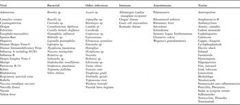
The multiple aetiologies of myocarditis are summarised in Table 1. Infectious exposures, especially viruses, account for the vast majority of paediatric myocarditis in Canada and the United States, whereas globally the most common cause is Trypanosoma cruzi (Chagas disease).Reference Whitton and Feuer10 The majority of paediatric cases are believed to be due to adenoviruses and enteroviruses, such as coxsackieviruses A and B, parvovirus, echovirus, and poliovirus.Reference Bowles and Towbin11–Reference Woodruff17 The relative frequency of cases related to these viruses is variable and there is much discussion as to which is more common.
Aetiologies of myocarditis.

In the past, enteroviruses, especially the coxsackie viruses, were considered the most common aetiology. A recent study suggested that adenovirus may be a more significant pathogen than previously recognised.Reference Bowles, Ni and Kearney18 Among patients given a clinical diagnosis of myocarditis, those with positive adenovirus polymerase chain reaction were less likely to have a corresponding histologic diagnosis of acute myocarditis compared with the enterovirus group (40% versus 79%).Reference Bowles, Ni and Kearney18 This suggests that adenovirus is associated with a lesser degree of inflammation in the myocardium. This may suggest a higher prevalence of adenovirus than suspected.
A subsequent study reviewed aetiologies for biopsy-confirmed myocardial inflammation and found parvovirus B 19 to be the most common, followed by enterovirus, human herpes virus 6, and then adenovirus.Reference Kuhl, Pauschinger and Seeberg19
Following the H1N1 Influenza A pandemic, there were case reports of associated myocarditis. Of the 80 confirmed viral infections reported by one group, there were four cases of associated myocarditis, three of which presented with fulminant myocarditis:Reference Bratincsak, El-Said and Bradley20 one patient had a fatal outcome due to acute atrioventricular block and two required extracorporeal membrane oxygenation support due to severely reduced left ventricular systolic function. Extracorporeal membrane oxygenation is implemented in many paediatric tertiary centres for either respiratory failure or cardiac compromise in cases of H1N1 infection. Further population data are required to clarify the clinical course of H1N1-associated myocarditis.
Pathology and pathophysiology
There are several proposed mechanisms of myocardial injury in viral myocarditis.Reference Esfandiarei and McManus4 These include the excessive immune-mediated destruction of the myocardium by infiltrating immune cells targeting the infected cardiomyocytes; autoimmune-mediated destruction of cardiac cells by circulating autoantibodies; and direct virus-induced cardiac myocyte injury.
It is proposed that three phases of viral myocarditis exist, beginning with a period of viral replication, followed by acute injury to the myocytes, and subsequent evolution of dilated cardiomyopathy, associated with profound alteration in the extracellular matrix of heart muscle.Reference Esfandiarei and McManus4 Up to 27% of dilated cardiomyopathy cases in children may be attributable to viral myocarditis.Reference Leonard21
Clinical presentation
The diagnosis of paediatric myocarditis is especially challenging because of the variable clinical presentation, ranging from asymptomatic patients with only subtle findings on an electrocardiogram to fulminant cardiac failure and sudden death. Diagnosis remains largely based on clinical judgement, supported by ancillary tests; therefore, a high index of suspicion is necessary.
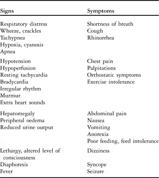
Clinical features are often those of congestive cardiac failure and myocarditis is identified as the most common cause of new onset cardiac failure in previously well children.Reference Calabrese, Rigo and Milanesi22 Even patients with mild symptoms are at risk of deterioration, and therefore early diagnosis is important in establishing appropriate monitoring and supportive care. Common symptoms and signs are listed in Table 2.
Signs and symptoms of myocarditis.

There are several paediatric studies that have examined the most common signs and symptoms of patients with myocarditis. Shortness of breath, respiratory distress, or an abnormal respiratory exam were commonly observed.Reference Freedman, Haladyn and Floh5, Reference Durani, Egan and Baffa23 In two studies, 84% of patients required more than one visit to a physician within 14 days before the diagnosis of myocarditis or dilated cardiomyopathy was made.Reference Durani, Egan and Baffa23 Children were often given an initial diagnosis of asthma or pneumonia.Reference Freedman, Haladyn and Floh5 Resting tachycardia is also an important subtle feature, and although this is not always a consistent finding, it may be the only finding of mild disease.Reference Freedman, Haladyn and Floh5, Reference Durani, Egan and Baffa23 Isolated vomiting is observed and this may be due to gut ischaemia secondary-to-low cardiac output. Hepatomegaly is a common feature on physical exam and may present as non-specific abdominal pain.Reference Freedman, Haladyn and Floh5, Reference Durani, Egan and Baffa23
In contrast to adults with myocarditis, chest pain is seldom reported in younger children though it may be a more common feature in adolescents. A case series of several teenage patients diagnosed with myocarditis described symptoms of severe chest pain, in combination with elevated cardiac troponin-I, ST-segment, and T-wave changes.Reference Kern, Modi and Atalay24
Unfortunately, sudden cardiac death may be the presenting feature of myocarditis, and in these cases the diagnosis is often made histologically. It is suggested that some cases may be the result of inflammatory infiltrates that act as arrhythmogenic foci, leading to fatal arrhythmia.Reference Ellis and Di Salvo25
Despite the existence of echocardiographic and endomyocardial biopsy criteriaReference Aretz26 for myocarditis and resulting cardiomyopathy, expert opinion currently suggests that the diagnosis of “probable” myocarditis is based on clinical judgement.Reference Grenier, Osganian and Cox27, Reference Batra and Lewis28
A broad clinicopathological classification system can be used in which there are four categories of myocarditis in the setting of viral illness.Reference Esfandiarei and McManus4
“Fulminant myocarditis” is associated with symptoms of significant left ventricular dysfunction and unexpected cardiac failure within 2–3 weeks of the onset of viral infection. “Subacute myocarditis” can be described as moderate left ventricular dysfunction and has less distinct clinical features of the disease. “Chronic active myocarditis” is associated with moderate left ventricular dysfunction, indistinct symptoms, and endomyocardial biopsy reveals ongoing inflammation, myocardial damage, and active scar tissue. “Chronic persistent myocarditis” is described in the setting of normal left ventricular function and palpitations or atypical chest pain, with persistent inflammation on biopsy.
Investigations
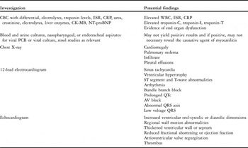
Despite many diagnostic modalities, suspicion of myocarditis is mainly based on clinical criteria. Diagnostic work-up should include baseline investigations listed in Table 3.
Initial investigations in suspected myocarditis.

AV = atrioventricular; CBC = complete blood count; CK-MB = creatine kinase muscle, brain; CRP = C-reactive protein; ESR = erythrocyte sedimentation rate; NT-proBNP = N-terminal B-type natriuretic peptide prohormone; PCR = polymerase chain reaction; QTc = corrected QT interval; WBC = white blood count
Abnormal troponin levels may support a diagnosis of myocarditis, but there are limited data in the current paediatric literature.Reference Durani, Egan and Baffa23, Reference Lippi, Salvagno and Guidi29 An elevated creatine kinase level may also be observed in myocarditis, although it is a non-specific marker. It has been suggested that, in comparison with creatine kinase, cardiac troponin-T may provide better sensitivity for detecting micronecrosis in myocarditis because of a proportionally higher and longer-lasting elevation of serum levels.Reference Franz, Remppis and Kandolf30 In the Myocarditis Treatment Trial, elevations of cardiac troponin I occurred more frequently than did elevations of creatine kinase muscle and brain subunits in patients with biopsy-proven myocarditis.Reference Smith, Ladenson and Mason31 There was one paediatric study that illustrated a cut-off point for a cardiac troponin T level of 0.052 nanograms per millilitre for diagnosing acute myocarditis with a sensitivity of 71% and specificity of 86%, although there was no correlation between cardiac troponin T level and ejection fraction.Reference Soongswang, Durongpisitkul and Nana32
Use of the N-terminal segment of the B-type natriuretic peptide prohormone levels may be helpful in the setting of myocarditis, especially with respect to recovery. In one study, elevated N-terminal B-type natriuretic peptide levels correlated well with clinical status and echocardiographic findings in patients with dilated cardiomyopathy or myocarditis.Reference Nasser, Perles and Rein33 N-terminal B-type natriuretic peptide levels had 78% sensitivity and 100% specificity for the diagnosis of persistent left ventricular dysfunction. Patients who recovered had N-terminal B-type natriuretic peptide levels that were within normal limits.Reference Nir, Bar-Oz and Perles34
Chest X-ray findings often include cardiomegaly, and may demonstrate pulmonary oedema, infiltrates, or pleural effusions.Reference Freedman, Haladyn and Floh5, Reference Durani, Egan and Baffa23 An abnormal electrocardiogram is very common, found in over 93% of patients.Reference Freedman, Haladyn and Floh5, Reference Durani, Egan and Baffa23 The most common abnormalities include sinus tachycardia, criteria for ventricular hypertrophy, ST segment, and T-wave abnormalities. Less commonly, arrhythmia, bundle branch block, prolonged corrected QT interval, and atrioventricular block are observed.Reference Durani, Egan and Baffa23 Abnormal QRS axis and low voltage QRS may be present. The combination of an electrocardiogram with chest X-ray in the setting of possible myocarditis has very good sensitivity for the diagnosis.Reference Freedman, Haladyn and Floh5
Echocardiographic findings may include increased ventricular end-systolic or diastolic dimensions, reduced shortening or ejection fractions, atrioventricular valve regurgitation, and regional wall motion abnormalities.Reference Checchia and Kulik35 Echocardiography is also important to rule out coronary abnormalities as a cause of depressed myocardial function. Abnormal right ventricular function on echo may be associated with increased likelihood of adverse outcomes including cardiac transplantation or death.Reference Mendes, Dec and Picard36
Other methods of non-invasive imaging are being investigated in the setting of myocarditis. Nuclear medicine techniques have not been used widely in paediatrics, in part due to concerns of excessive radiation exposure for children.Reference Jeserich, Konstantinides and Pavlik37
Cardiovascular magnetic resonance imaging is a widely accepted tool to assess myocarditis. This technique provides not only detailed functional and morphological assessment of the heart, but also reliable visualisation of tissue markers of myocarditis including oedema, inflammation, and fibrosis.Reference Abdel-Aty, Boye and Zagrosek38–Reference Mahrholdt, Goedecke and Wagner41 Previously, oedema could not be well imaged; however, cardiac magnetic resonance is now able to visualise myocardial oedema without using radiation or contrast agents.Reference Friedrich44 Currently, cardiac magnetic resonance may be considered the single best non-invasive imaging study for this indication, and standardised protocols for cardiac magnetic resonance imaging of myocarditis are available.Reference Skouri, Dec and Friedrich42, Reference Friedrich, Sechtem and Schulz-Menger43
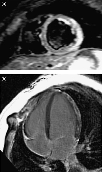
In acute myocarditis, cardiac magnetic resonance assessment of myocardial oedema is performed using T2-weighted short TI inversion recovery imaging in which regions of myocardial oedema appear bright, whereas fat and blood signals are suppressed (Fig 1).Reference Abdel-Aty, Simonetti and Friedrich45 The sensitivity and specificity of this technique were 78% and 91%, respectively.Reference Jeserich, Konstantinides and Pavlik37 In general, the presence of myocardial oedema may reflect the acuity of myocardial injury and can become evident within the first 2–3 weeks from the insult.Reference Cocker and Friedrich46
Cardiovascular magnetic resonance imaging of acute myocarditis in a teenage male patient. (a) T2-weighted short TI inversion recovery image of the left ventricle in short axis demonstrating bright signal intensity in the regions of myocardial oedema. (b) Late enhancement (T1-weighted gradient echo sequence with myocardial nulling) of fibrotic areas of the myocardium (bright signal) in a four-chamber orientation 10 minutes after intravenous gadolinium administration.

Gadolinium contrast-enhanced cardiac magnetic resonance may be used to assess myocardial hyperaemia.Reference Friedrich, Strohm and Schulz-Menger40 An increased ratio of myocardial signal intensity post-contrast compared to pre-contrast and normalised to skeletal muscle signal is indicative of myocardial hyperaemia and capillary leakage. The sensitivity and specificity of T1-weighted images in patients with suspected chronic myocarditis were 62% and 86%, respectively, using immunohistological methods as the gold standard.Reference Gutberlet, Spors and Thoma39 Similar to myocardial oedema, early enhancement normalises approximately 4 weeks after clinical presentation and therefore may be used as a marker of the acuity of myocardial injury.Reference Wagner, Schulz-Menger and Dietz47 In a small proportion of patients, early enhancement may persist, and is correlated to reduced cardiac function. It is not currently known whether persistently elevated early enhancement truly reflects ongoing viral infection.
The cardiac magnetic resonance technique of gadolinium late enhancement allows visualisation of myocardial necrosis and fibrosis associated with myocarditis. Gadolinium accumulates in regions of fibrosis, leading to increased signal intensity in late enhancement images (Fig 1). In the setting of myocarditis, the pattern of myocardial fibrosis varies considerably but typically involves patchy focal fibrosis of the sub-epicardium or mid-myocardial wall, rarely extending to the sub-endocardium.Reference Mahrholdt, Goedecke and Wagner41
The presence of myocardial fibrosis detected by cardiac magnetic resonance late enhancement is more common in males and younger patients, and the pattern of myocardial injury sustained in young patients tends to be more regional and more severe, with a higher incidence of irreversible myocardial scarring.Reference Cocker, Abdel-Aty and Strohm48 This pattern of myocardial injury may explain why younger patients are at risk for adverse cardiac outcomes. The presence of gadolinium late enhancement 4 weeks after acute presentation is also correlated with reduced left ventricular ejection fraction and clinical symptomatology at 30-month follow-up.Reference Wagner, Schulz-Menger and Dietz47
To standardise cardiac magnetic resonance imaging to diagnose myocarditis, the International Consensus Group on Cardiovascular Magnetic Resonance created new criteria, known as the “Lake Louise Criteria”.Reference Friedrich, Sechtem and Schulz-Menger43 These recommendations describe indications for cardiac magnetic resonance in patients with suspected myocarditis, cardiac magnetic resonance protocol standards, terminology for reporting cardiac magnetic resonance findings, and diagnostic cardiac magnetic resonance criteria for myocarditis. These guidelines report that the greatest balance of diagnostic sensitivity and specificity for acute myocarditis is achieved with the presence of any two or more of the following three criteria: myocardial oedema (T2 short TI inversion recovery), hyperaemia/capillary leak (early enhancement), or myocardial fibrosis (late gadolinium enhancement).
The gold standard for diagnosis of myocarditis remains endomyocardial biopsy; however, this approach has significant limitations.Reference Aretz26 According to the Dallas Criteria, endomyocardial specimens are considered diagnostic of active myocarditis, if routine light microscopy reveals infiltrating lymphocytes and myocytolysis.Reference Aretz26 Borderline or ongoing myocarditis is defined as lymphocytic infiltration in the absence of myocytolysis. Biopsy is considered negative, if both lymphocytic infiltration and myocytolysis are absent.
A potential reason to proceed with biopsy is that the presence of immune complexes and complement deposits supports an immune-mediated process; therefore, the management may differ from that of the case of viral infection. For example, immune suppression is not indicated in cardiomyopathy but may be worthwhile in viral myocarditis. Biopsy may be useful in the setting of eosinophilic infiltrates, which could suggest a hypersensitivity response to an exogenous agent. In this case, once the histology is confirmed, the offending agent can be removed and the clinical picture may improve.
A significant limitation of cardiac biopsy is that myocarditis affects the myocardium in a patchy fashion, and therefore negative biopsy may not exclude disease. There remains controversy as to whether or not to proceed with biopsy, especially since biopsy can be associated with arrhythmia and perforation of cardiac tissue. Paediatric literature also suggests that the highest rate of complication occurs in patients being evaluated for myocarditis, as well as those requiring inotropic support.Reference Pophal, Sigfusson and Booth49 Despite the overall complication rate being relatively low (1–6%), with mortality between 0.0% and 0.4%, risk-benefit analysis occurs on a case-by-case basis.Reference Friedrich, Sechtem and Schulz-Menger43, Reference Pophal, Sigfusson and Booth49–Reference Holzmann, Nicko and Kuhl52
Treatment
Despite improved understanding of the pathogenesis of myocarditis, treatment strategies remain controversial and the general approach is supportive. Stabilisation of children who present with haemodynamic compromise and early involvement of paediatric cardiology specialists are crucial.
Treatment of congestive cardiac failure may include diuretics, afterload reduction, inotropic support, anticoagulation, arrhythmia management, and ventilatory support. Aggressive administration of intravenous fluids initially may exacerbate cardiac failure. Transfusions of packed red blood cells may be required to optimise oxygen carrying capacity. The use of inotropes such as dopamine, dobutamine, and milrinone may be necessary. Afterload reduction, if it can be tolerated, with phophodiesterase inhibitors such as milrinone can be extremely useful. It is also suggested that the early introduction of angiotensin-converting enzyme inhibitors, as well as beta-blockers, once the acute decompensatory phase has begun to resolve, may be beneficial. These medications may help prevent the remodelling that evolves to dilated cardiomyopathy as well as relieving vasospasm to some degree.Reference Ellis and Di Salvo25, Reference Tominaga, Matsumori and Okada53 In paediatric patients, carvedilol was suggested in this regard but has not been studied extensively.Reference Bruns, Chrisant and Lamour54 Anticoagulation may be recommended if the ejection fraction is severely decreased or in the setting of atrial arrhythmias.Reference Gunthard, Stocker and Bolz55
Some patients may require ventilatory support as a means of reducing cardiac demand. Ventilation and oxygenation may be best managed with continuous positive airway pressure or other non-invasive methods, for several reasons. Increased intrathoracic pressure via nasal or mask continuous positive airway pressure may help to reduce left ventricular afterload by reducing transmural myocardial wall tension, without causing the hypotension associated with pharmacologic afterload reduction. Intubation medications can cause hypotension and acute cardiovascular collapse; therefore, continuous positive airway pressure may also circumvent this and is an excellent adjunctive therapy for decompensated cardiac failure.Reference Bradley, Holloway and McLaughlin56–Reference Bohn and Benson58
Those with more severe cases of myocarditis may require circulatory support in the form of extracorporeal membrane oxygenation or ventricular assist devices. Extracorporeal membrane oxygenation may also be used to support patients through to recovery from acute myocarditis, or it may serve as a bridge to transplant in dilated cardiomyopathy.Reference Sezai, Hata and Niino59 Patients over 15 kilograms may respond best to pulsatile ventricular assist devices.Reference Potapov, Stiller and Hetzer60 There have been case reports of prolonged support using a ventricular assist device as a bridge to recovery or transplant.Reference Jones, Cassidy and Kirk61
Immune suppression
An area of significant controversy involves the use of immune suppression and immune modulators in the context of myocarditis. There are large-scale adult studies with respect to immune suppression, and overall the data have shown some limited, short-term response to these therapies.
In two large adult prospective studies, immunosuppression including prednisone with or without azathioprine or cyclosporine was used.Reference Parrillo, Cunnion and Epstein62, Reference Mason, O'Connell and Herskowitz63 With prednisone alone, there was modest improvement in left ventricular function in patients with evidence of inflammation on biopsy; however, this improvement was not sustained.Reference Parrillo, Cunnion and Epstein62 The Myocarditis Treatment Trial found no significant improvement in function and no difference in survival when compared with controls.Reference Mason, O'Connell and Herskowitz63 Despite the existence also of a handful of uncontrolled studies that showed benefit with various immunosuppressive regimes, some meta-analyses of adult studies did not show a significant beneficial effect of immunosuppression.Reference Jones, Herskowitz and Hutchins64–Reference Garg, Shiau and Guyatt69
It is always difficult to extrapolate the findings of adult studies to the paediatric population. Paediatric studies are limited and there are no randomised controlled trials. Many used prednisolone and did find some benefit, though not always statistically significant, by adding a second agent. In one trial, patients were stratified into either standard treatment, or given one of three immunosuppressive therapies, prednisolone, prednisolone and azathioprine, or prednisolone and cyclosporin A.Reference Camargo, Snitcowsky and da Luz70 The groups receiving immunosuppressive treatment with a second agent, in addition to the prednisolone, showed improved haemodynamic parameters, as well as histological improvement in inflammation.Reference Camargo, Snitcowsky and da Luz70 Some small case series suggested some clinical benefit to immunosuppressive treatment, again using corticosteroids with an additional agent; however, most were small, uncontrolled studies.Reference Chan, Iwahara and Benson71–Reference Kleinert, Weintraub and Wilkinson75 A meta-analysis of paediatric studies between 1984 and 2003 systematically reviewed the impact of immunosuppressive therapy in children with myocarditis and overall found no consistent benefit.Reference Hia, Yip and Tai76 This included nine paediatric studies, all of which were small and very heterogeneous in design. Many of the studies were retrospective and uncontrolled, introducing significant bias.
Intravenous immunoglobulin was also examined as a potential immunomodulating therapy to potentially decrease inflammation. Despite some studies observing a trend towards improved cardiac status following treatment, there are no randomised controlled trials using intravenous immune globulin in children with myocarditis. Robinson et alReference Robinson, Hartling and Crumley77 conducted a comprehensive multi-database search including patients of all ages with myocarditis treated with intravenous immune globulin. They concluded that if there is evidence that ongoing, active infection may be causing persistent cardiac dysfunction, intravenous immune globulin may be helpful. Even if post-infectious inflammation is the most likely aetiology, intravenous immune globulin may still be beneficial by preventing formation of cytokines.Reference Kishimoto, Shioji and Kinoshita78 There is one multi-centre adult randomised controlled trial using intravenous immune globulin.Reference McNamara, Holubkov and Starling79 They found no significant difference in rates of survival, cardiac transplantation, use of ventricular assist devices, improvement in ventricular function, or functional capacity at 1 year of follow-up. A Cochrane review of intravenous immune globulin in dilated cardiomyopathy or myocarditis showed no benefit in adults; however, some case reports and case series suggested that adults treated with intravenous immune globulin did exhibit improved cardiac function.Reference McNamara, Rosenblum and Janosko66, Reference Robinson, Hartling and Crumley77–Reference McNamara, Holubkov and Starling79, Reference Goland, Czer and Siegel80–Reference Abe, Okura and Hoyano86
Some case reports and series in children have shown improved ventricular function and clinical improvement after high-dose intravenous immune globulin, however with small numbers.Reference Drucker, Colan and Lewis87–Reference Kim, Sohn and Park91 Overall, it was felt that the validity of many of these studies was compromised due to lack of control groups. There was one case report of coxsackie virus A19 myocarditis in which intravenous immune globulin was not effective and another case series found no additional benefit by adding intravenous immune globulin to steroid treatment.Reference Wang, Li Lu and Wu92, Reference English, Janosky and Ettedgui93
Studies using immunosuppressive or immunomodulating treatments, therefore, were difficult to translate into an effective, routine therapy for children or adults with myocarditis. Many patients with myocarditis have spontaneous improvement. It is difficult to know if the observed improvement after treatment with immunosuppression or intravenous immune globulin is attributable to treatment versus the natural course of disease. Potentially, some aetiologic types of myocarditis respond differently and certain histologic characteristics may make specific patients better responders than others.Reference Balaji, Wiles and Sens73, Reference Kleinert, Weintraub and Wilkinson75, Reference Parrillo94 Further studies are needed to determine the possible utility of immunosuppressive or immunomodulating therapy.
Outcomes and prognosis
As described, it is difficult to estimate the true incidence and prevalence of myocarditis because of the broad spectrum of symptoms. It is said that the prognosis for children with viral myocarditis tends to be more positive than the outcomes with dilated cardiomyopathy.Reference Chan, Iwahara and Benson71, Reference Drucker, Colan and Lewis87, Reference Lee, McCrindle and Bohn95 Survival rates for paediatric patients with myocarditis can be as high as 93%.Reference Lee, McCrindle and Bohn95–Reference Duncan, Bohn and Atz97 However, a large, multi-centre study including all age groups showed that there was a significant mortality in neonates and infants (33–45% survival, 23–32% improvement) with better outcomes in children between 1 and 18 years of age (78–80% survival, 46–67% improvement).Reference Bowles, Ni and Kearney18 A study of 28 children with a diagnosis of myocarditis also observed that only 17 survived to hospital discharge with variable degrees of improved heart function, whereas the remaining 11 patients developed intractable cardiac failure leading to cardiac transplant listing (seven cases) or death (four cases).Reference Kuhn, Shapiro and Walls98 Predictors of a poor outcome were found to be ejection fraction less than 30%, shortening fraction less than 15%, left ventricular dilatation, and moderate-to-severe mitral valve regurgitation.Reference Kuhn, Shapiro and Walls98
The authors of several case series involving children requiring mechanical support for myocarditis have reported survival rates of 67–83%.Reference Lee, McCrindle and Bohn95, Reference Duncan, Bohn and Atz97, Reference Lin, Chang and Li99–101 An updated review of the extracorporeal membrane oxygenation registry revealed a survival rate of 61% for children supported through acute myocarditis.Reference Rajagopal, Almond and Laussen102 This is comparable to, and perhaps better than, survival rates of children supported with extracorporeal membrane oxygenation after repair or palliation of congenital cardiac disease, in which survival rates range between 37% and 42%.101, Reference Kolovos, Bratton and Moler103–Reference Morris, Ittenbach and Godinez105 Hetzer reported that of 21 children supported with the Berlin Heart Excor device for myocarditis or cardiomyopathy, 90% survived to hospital discharge.Reference Hetzer and Stiller106 Collective experience with ventricular assist devices in this setting, however, is quite limited.
The prognosis in biopsy-proven myocarditis may depend on the severity of symptoms, histologic classification, and biomarkers. Acute fulminant myocarditis is associated with better overall survival.Reference Amabile, Fraisse and Bouvenot107–Reference Chau, Chow and Chiu109 Giant cell myocarditis, although rare, is associated with especially poor outcomes and a reported median duration of survival, if untreated, of 5.5 months.Reference Cooper, Berry and Shabetai110 It also carries an 89% rate of death or transplant.
Myocarditis may account for nearly half of all children with dilated cardiomyopathy.Reference Liu and Mason111, Reference Towbin, Lowe and Colan112 The outcomes for patients with acute viral myocarditis are much better than cases with established dilated cardiomyopathy.Reference Chan, Iwahara and Benson71, Reference Drucker, Colan and Lewis87, Reference Lee, McCrindle and Bohn95 For this reason, a high index of suspicion for the disease, and early effective supportive care are crucial. Myocarditis remains a relatively common indication for transplant listing. Overall, 1–8% of patients with acute myocarditis eventually go on to transplant.Reference Ellis and Di Salvo25 Owing to the potential for recovery, even with severe disease at presentation, patients are not typically listed for cardiac transplant unless recovery is considered extremely unlikely despite reasonable management and a period of observation.
Conclusions
Acute myocarditis comprises a wide clinical spectrum including the asymptomatic patient with subclinical myocardial dysfunction to severe cardiac failure or sudden cardiac death. In paediatrics, the symptoms and signs can mimic many other common diseases and a high index of clinical suspicion is imperative. Treatment of myocarditis remains supportive and should be initiated even before solidifying the diagnosis. Biopsy remains the gold standard for diagnosis; however, it has significant limitations. Cardiac magnetic resonance imaging has become a widely accepted non-invasive imaging modality for myocarditis and standardised protocols are now available for myocarditis. There exist controversial data and no consensus with respect to immunosuppressive or immunomodulating therapy for myocarditis. Similarly, there is no foundation of evidence to indicate a routine treatment strategy other than standard cardiac failure management for these patients. Most patients with myocarditis have spontaneous improvement in their cardiac function and symptoms; however, there remains significant morbidity and mortality associated with this condition. Additional studies are a necessity to clarify the most appropriate management options in paediatrics to improve outcomes.
Acknowledgement
Sources of funding: None. Conflicts of Interest: None.