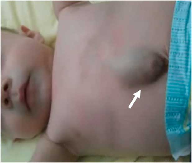
A13-day-old, full-term, male neonate was referred to our institution with mild signs of cardiac failure. On admission, physical examination showed a subcutaneous pulsatile mass in the epigastrium synchronous with the cardiac cycle (Fig 1). Cardiac auscultation revealed rhythmic heart sounds with 3/6 systolic murmur. The electrocardiogram of the patient showed normal sinus rhythm.
Photograph of the baby showing a subcutaneous pulsatile mass (arrow) in the epigastrium.

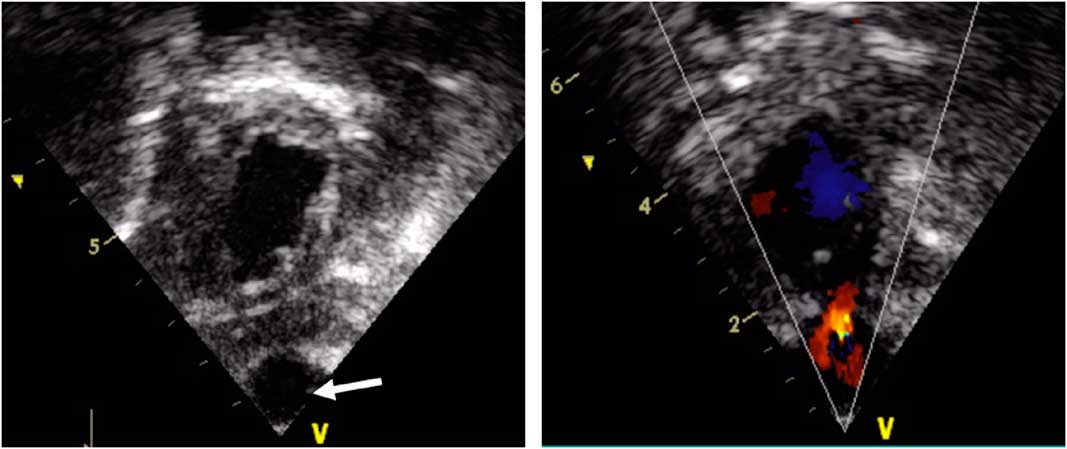
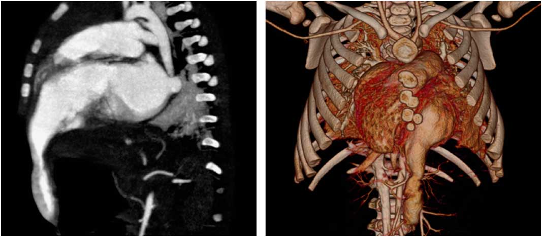
Echocardiography revealed a diverticular formation arising from the left ventricular anterior wall extending into the subcutaneous plane in the anterior abdominal wall (Fig 2, left). In addition, a large interventricular septal defect and a patent ductus arteriosus were noted. Color Doppler revealed turbulent blood flow between the left ventricle and the diverticular lesion (Fig 2, right). Cardiac CT confirmed a 4-cm-long, thin-walled diverticular lesion arising from the anterolateral wall of left ventricle extending downwards through the diaphragm reaching the epigastric region (Fig 3). No thrombus was seen in the left ventricle, or inside the lesion.
Left: Echocardiography demonstrating an isolated left ventricular diverticulum (arrow). Right: Color Doppler image showing flow within a left ventricular diverticulum.

Left: Computed tomography (sagittal reconstruction) showing a protrusion of the left ventricle into the epigastric region. Right: 3D Computed tomography image showing a diverticulum originating from the antero-lateral wall of left ventricle.

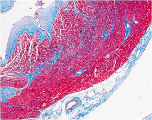
Given the potential risks for rupture and systemic embolism, the patient was scheduled for surgery. At 4 months of age, the diverticular formation was surgically excluded by closure of the orifice of the free left ventricular wall; furthermore, the subaortic ventricular septal defect was repaired using a bovine pericardial patch, and the patent ductus arteriosus was surgically closed. Histological examination of the resected pouch revealed the presence of preserved myocardial layers throughout the lesion (Fig 4).
Histological section showing the presence of epicardium, myocardium and endocardium.

Congenital ventricular diverticulum is an extremely rare cardiac abnormality, accounting for ~0.05% of all congenital heart malformations. It is currently believed that it occurs as a result of impaired development of the endocardial tube during the 4th week of embryologic development.1 Although it may present as an isolated disorder, this entity is often associated with other cardiac abnormalities or with Cantrell pentalogy in around 70% of cases.
Financial Support
This research received no specific grant from any funding agency or commercial or non-for-profit sectors.
Conflicts of Interest
None.
Supplementary material
To view supplementary material for this article, please visit https://doi.org/10.1017/S1047951117000245