Case
A male child with hypoplastic left heart syndrome with mitral stenosis and aortic atresia underwent neonatal stage 1 Norwood palliation with a Sano shunt, and subsequently a right-sided bidirectional Glenn shunt – superior cavopulmonary connection – at 4 months of age.
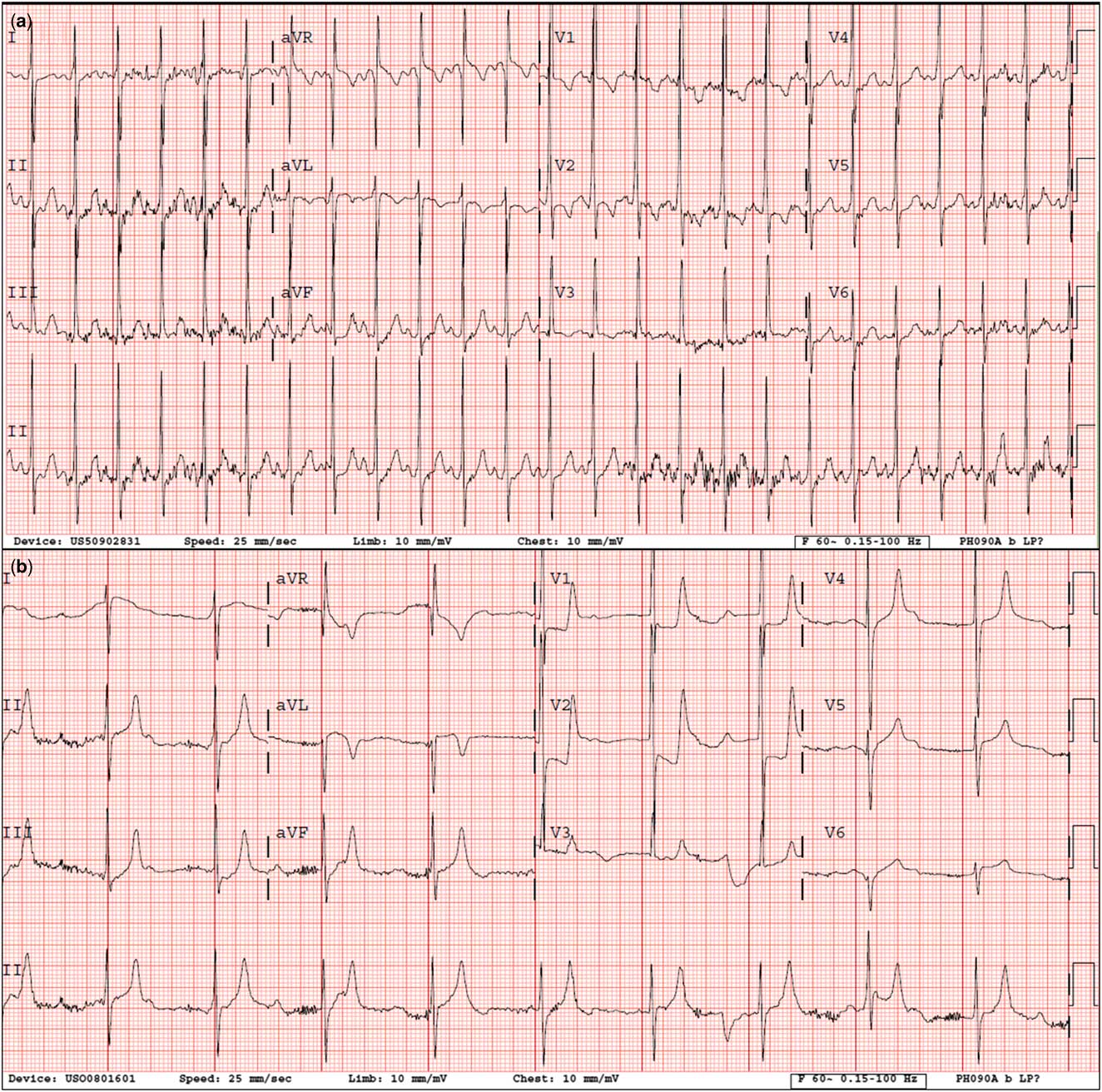
At 2 years of age, in the setting of 3 days of runny nose and congestion, he acutely developed diarrhoea and multiple episodes of emesis, and within an hour he became listless and was presented to the emergency department. In the emergency department, he was bradycardic (~50 bpm), hypoxaemic – room air saturations in the 50’s – poorly perfused, and minimally responsive, consistent with cardiogenic shock. An electrocardiogram (Fig 1) revealed complete heart block with a junctional escape, minimal leftward forces in V6, ST depression isolated to leads V1, V2, and possibly aVR, and likely subtle ST elevation in V5 and possibly V6. Isoproterenol improved the junctional rate and perfusion at the time of transfer to the cardiac intensive care unit.
Baseline 12-lead electrocardiogram ( a ) and at presentation ( b ). Baseline electrocardiogram demonstrates sinus rhythm and right ventricular hypertrophy. Electrocardiogram at presentation demonstrates complete heart block with narrow QRS escape, right ventricular hypertrophy, and decreased leftward forces in V6. ST depression is seen in V1, V2, and possibly aVR. Subtle ST elevation is seen in V5 and possibly V6.

Transthoracic echocardiography revealed diminished global function in the setting of significant acidosis, with further reduced left ventricular free wall, septal, and right ventricular posterior wall motion. Suboptimal echocardiographic windows limited the visualisation of the atretic native aortic root. Although right coronary artery flow was demonstrated by colour Doppler mapping, a poor angle of insonation limited assessment of left coronary artery flow. A milrinone infusion was initiated for depressed cardiac function.
Laboratory examinations revealed a troponin I level of 31.8 ng/ml (nl 0–0.119 ng/ml) and multiple viral infections as follows: Adenovirus, Human Metapneumovirus, Rhinovirus, and Parainfluenza type 4. Leading differential diagnoses included viral myocarditis and myocardial infarction involving conduction tissue.
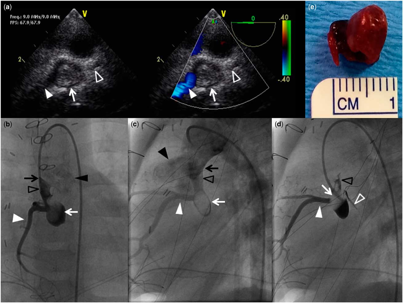
The patient subsequently developed unstable ventricular tachycardia requiring chest compressions and synchronised cardioversion. To restore atrioventricular synchrony with temporary pacemaker wires, he was urgently taken to the operating room. There, a trans-oesophageal echocardiogram demonstrated exacerbated segmental dysfunction, prominent right coronary artery flow, absent left coronary artery flow, and likely thrombus within the native aortic root (Fig 2, Supplementary Video S1). To confirm the above findings, he was transferred to the catheterisation laboratory, where angiography documented a left-dominant coronary pattern and a large mobile thrombus in the native aortic root obstructing the left main coronary ostium (Fig 2, Supplementary Video S2). Given the size and mobility of the thrombus, to avoid catastrophic embolisation or occlusion of the right coronary ostium, he underwent immediate surgical removal of the 1-cm thrombus (Fig 2), and subsequently a permanent epicardial pacemaker was placed.
Diagnostic images (counter-clockwise): ( a ) Trans-oesophageal echocardiogram two-dimensional (2D) and colour compare still frame where a cross-section of the native aortic root demonstrates a thrombus within the native aortic root (white arrow), prominent flow into the right coronary artery (solid white arrow head), and absent left coronary artery flow (empty white arrow head). Cardiac catheterisation images ( b – d ): Aortic angiogram from straight anterior-posterior (AP) ( b ) and lateral ( c ) projections. Contrast fills the native ascending aorta (empty black arrow heads) and the neo-ascending aorta (black arrow heads), demonstrating the Damus–Kaye–Stansel anastomosis (black arrow). A thrombus is visualised within the native aortic root (white arrow). A posterior descending coronary artery is not seen arising from the right coronary system (white arrow head), consistent with a left dominant coronary artery pattern. The left coronary artery, obstructed by a thrombus, does not fill with contrast. ( d ) The same lateral projection in ( c ) is demonstrated again, but with the catheter deeper in the native aortic root, injection of contrast shifts the thrombus cephalad, away from the left coronary ostium, allowing the left main coronary artery (empty white arrow head) to briefly fill with contrast, demonstrating its patency.( e ) Surgical specimen of the thrombus.

The patient has been anticoagulated with warfarin and listed for orthotopic heart transplantation due to persistent ventricular dysfunction along with inotrope dependence.
Discussion
Thrombosis of the native aorta in hypoplastic left heart syndrome is rare.Reference Janssen, Ohmstede, Liske, Parra, Drinkwater and Kavanaugh-McHugh 1 The coronary pattern seen in our patient is also uncommon. In the general population, coronary distribution is right dominant in ~70%, shared in 20%, and left dominant in only 10%.Reference Hutchins, Nazarian and Bulkley 2 , Reference Kirklin and Barrett-Boyes 3 Left dominance is more common with aortic stenosis (20%), bicuspid aortic valve (29%), and all-comer hypoplastic left heart syndrome (37%);Reference Hutchins, Nazarian and Bulkley 2 , Reference Hansen, Uebing, Scheewe, Kramer and Fischer 4 however, within the sub-population of patients with mitral stenosis and aortic atresia, as seen in our patient, left dominance occurs in only 15%.Reference Hansen, Uebing, Scheewe, Kramer and Fischer 4
Typically, a branch of the dominant coronary system, the atrioventricular nodal artery, arises from the proximal posterior descending artery near the crux of the heart and supplies the atrioventricular node.Reference Kirklin and Barrett-Boyes 3 Here, occlusion of the left coronary ostium resulted in ischaemia of the the atrioventricular node and complete heart block. Atrioventricular synchrony plays a significant role in generating stroke volume in single ventricle physiology. The absence of atrioventricular synchrony in our patient contributed to reduced cardiac output and cardiogenic shock.
Coronary ischaemia manifesting with complete heart block has not been previously reported in patients with hypoplastic left heart syndrome. Although myocardial infarction in adults frequently presents with acute complete heart block with ventricular dysfunction, atrioventricular node ischaemia is rare in children. This fact delayed our full consideration of the correct diagnosis, which was only definitively made with multi-modality imaging.
Acknowledgements
None.
Financial Support
This research received no specific grant from any funding agency, commercial, or not-for-profit sectors.
Conflict of Interest
None.
Supplementary materials
To view supplementary materials for this article, please visit http://dx.doi.org/10.1017/S1047951114002558