Significant outcomes
-
∙ Lithium increased cell viability in SH-SY5Y neuroblastoma culture.
-
∙ Lithium may protect against increased oxidative stress (OxS), while the lithium/haloperidol combination may induce peroxidation.
-
∙ The neuroprotective effects of lithium may be attributed, in part, to its OxS-scavenging activities.
Limitations
-
∙ Low statistical power (<0.8) of the study.
-
∙ Difficulty in extrapolating these findings in vivo.
-
∙ The short time of incubation of the cell line samples with the active substances.
Introduction
Lithium should be used as a first-line treatment for acute mania and for maintenance therapy of adults with bipolar disorder (BD). Lithium should also be a second-line option for acute bipolar depression (Reference Connolly and Thase1). There are also many other indications for the use of lithium: schizoaffective disorder-bipolar type, cyclothymia, or as supplemental therapy in treatment-resistant major depression. The fact that long-term lithium treatment prevents suicide lends it an additional advantage over other mood stabilisers.
The specific pharmacological mechanisms of lithium are not entirely clear. In terms of neurotransmitter activity, lithium appears to regulate the dopaminergic system as well as enhance serotoninergic and cholinergic neurotransmission. Long-term lithium treatment increases GABA-ergic inhibition and reduces excitatory glutamatergic neurotransmission. Nowadays the focus has shifted to the regulation of signal transduction mechanisms. Lithium is an inhibitor of inositol monophosphatase (IMPase) and inositol polyphosphate 1-phosphatase (IPPase) and it is a regulator of adenylyl cyclase (Reference Shaldubina, Agam and Belmaker2,Reference Lenox and Frazer3).
In the last 2 decades, the neuroprotective properties of lithium have been extensively studied. Neuroprotection induced by lithium is probably mainly related to its potent inhibition of the enzyme glycogen synthase kinase-3β (GSK-3β) and its downstream effects, including antioxidant processes (Reference King and Jope4). Several additional ways have been proposed: increased expression of neuroprotective factors, protein bcl-2 (B-cell lymphoma/leukemia-2), factor BAG-1, and brain-derived neurotrophic factor (BDNF) and its receptor; decreased expression of p53 and bax proteins, inhibition of the caspase-3 pathway, post-translational modifications of stress proteins, or changes in gene expression. Moreover, lithium may enhance neurogenesis and reduce proinflammatory status (Reference Rybakowski5).
Finally, there has been a proposal that long-term lithium treatment has disease-modifying properties in some neurodegenerative diseases: Parkinson disease, Huntington disease, amyotrophic lateral sclerosis, and finally, Alzheimer disease (Reference Forlenza, De-Paula and Diniz6,Reference Scheuing, Chiu, Liao, Linares and Chuang7). Several studies suggested that continuous lithium treatment could reduce the prevalence of dementia, especially Alzheimer disease, in patients with BD (Reference Nunes, Forlenza and Gattaz8,Reference Gerhard, Devanand, Huang, Crystal and Olfson9). Similarly, it was found that patients on chronic lithium treatment showed lower rates of cognitive decline (Reference Terao, Nakano, Inoue, Okamoto, Nakamura and Iwata10,Reference Forlenza, Diniz, Radanovic, Santos, Talib and Gattaz11). This is in accordance with reports that lithium inhibits crucial processes in the pathogenesis of Alzheimer disease (Reference Hernandez, Lucas and Avila12).
In neuroimaging studies in subjects with BD, it was found that lithium therapy could cause cortical thickening, and an increase in the volume of the hippocampus and amygdala. Additionally, lithium treatment was associated with increased N-acetylaspartate and myo-inositol levels in magnetic resonance spectroscopy of the brain (Reference Phillips, Travis, Fagiolini and Kupfer13). Moreover, a significant increase in plasma BDNF levels after lithium treatment and its correlation with treatment response in acute mania was reported (Reference de Sousa, Zarate and Zanetti14).
Oxidative stress (OxS) is a state of imbalance between oxidant processes and antioxidant defences, potentially leading to damage (Reference Sies15). Increased OxS induces peroxidation in lipids, proteins, and DNA (Reference Mahadik and Mukherjee16). The level of thiobarbituric acid reactive substances (TBARs) and protein carbonyls are a direct index of cell lipid and protein peroxidation, respectively (Reference Rice-Evans, Diplock and Symons17). On the other hand, the measure of total antioxidant capacity (TAC) takes into account the cumulative action of all the antioxidants present in plasma and body fluids (Reference Re, Pellegrini, Proteggente, Pannala, Yang and Rice-Evans18).
OxS likely plays an important role in the pathophysiology and disease course of BD (Reference Berk, Kapczinski and Andreazza19). Several studies have reported increased levels of peroxidation products in BD patients, both in manic and depressive episodes, as well as in euthymia (Reference Tsai and Huang20–Reference Bengesser, Lackner and Birner23). Additionally, a negative correlation between serum TBARs and BDNF levels was demonstrated (Reference Kapczinski, Frey, Andreazza, Kauer-Sant'Anna, Cunha and Post24). An imbalance between the main antioxidant enzymes in the plasma of bipolar patients was also reported (Reference Andreazza, Cassini and Rosa25). There is also evidence that BD is associated with mitochondrial dysfunction that would greatly contribute to the neural loss seen in BD (Reference Marazziti, Baroni and Picchetti26). Additionally, oxidative damage to membranes leads to alterations in fluidity what may induce cell death (Reference Mahadik, Evans and Lal27). OxS-induced DNA damage may also activate several intracellular signalling pathways, leading to apoptosis (Reference Buttner, Bhattacharyya, Walsh and Benes28). Thus, increased OxS may be partly responsible for neurodegenerative changes in BD patients.
Mood stabilisers have been reported to render antioxidant effects that may contribute to their neuroprotective properties. There has been quite a lot of research on the influence of lithium on the OxS status in animal models and cell cultures; however, the results have been mixed (Reference Shao, Young and Wang29–Reference Chadha, Bhalla and Dhawan32). To date, there have been few such studies on humans. It was demonstrated that TBARs levels, increased in unmedicated manic patients, were reduced by acute treatment with lithium. Their antioxidant enzymes activity ratio (SOD/CAT) was normalised as well (Reference Machado-Vieira, Andreazza and Viale21). Similarly, plasma TAC in bipolar subjects was significantly increased with lithium treatment (Reference Aliyazicioglu, Kural, Colak, Karahan, Ayvaz and Deger33). Another study showed that a significant reduction in plasma lipid peroxidation (LP) after lithium treatment was associated with an improvement in erythrocyte Na+-K+-ATPase activity (Reference Banerjee, Dasgupta, Rout and Singh34). Antioxidant properties of lithium were also shown in non-pathological conditions, in healthy subjects (Reference Khairova, Pawar and Salvadore35).
The influence of antipsychotic medications on OxS parameters are under intense scrutiny. There is a lot of data that chronic treatment with typical antipsychotics, especially haloperidol, can induce OxS (Reference Parikh, Khan and Mahadik36–Reference Dietrich-Muszalska, Kontek and Rabe-Jabłońska38). Haloperidol was also demonstrated to significantly reduce expression of genes encoded for antioxidant enzymes (Reference Schmidt, Hemmeter, Krieg, Vedder and Heiser39). However, a systematic review by Lepping et al., covering 42 studies on the impact of neuroleptics on OxS parameters in human cells, did not give a clear conclusion (Reference Lepping, Delieu, Mellor, Williams, Hudson and Hunter-Lavin40).
The aim of this research was to estimate the effect of lithium on LP in neuroblastoma cell culture and to assess if lithium could influence haloperidol-induced OxS changes. As OxS may cause neurotoxicity, we also evaluated the impact of lithium alone and in combination with haloperidol on the metabolic activity of neuronal cell lines under OxS conditions.
Materials and methods
Materials
All the reagents and chemicals used in these experiments were of the highest analytical grade of purity. Lithium carbonicum and haloperidol were obtained from commercial sources (Sigma-Aldrich, St. Louis, Missouri, USA) in the form of active substances. Lithium was dissolved in water and haloperidol in dimetylsulfoxide (DMSO) so as to produce stock solutions. All the elements used in neuroblastoma culture were purchased from commercial sources: Ham’s F-12 – Sigma-Aldrich Co, EMEM (EBSS) – Sigma-Aldrich Co., FBS, gentamicin – Genos, trypsin-EDTA, glutamin, MTT, MEM-NEAA (100×) – Sigma-Aldrich Co., PBS – ALAB.
Cell culture
Human neuroblastoma SH-SY5Y cells were purchased from Sigma-Aldrich Co. They originated from the European Collection of Cell Cultures (ECACC). Cell lines were cultured in a minimum essential medium/nutrient mixture, F-12 Ham’s (1 : 1) medium supplemented with 10% fetal bovine serum, gentamicin (5 µg/ml), glutamine (4 mM), and MEM-NEAA (1×). The cultures were maintained in a humidified atmosphere of 5% CO2/95% air at 37°C. Cells were seeded in a 24-well plate at a density of 40×103 cells per well and then pre-incubated for 24 h with various concentrations of lithium and haloperidol. The final lithium concentration was 70% of the plasma concentration achieved in the prophylaxis and treatment of BD (Reference Soares, Boada and Keshavan41): 0.5 and 0.7 mmol/l, respectively. The final haloperidol concentration was 200 ng/ml (Reference Kornhuber, Schultz and Wiltfang42). Lithium carbonate was dissolved in water and haloperidol in DMSO so as to attain a final concentration of DMSO of 0.01%.
Some of the cultured cells were then exposed to hydrogen peroxide (H2O2) (final concentration 150 µmol/l) for another 24 h to achieve OxS conditions. It was previously shown to be a good model of OxS-induced cell death (Reference Zhao, Liu, Wei, Yuan, Yuan and Xiao43).
Lipid peroxidation
After 48 h of incubation, the cells were washed and dispersed in phosphate buffered saline and then lysed by sonication. LP was measured as the concentration of TBARs according to the Rice-Evans method (Reference Rice-Evans, Diplock and Symons17). A 0.5 ml of the homogenate was shaken with 0.5 ml 0.37% thiobarbituric acid (TBA) in 0.25 M HCl and 0.5 ml 15% trichloroacetic acid (TCA) in 0.25 M HCl. The mixture was immersed in a bath of boiling water for a period of 10 min., followed by rapid cooling and centrifugation (15 min at 13,400 rpm) to obtain a supernatant. The absorbance values of the supernatants were measured at a wavelength of 535 nm (Specol, 1 cm cuvettes). TBARs content was calculated based on a standard curve prepared under identical conditions from the final product of the lipid peroxidation, MDA (1,1,3,3-tetraethoxypropane, Sigma-Aldrich). The TBARs values were expressed in µmol/l. All the experiments were performed in triplicate.
MTT test
Cell viability was measured according to the Mossmann method (Reference Mosmann44). 3-(4,5–dimethylthiazol–2–yl)-2,5-diphenyl tetrazolium bromide (MTT) was added to each well and this was followed by incubation for 2 h at 37°C. MTT was reduced by viable cells to a coloured formazan salt. Absorbance was measured in a conventional ELISA plate reader at a wavelength of 570 nm. The experiments were carried out in triplicate.
Statistical analysis
The results were subjected to statistical analysis. All calculations were made using the software application Statistica v. 10.0 by Statsoft Inc. (StatSoft, Inc., Tulsa, Oklahoma, USA). All measurements were converted to percentages, with control samples being 100%. Mean values, standard deviation (SD) and standard error of the mean (SEM) were calculated. The data were analysed with the one-way variance analysis (ANOVA). Statistical significance was set at 5% (α=0.05). Post hoc comparisons were carried out with the Tukey test. The results are shown in bar graphs.
Results and discussion
Lipid peroxidation
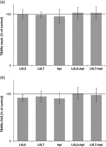
TBARs amounts in controls, lithium, haloperidol, or combination samples did not differ significantly from one another, both in neutral and OxS environment (Fig. 1). However, a trend of decreasing LP levels was observed in the lithium samples compared with controls in pro-oxidative conditions.
Lipid peroxidation in cell cultures (a) in neutral (n=4) and (b) in oxidative stress (OxS) conditions (n=3) shown as TBARs concentration. The results are expressed as percentages of those of the controls and shown as mean±SD. TBARs, thiobarbituric acid reactive substances.

In agreement with the results of that part of our study regarding neutral conditions, it was no influence demonstrated of lithium treatment on LP in erythrocytes and brain tissue of healthy rats (Reference Abdalla and Bechara45), nor in the liver and kidneys of mice (Reference Nciri, Allagui, Vincent, Murat, Croute and El Feki46). Consistently, Shin et al. (Reference Shin, Cho and Lim31) showed that, admittedly, lithium blocked the apoptosis of motor neurons in amyotrophic lateral sclerosis mice, but did not prevent OxS generation. Moreover, according to Vasconcellos et al. (Reference Vasconcellos, Nieto and Crema30), lithium treatment decreased the generation of reactive oxygen species in the hippocampus of rats, but it was not able to prevent a stress-induced increase in damage to macromolecules. On the other hand, the influence of lithium administration on the oxidative status of rats was protective but only in brain tissue; no changes were seen in the blood, liver or kidneys (Reference Kiełczykowska, Pasternak, Musik, Wrońska-Tyra and Hordyjewska47).
Similarly to our results, many studies have already demostrated at least a partial attenuation of elevated OxS under lithium treatment. It was found that chronic treatment with lithium inhibited the glutamate-induced (Reference Shao, Young and Wang29) and aluminium-induced (Reference Bhalla and Dhawan48), but not basal, increase of oxidation markers in rat cerebral cortical cells. Lithium was also found to ameliorate OxS induced by acute ketamine in a mouse model of stress (Reference Chiu, Scheuing and Liu49). Additionally, it was shown that SH-SY5Y cells under chronic lithium treatment displayed higher cell growth rates, lower basal levels of TBARs and improved tolerance to high lithium concentration-induced OxS (Reference Allagui, Nciri, Rouhaud, Murat, Feki and Croute50). Similar results were demonstrated in animal models of some diseases involving high level of OxS. Chronic lithium treatment normalised catalase (CAT) and superoxide dismutase activity in the rat model of depression (Reference Song, Killeen and Leonard51) and decreased TBARs formation (Reference Frey, Valvassori and Réus52,Reference Macêdo, de Lucena and Queiroz53) or increased CAT activity (Reference Jornada, Valvassori and Steckert54) in an animal model of mania. Moreover, similar results of OxS parameters under lithium treatment were shown in an animal model of sepsis (Reference Albayrak, Halici and Polat55).
However, several studies have been published demonstrating the pro-oxidative activity of lithium. It was shown in liver tissue (Reference Chadha, Bhalla and Dhawan32), in the kidneys (Reference Nciri, Allagui, Murat and Croute56), in the erythrocytes (Reference Malhotra and Dhawan57), and in lung tissue (Reference Sahin, Sulak and Yavuz58) of rats.
A number of different factors may contribute to the heterogeneity of the results observed in the literature of the influence of lithium on OxS parameters. They likely include: lithium concentration, duration of treatment, primary state of oxidative balance and type of cells/tissue examined. In summary, lithium may prevent peroxidation in neuronal cells under long-term treatment at therapeutic concentration and against induced OxS, confirming the trend seen in our study.
A statistically insignificant yet favourable trend was also observed in our study with regard to increased level of TBARs in the combination of lithium and haloperidol samples compared with other samples in neutral (Fig. 1a) and compared with lithium or haloperidol samples in pro-oxidative conditions (Fig. 1b). Despite the fact that both medications were used in therapetic concentrations and none of them alone caused increased LP, the combination should be utilised cautiously in the treatment setting. This trend is consistent with our previous study (unpublished data) which showed that lithium in combination with haloperidol significantly increased LP in human plasma in vitro compared with controls.
Neurotoxicity associated with the combined use of lithium and high-potency neuroleptics has been reported since 1974 (Reference Cohen and Cohen59). The precise mechanism by which the lithium/haloperidol combination induces toxicity is, however, poorly understood. As long ago as the 1970s and 1980s, the hypothesis of OxS underlying haloperidol/lithium toxicity was proposed (Reference Guynn and Faillace60,Reference Sawas and Gilbert61). To the best of our knowledge, there have not been any publications since on the possible mechanism of lithium/haloperidol toxicity in terms of OxS.
Cell viability
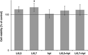
In spite of the higher absorbance measurements in the lithium samples in the MTT test, no differences were observed based on the ANOVA. Because of low statistical power (<0.8), statistical analysis of the results only from the lithium samples and controls were undertaken with the ANOVA and evaluated further with the Tukey test. The analysis showed significantly higher cell metabolic activity in therapeutic lithium samples compared with control values (Fig. 2), but the MTT assay did not directly answer the question if the change of cell activity resulted from attenuation of OxS or lithium-induced cell proliferation.
Cell viability in SH-SY5Y culture shown as absorbance (λ=570 nm) measurements. The results are expressed as percentages of those of the controls and shown as mean±SD; n=4; *p<0.05 compared with controls.

Given the evidence from previous studies, we concluded that the neuroprotective effects of lithium may be attributed, in part, to its OxS-scavenging activities. Similarly to our results, Lai et al. (Reference Lai, Zhao, Warsh and Li62) found a reduced H2O2-induced cytotoxicity in SH-SY5Y, but not in glial, cells pretreated with lithium. It was also shown that chronic exposure to lithium induces adaptive changes in the metabolism of SH-SY5Y cells involving a higher cell growth rate and a better resistance to OxS (Reference Allagui, Nciri, Rouhaud, Murat, Feki and Croute50,Reference Nciri, Desmoulin and Allagui63). Another study found that lithium, via the inhibition of GSK-3β, blocked intrinsic, but not H2O2-induced OxS that resulted in apoptosis (Reference King and Jope4). Thus, neuroprotection against extrinsic OxS possibly involves different mechanisms. In a recent study, lithium was found to ameliorate neural cell apoptosis/necrosis through a decrease in level of intracellular calcium (Reference Scola, Laliberte and Kim64). Several examples of the molecular mechanisms of action of lithium, except attenuation of OxS that result in its neuroprotective function can be listed, as described in the Introduction section.
In summary, H2O2, as expected, increased SH-SY5Ycell mortality through apoptotic and necrotic pathways. Lithium worked well against H2O2 treatment, which might be due, in part, to its effective ability to scavenge OxS-related molecules or enhance the generation of antioxidants.
Limitations
The main limitation of our study is its low statistical power (<0.8). Another significant shortcoming of our experimental design is the inherent difficulty in being able to extrapolate these findings in vivo. Moreover, an apparent limitation of the present study is the short time of incubation of the cell line samples with the active substances. In addition, some limitation of the MTT assay exist – it measures metabolic activity, rather than exact viability. Because of the limitations it is important to validate our results.
Summary
In conclusion, the main finding of this study was the increased cell viability in the lithium samples. Our present study also suggests that lithium may protect against increased OxS, while the lithium/haloperidol-induced neurotoxicity may be due to peroxidation. The OxS results are not, however, statistically significant. The connection between OxS changes and cell viability is not so clear, thereby necessitating further studies to clarify these aspects. Nevertheless, the results may prove significant in future studies on potential candidates as a novel therapeutic agents for BD.
Acknowledgement
The authors are thankful to the Head of the Department of Pharmaceutical Biochemistry, Medical University of Lodz, Prof. Marek Mirowski and the Head of the Department of Medicinal Chemistry, Medical University of Lodz, Prof. Anna Janecka for providing the facilities required to carry out the research work.
Authors Contributions
The work presented here was carried out in collaboration between all authors. O.G.K., J.R.B., and D.S. defined the research theme. O.G.K. and W.M. designed methods and experiments and carried out the laboratory experiments. O.G.K. and J.R.B. analysed the data and interpreted the results. O.G.K. and J.L. wrote the paper.
Financial Support
Supported by grant 502-11-756 from the Medical University of Lodz, Poland, and by grant N402 456138 from the Polish Ministry of Science and Higher Education.
Conflicts of interest
None.