Introduction
Personal appearance contributes to the perception of one's image and has enormous social significance in most cultures. In anorexia nervosa (AN), a severe eating disorder, physical appearance becomes an obsession; although pathologically underweight, anorexics see themselves as just normal or even fat. Such overestimation of body size has long been considered as primarily due to psycho-affective causes. Consequently, most studies on body image in AN principally addressed the related emotional aspects (Cooper et al. Reference Bradley, Taylor, Rovet, Goldberg, Hood, Wachsmuth, Azcue and Pencharz1987; Rosen et al. Reference Kerkhoff1991; Smeets et al. Reference Lacquaniti, Boller and Grafman1997; Benninghoven et al. Reference Benninghoven, Raykowski, Solzbacher, Kunzendorf and Jantschek2007; Surgenor et al. Reference Surgenor, Maguire, Russell and Touyz2007; Abraham et al. Reference Abraham, von Lojewski, Anderson, Clarke and Russell2009) overlooking possible contributions of the neural mechanisms supporting body representation, although some evidence for this is now emerging (Kinsbourne & Bemporad, Reference Grunwald, Ettrich, Assmann, Dähne, Krause, Busse and Gertz1984; Braun & Chouinard, Reference Benninghoven, Raykowski, Solzbacher, Kunzendorf and Jantschek1992; Bradley et al. Reference Audenaert, Van Laere, Dumont, Vervaet, Goethals, Slegers, Mertens, van Heeringen and Dierckx1997; Maggia & Bianchi, Reference Heilman, Watson, Valenstein, Heilman and Valenstein1998; Grunwald et al. 2001 a, b, Reference Frank, Bailer, Henry, Wagner and Kaye2002; Audenaert et al. Reference Audenaert, Van Laere, Dumont, Vervaet, Goethals, Slegers, Mertens, van Heeringen and Dierckx2003; Chowdhury et al. Reference Chowdhury, Gordon, Lask, Watkins, Watt and Christie2003; Wagner et al. Reference Rosen, Srebnik, Saltzberg and Wendt2003; Frank et al. Reference Frank, Bailer, Henry, Wagner and Kaye2004; Kojima et al. Reference Grunwald, Ettrich, Busse, Assmann, Dähne and Gertz2005; Lask et al. Reference Lask, Gordon, Christie, Frampton, Chowdhury and Watkins2005).
Neuropsychology distinguishes multiple functional levels of body knowledge, including representations derived from sensorimotor integration that support basic motor activities (i.e. catching/avoiding a moving target), and ‘sense of self’, which enables us to feel that we inhabit our body (Sirigu et al. Reference Krieg, Holthoff, Schreiber, Pirke and Herholz1991; Gallagher, Reference Clower, Dum and Strick2000). Converging data from lesion studies, direct cortical stimulation and functional imaging emphasize the role of parietal lobes in the integration and synthesis of multiple sources of sensory information for the establishment and maintenance of a coherent representation of the body and peri-personal space (Graziano & Gross, Reference Epstein, Wiseman, Sunday, Klapper, Alkalay and Halmi1995; Karnath & Their, Reference Gold and Shadlen1997). Damage to these structures causes anomalous body experiences, including denial of motor deficits, delirious beliefs about the body, and metric disturbances of body schema. These deficits generally involve the body-half opposite to the damaged hemisphere and commonly follow right parietal lesions (left parietal areas being more concerned with conceptual aspects of body knowledge; Sirigu et al. Reference Krieg, Holthoff, Schreiber, Pirke and Herholz1991). Indeed, the most common consequence of right parietal damage is unilateral neglect, which, in addition to defective exploration of the left side of egocentric space, can produce a rightward shift of the subjective body midline (Heilman et al. Reference Geary, Saults, Liu and Hoard2003; Adair & Barrett, Reference Adair and Barrett2008).
It is unclear whether these elementary, sensory-based aspects of body representation are affected in AN, and whether their involvement contributes to the phenomenology of the disorder. A probable reason for this gap is the difficulty in avoiding potential confounds due to emotional/attitudinal components towards the body. To date, available measures of body representation include self-report questionnaires [satisfaction/dissatisfaction indices, as in the Body Shape Questionnaire (BSQ); Cooper et al. Reference Bradley, Taylor, Rovet, Goldberg, Hood, Wachsmuth, Azcue and Pencharz1987; Cuzzolaro et al. Reference Cuzzolaro, Vetrone, Marano and Garfinkel2006] that concentrate on the emotional issue, and few perceptual techniques. The latter require patients to estimate their body (or body parts) size directly, or by selecting the matching prototype among alternatives (Benson et al. Reference Benson, Emery, Cohen-Tovée and Tovée1999; Harari et al. Reference Harari, Furst, Kiryati, Caspi and Davidson2001; Shafran & Fairburn, Reference Shafran and Fairburn2002; Letosa-Porta et al. Reference Letosa-Porta, Ferrer-Garcia and Gutiérrez-Maldonado2005; for a review of earlier studies see Skrzypek et al. Reference Skrzypek, Wehmeier and Remschmidt2001). By inquiring directly about body image, these measures discriminate poorly between top-down influences induced by emotions/attitudes towards the body (Smeets et al. Reference Lacquaniti, Boller and Grafman1997) and disturbances of body-size perception due to proprioceptive disorders/distorted body schema (Epstein et al. Reference Epstein, Wiseman, Sunday, Klapper, Alkalay and Halmi2001).
However, the possibility for a neurophysiological basis to body misperception in AN exists; converging evidence indicates a profound involvement of a multi-modal area, the parietal cortex, in supporting body representation (Sirigu et al. Reference Krieg, Holthoff, Schreiber, Pirke and Herholz1991; Graziano & Gross, Reference Epstein, Wiseman, Sunday, Klapper, Alkalay and Halmi1995; Karnath & Their, Reference Gold and Shadlen1997; Gallagher, Reference Clower, Dum and Strick2000). Parietal malfunctioning emerges in anorexics with respect to haptic perception (Grunwald et al. 2001 a, b, Reference Frank, Bailer, Henry, Wagner and Kaye2002) or spatial processing tasks (Kinsbourne & Bemporad, Reference Grunwald, Ettrich, Assmann, Dähne, Krause, Busse and Gertz1984; Braun & Chouinard, Reference Benninghoven, Raykowski, Solzbacher, Kunzendorf and Jantschek1992; Bradley et al. Reference Audenaert, Van Laere, Dumont, Vervaet, Goethals, Slegers, Mertens, van Heeringen and Dierckx1997; Maggia & Bianchi, Reference Heilman, Watson, Valenstein, Heilman and Valenstein1998). In addition, atypical patterns of activation have been documented in the parietal regions of the anorexics' brain when observing a digitally distorted image of their own body (Wagner et al. Reference Rosen, Srebnik, Saltzberg and Wendt2003), but also after changes in body mass due to weight gain (Kojima et al. Reference Grunwald, Ettrich, Busse, Assmann, Dähne and Gertz2005). The hypothesis of a specific dysfunction in somatosensory integration in the right parietal cortex of anorexics has been proposed explicitly, mainly based on the difficulties these individuals show in reproducing spatial arrays learnt through haptic exploration (Grunwald et al. 2001 a, b). This suggests that AN patients may be impaired in integrating the incoming somatosensory information required to build body representation. Accordingly, a neuropsychological component would possibly add to the well-known psycho-affective dimension of the disorder.
In the current study we assessed the possibility that body distortions in AN reflect an impairment in the processes of visual–spatial integration typically supported by the right parietal lobe. Should this be the case, anorexics should show difficulties in tasks tapping these functions (namely when the body is used as a spatial reference), similarly to what happens in neurological patients suffering from damage to the right parietal lobe. We postulated that the disturbances would affect to a larger extent the left part of the body, in line with neuropsychological findings (Heilman et al. Reference Geary, Saults, Liu and Hoard2003; Adair & Barrett, Reference Adair and Barrett2008). Conversely, a pure psycho-affective disorder should produce a symmetrical pattern of perturbations, affecting both sides of corporeal space equally. We tested body representation in clinically diagnosed anorexics, healthy volunteers and stroke patients with selective parietal lesions, using a psychophysical procedure based on the perception of approaching visual stimuli. This method reproduces the common situation of catching/avoiding approaching items, and assesses perceived body dimensions based on pragmatic body knowledge supporting elementary motor activities. Being similar to natural conditions, this ecological approach should be less susceptible to the influence of attitudes/emotions towards the body than judgments obtained using self-report questionnaires or optically distorted silhouettes (Skrzypek et al. Reference Skrzypek, Wehmeier and Remschmidt2001).
Method
Participants
According to the Declaration of Helsinki and local ethical guidelines (Centre Léon Bérard, Lyon), all participants gave informed consent to participate.
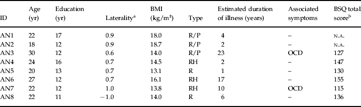
Eight female anorexics [AN patients, mean age 23.4±4.0 years; mean body mass index (BMI) 15.3±2.1] were recruited among patients referring to Clinique Saint Vincent de Paul, Lyon for treatment (Table 1). AN was the primary diagnosis in all cases (DSM-IV F 50.0; APA, 1994). None reported a history of neurological disorders, or current/past substance dependence/abuse. All had normal/corrected-to-normal vision. Perceived body image was assessed using the BSQ (Cooper et al. Reference Bradley, Taylor, Rovet, Goldberg, Hood, Wachsmuth, Azcue and Pencharz1987; Rousseau et al. Reference Kinsbourne, Bemporad, Fox and Davidson2005). Neuropsychological tests confirmed the integrity of the perceptual processes, body schema and attention. Subject AN8 suffered from severe anxiety and her neuropsychological assessment could not be completed.
Summary of demographic and clinical data for the group of patients suffering from anorexia nervosa
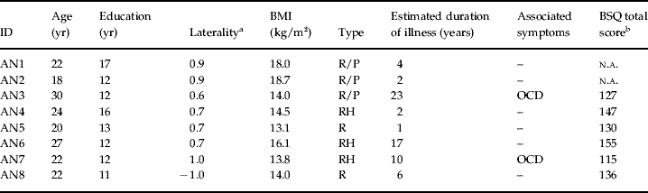
AN, Anorexia nervosa; BMI, body mass index; BSQ, Body Shape Questionnaire; R, restrictive; P, purging; RH, restrictive-hyperactive; OCD, obsessive–compulsive-disorder; n.a., not applicable.
a Laterality according to the Edinburgh Inventory.
b Range 34–204, mean score 75.8 (normative French sample n=242).
We also tested four right-handed patients suffering from selective vascular lesion affecting the right (RP patients, one 42-year-old male, one 64-year-old female) or left parietal cortex (LP patients, two males, aged 36 and 57 years), as assessed by magnetic resonance imaging (MRI). Post-treatment images were co-registered to the Bancaud–Talairach atlas to locate ischemic areas according to Brodmann area (BA). Angular and supramarginal gyri (BA 39, BA 40) were damaged in all cases, with possible extensions into the middle temporal cortex (RP2). All patients showed unimpaired elementary sensory/motor functions and scored within normal limits at standard neuropsychological assessment.
Control participants for anorexics were 11 right-handed healthy individuals (six females, mean age 29.4±7.0 years; normal/corrected-to-normal vision), naive about the aim of the research. Although the male brain is more specialized for visual–spatial functions (Geary et al. Reference Cooper, Taylor, Cooper and Fairburn2000) and females exhibit a cultural bias towards greater attention to body size/appearance, no gender differences emerged for the measures tested here; hence, this was considered as a unique group.
Four older right-handed volunteers (two females, mean age 54.5±7 years) with no history of neurological/psychiatric disorders were tested separately as age-matched controls for parietal patients.
Apparatus and procedure
Participants sat in a quiet darkened room, head/body displacements prevented by straps positioned over the forehead and seatbelts crossing the chest. A robotic arm, equipped with a green light-emitting diode (LED) at the fingertip, was located in front of the participant, 40 cm away from the anatomical landmark of interest. The mechanical arm moved the LED along the horizontal plane at constant speed (20 cm/s) towards subjects, stopping at 16 cm from their body (Fig. 1). Goggles equipped with translucent liquid crystals shutters synchronized with the robotic arm allowed vision only during the LED's displacement. Trials started with a warning tone and the room darkening; the LED was switched on, the mechanical arm started to move, and the shutters on the goggles opened synchronously. Participants were instructed to track the moving LED visually and, when it was turned off and the shutters closed, to mentally complete its trajectory and decide whether it would have eventually hit their body. Verbal responses (yes/no) were recorded.
Schematic view of the apparatus. An ecological procedure was used to assess the location of subjective body boundaries in anorexic patients, healthy volunteers and patients suffering from parietal brain damage. In the dark, participants were required to mentally extrapolate the trajectory of an approaching light-emitting diode (LED), mounted on a robotic arm (Model A-225, CRS Robotics, Canada, running on Robcom for Windows software, version 4.32), which was turned off halfway between its starting position and the observer's body. Participants wore goggles equipped with translucent liquid crystals shutters (Translucent Technologies Inc., Canada) synchronized with the robotic arm, allowing vision only during displacement of the LED. Trunk and head movements were prevented by straps positioned over the forehead and seatbelts crossing the chest; arms were unrestrained but participants were asked to keep them close to the chest on armrests. Subjects reported verbally whether the LED would have eventually impacted on their body, had it continued to move. Linear trajectories were randomly distributed around one of four different anatomical references; participants were left unaware of these anatomical landmarks throughout the task.

The selected landmarks for testing the contour of the body corresponding to its maximal width (i.e. body boundaries) were the upper proximal joints (shoulders), a reliable marker of trunk extension (specifically, the point on the edge of the left/right deltoid muscles, 5 cm below a horizontal plane passing through the subjects' jugular notch). A second, control, landmark was chosen at head level, on the cheekbones (precisely, the edge of the zygomatic bones at the level of a horizontal plane touching the subject's nostrils). Participants were left unaware of these anatomical landmarks throughout the task.
Seventeen trajectories were arranged symmetrically around each landmark. The Ø trajectory was defined before each session by aligning a laser beam mounted on the mechanical arm with the chosen landmark. The remaining 16 trajectories were distributed at 0.5, 1, 1.5, 2, 2.5, 3, 4, and 5 cm on either side of the Ø trajectory. Each trajectory was presented five times in a randomized sequence (85 trials). Perception of body boundaries at the level of each landmark (left/right cheekbone; left/right shoulder) was tested in separate sessions (each preceded by five familiarization trials) run on the same day. The order of the sessions was counterbalanced within participants.
Data collection and statistical analysis
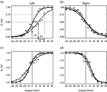
For each participant, manually recorded ‘hit’ and ‘miss’ responses were converted into proportions and fitted with a logistic regression equation (Fig. 2) of the general form

where P is the probability of ‘hit’ responses, w the trajectory endpoints, and b 1 and b 2 the fitted parameters of the equation representing the horizontal offset and the slope of the sigmoid-shaped function respectively.
Body boundary estimates in healthy participants (–▪–, n=11) and anorexic patients (–○–, n=7). Performance is expressed as the frequency of ‘hit’ reports as a function of trajectory endpoints toward the shoulders (a, b) and head (c, d). Responses were converted into proportions and fitted with a logistic regression equation of the general form p=1/[1+exp[–b 1 – b 2w)], where p is the probability of ‘hit’ responses, w the trajectory endpoints, and b 1 and b 2 the fitted parameters of the equation representing the horizontal offset and the slope of the sigmoid-shaped function respectively. Panel (a) shows the computation method for the perceived boundary location (BL), defined as the location where ‘hit’ and ‘miss’ responses are equally probable (p=0.5, point of subjective equality). Positive deviation indicates a rightward deviation of the perceived boundary. An index of boundary uncertainty (BU) was computed as the distance on the abscissa between the point of subjective equality and the location where 75% ‘hit’ responses were recorded. Logistic regression curves were fitted to the average performance of the healthy controls and anorexic participants. Significant differences between groups were found on the left side (t test p<0.01, lines: standard error).

Perceived body boundary location (BL) was defined as the point of subjective equality, that is the point having equal probability to elicit either response. Perceived boundary uncertainty (BU) was defined as the distance between the points having 50% and 75% probabilities to elicit a ‘hit’ response. Thus, each subject's performance was characterized by a BL and a BU value for each landmark. Separate two-tailed t tests were used to compare BU values across landmarks within each group. In addition, BL data (for each landmark) for anorexics were compared to those for controls by separate t tests. Because of the sample size, data from parietal patients were compared individually to those of controls using confidence intervals for abnormal performance [set at 3 standard deviations (s.d.) from controls' performance]. Only results exceeding these limits are reported.
Control task 1
This task assessed the integrity of the processes involved in tracking visual stimuli in AN patients (subjects AN3–8, Table 1). Subjects were instructed to (1) fixate a cross in the center of the screen for 1 s, (2) move their eyes as fast as possible toward a square presented at one of 12 peripheral locations in the left/right hemi-fields, (3) fixate the stimulus until it disappeared, and (4) return to fixation of the cross. Each square was displayed for 1.5 s, and each location was tested five times. Eye movements were recorded at 60 Hz with a video eye-tracker (Tobii, Sweden).
Control task 2
This task assessed the capacity of predicting the virtual impact of an approaching object per se, that is aiming at an external object rather than at one's body. AN patients and five healthy female volunteers (mean age 31.6±5.7 years, mean BMI 18.6±1.1) were tested. Subjects sat at approximately 60 cm from a screen, on which a white bar (3.75 cm×0.5 cm) on a black background was displayed in the lower left (left block) or lower right (right block) corner (depending on the block). A single dot (ø 0.2 cm) appeared 18 cm above the bar, aligned with its external edge, and immediately started to move linearly towards the bar (at ∼3 cm/s). The dot disappeared at 56% of its trajectory (10 cm). Subjects decided whether it would have eventually hit the bar had it kept moving, and pressed a key accordingly. Seven possible trajectories were tested: the Ø trajectory aimed straight at the bar's edge, the others toward virtual impact points located at 0.45, 0.75 and 1 cm to the left/right of the edge. Each trajectory was repeated five times per block (except for the Ø trajectory, which was repeated 10 times). Two right and two left blocks were run in pseudo-random order. Data were analyzed as for the main experiment to calculate the perceived BL and BU index for bars appearing on the left/right side of the screen.
Results
Healthy subjects
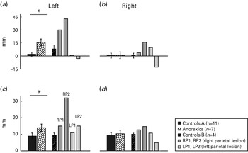
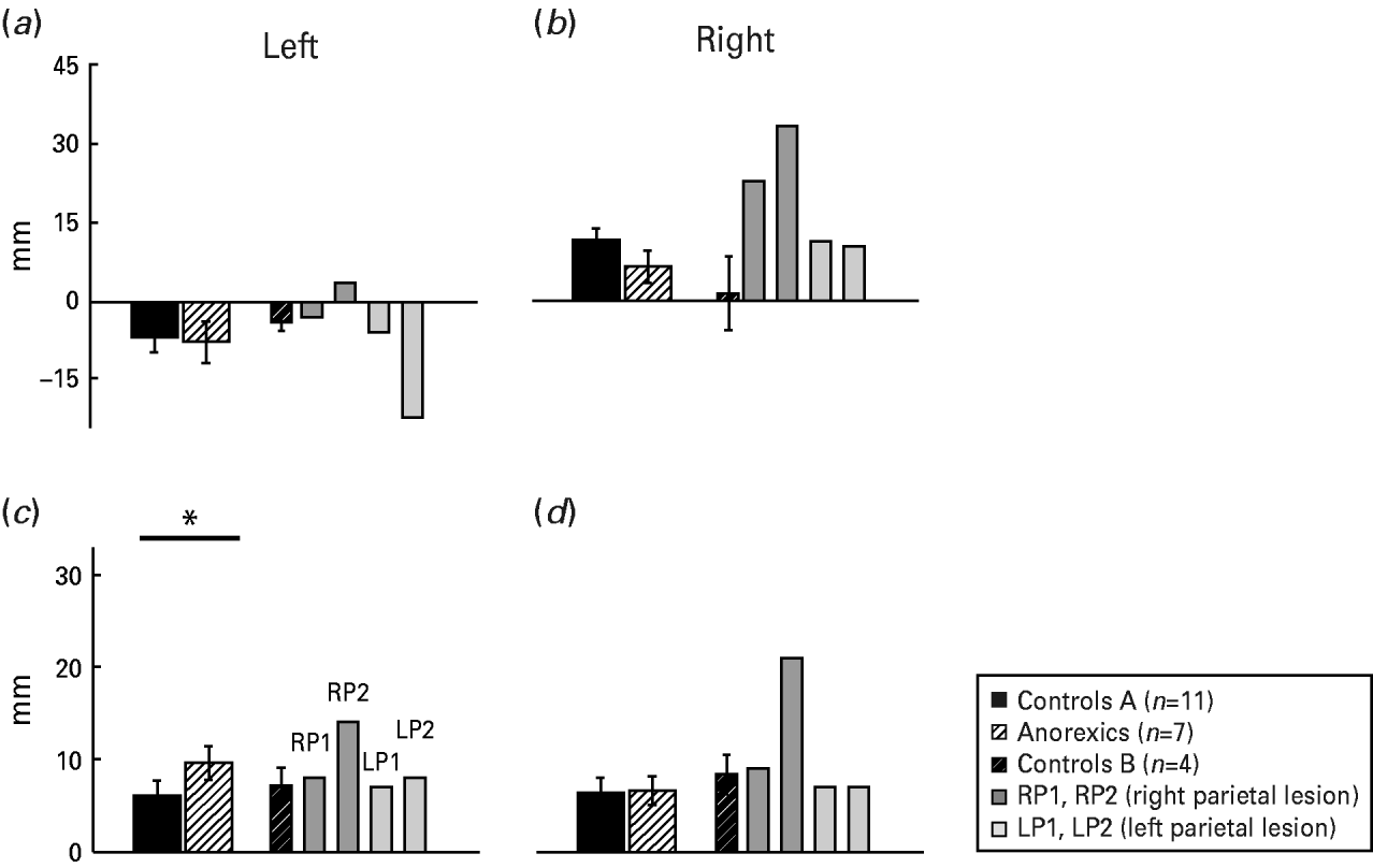
For trajectories directed at the shoulder's landmarks, healthy volunteers estimated body boundaries very close to the real ones [Fig. 2 (a, b); BL: left 2.1±2.4 mm, right 0.0±2.9 mm]. At the head level, boundaries were estimated slightly outside the edge of the zygomatic bone, the landmark used as reference [Fig. 2 (c, d); BL: left −7.0±2.9 mm, right: 12.2±2.3 mm]. This bias was not associated with greater BU; estimates obtained for the shoulder (mean BU 9.2±11.8 mm) were in fact larger than for the head (6.2±7.9 mm, p<0.01, Figs. 3 and 4).
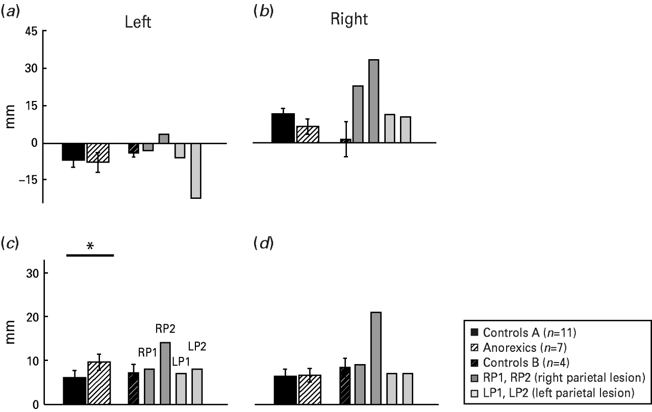
Shoulder boundary location (BL) and boundary uncertainty (BU). (a) and (b) show perceived BLs for 11 healthy subjects, seven anorexics, four healthy controls and four stroke patients with right or left parietal lobe lesions. Averaged data for the healthy and anorexic subjects are derived from individual logistic regression fits. On the y axis, 0 corresponds to the true anatomical landmark. Positive deviation indicates a rightward deviation of the perceived boundary. (c) and (d) show BU values: higher values correspond to greater uncertainty. Significant differences between healthy and anorexic patients were found on the left side (* t test p<0.01; lines, standard error).

Head boundary location (BL) and uncertainty (BU). (a) and (b) show perceived BLs for the left and right head landmark for 11 healthy subjects (controls A), seven anorexics, four older healthy subjects (controls B), and four stroke patients with right or left parietal lobe lesions. Averaged data for the healthy and anorexic subjects are derived from individual logistic regression fits. On the y axis, 0 corresponds to the true anatomical landmark. Positive deviation indicates a rightward deviation of the perceived boundary. (c) and (d) show BU values for the left and right head landmark. Higher values correspond to greater uncertainty (* t test p<0.01; lines, standard error).
Anorexic patients
The performance of the AN patients differed markedly from that of the controls but anomalies were limited to the left side of the body. The results for 7/8 patients [(Fig. 2 (a, c)] were characterized by flatter curves and a significant rightward shift of the left shoulder boundary, that is an inward displacement (Fig. 3a, 15.7±4.1 vs. 2.1±2.9 mm, p<0.01). Anorexics were significantly less precise than controls, in that they had greater BUs at both left anatomical landmarks (shoulder: 13.9±2.3 vs. 8.9±1.8 mm; head: 9.6±1.8 vs. 6.0±1.7 mm, both p<0.01; Figs. 3 and 4). By contrast, the two groups did not differ on right-side estimations [Fig. 2 (b, d)]. One patient (AN8) was dropped from the analyses because she was unable to estimate left body boundaries, responding ‘hit’ to all stimuli, a condition that she was aware of (and upset about) during testing. However, her performances were within the controls' range for all trajectories directed at the right face boundary (BL=–3.6 mm, BU=10.4 mm) and were deviated rightward but not random at the shoulder level (BL=34.0 mm, BU=16.1 mm). To a lesser extent, difficulties in dealing with the left boundary were reported spontaneously by four more patients (AN4–7).
Parietal lobe lesions
As expected, RP patients presented metric anomalies of the body schema, characterized by large rightward deviations compared to healthy subjects (Fig. 3), at all landmarks. The shift was largest at the level of the left shoulder, which they located deeply within the real body limits, and was highly specific. Estimates by LP patients were within controls' limits on most landmarks (Fig. 3).
Control tasks
No significant differences between left/right visual hemi-fields were found in AN patients for saccadic reaction times, amplitude and direction (all p's>0.17, paired t test). In addition, perceived BLs for an external object were comparable for anorexics and controls (p>0.5), and for left and right sides in anorexics (p>0.08).
Discussion
By testing body size perception implicitly, using collision judgments, we could eliminate potential confounds due to psycho-affective attitudes towards body weight and size in AN. This allowed us to uncover an unsuspected neuropsychological aspect of anorexia; consistent deviations of body schema emerged but, strikingly, not in the direction predicted by the phenomenology of the disorder, or by patients' reported feelings of being overweight (Table 1). By contrast, performance showed clear similarities with that of patients with right parietal damage.
Estimating the impact point of a moving target requires integration of visual and non-visual sensory information concerning the body. Bimodal neurons in the monkey parietal cortex and related structures perform this integration by responding to both somatosensory and visual information from a given body area and surrounding space (Fogassi et al. Reference Chowdhury, Gordon, Lask, Watkins, Watt and Christie1996; Duhamel et al. Reference Braun and Chouinard1998; Graziano & Cooke, Reference Duhamel, Colby and Goldberg2006). Of note, their firing rate increases as the visual stimulus approaches the tactile receptive field. Psychophysically, such properties provide a basis for the accurate performance of healthy participants, contributing to a modified spatial resolution of the region targeted by the approaching visual stimulus in anticipation of contact. In addition, the precise estimation at the shoulder's boundary is in line with the functional role played by this joint as a spatial reference for arm movements (Lacquaniti, Reference Harari, Furst, Kiryati, Caspi and Davidson1997) and as a dimensional reference for body displacements. The slight overestimation at head level could mean that observers (not informed of the zygomatic reference) correctly considered ears as the body boundary (the overestimation amounting approximately to the distance between the zygomatic landmark and projection of the ears from the head). Alternatively, observers could have implicitly applied a ‘safety margin’ around the head, an idea that finds an echo in studies of non-human primates (Graziano & Cooke, Reference Duhamel, Colby and Goldberg2006).
The performance of RP patients is in line with predictions derived from neuropsychological findings on neglect patients, who can show a shift of the subjective midline towards the side of the lesion (Kerkhoff, Reference Graziano, Gross and Gazzaniga2001). Of interest, at the time of testing, RP patients were asymptomatic, suggesting that this is a very sensitive test of residual right parietal deficit. The left-sided bias and imprecision observed in AN patients offer a compelling analogy with the performance of RP patients, indicating a non-emotional component of body misperception in AN. These results cannot be attributed to non-specific effects of malnutrition, as performance on neuropsychological tests was within normal limits. Furthermore, errors selectively affected judgments concerning the left side of the body.
The question must be raised, however, of the link between our findings and anorexics' behavior. One possibility is that dysfunctional eating might induce a progressive derangement of body perception. A distorted body image would represent the physiological outcome of malnutrition interfering with the brain neurochemistry of circuits involved in visual–spatial cognition. Alternatively, and in accord with the asymmetrical distribution of responses, the opposite causal relationship could be postulated: a primary distorted body representation could contribute to anomalous eating patterns by generating dissatisfaction with one's body image. Attitude toward food/weight could, in this perspective, correspond to a pathological attempt at restoring a coherent body schema.
Eating disorders in AN are often accompanied by obsessive–compulsive disorders (OCD; Swinbourne & Touyz, Reference Maggia and Bianchi2007). Anorexia, like OCD, may be linked to basal ganglia (BG) dysfunction (Krieg et al. Reference Grunwald, Ettrich, Krause, Assmann, Dähne, Weiss and Gertz1991; Steinglass & Walsh, Reference Lask, Gordon, Christie, Frampton, Chowdhury and Watkins2006), striatal dopamine dysfunction having been invoked to explain various AN symptoms (Kaye et al. Reference Graziano and Cooke1999; Wagner et al. Reference Platt and Glimcher2007). Studies in non-human primates show that the posterior parietal cortex receives, through the thalamus, major inputs from BG (Clower et al. Reference Benson, Emery, Cohen-Tovée and Tovée2005; Murayama et al. Reference Karnath and Thier2006). The parietal cortex is essential for development and maintenance of body representations whereas BG play a crucial role in adapting behavior to motivational/emotional needs (Schultz et al. Reference Kojima, Nagai, Nakabeppu, Muranaga, Deguchi, Nakajo, Masuda, Nozoe and Naruo2003). Motivation and reward also modulate the activity of parietal neurons (Platt & Glimcher, Reference Kaye, Frank and McConaha1999; Sugrue et al. Reference Letosa-Porta, Ferrer-Garcia and Gutiérrez-Maldonado2004; Gold & Shadlen, Reference Cuzzolaro, Vetrone, Marano and Garfinkel2007). Functional interactions between BG and parietal cortex may thus play a role in the development and regulation of body image. Anorexia might involve a dysfunction of such a putative parietal–basal ganglia circuit. Preliminary reports from our group on patients suffering from Parkinson's disease support this possibility (personal communication).
Although we are not questioning the role of environmental and cultural factors in AN, our findings introduce the possibility that neurophysiological vulnerability factors are involved and/or that intense preoccupation with weight and body image might itself lead to a dysfunction of key neural systems. Most AN patients tested were in the acute phase, hence we could not determine whether magnitude of body perception asymmetry related to severity and duration of symptoms. However, two patients (AN1 and AN2) who had almost completed their treatment and recovered body weight (BMI 18/18.7) still showed deviations comparable to those of patients in the acute phase of the disease; body perception anomalies might thus constitute a stable marker of vulnerability.
This newly disclosed neuropsychological component of body misperception in AN offers compelling implications for clinical treatment. Developing methods to compensate the neuropsychological body disturbance could facilitate the emergence of a correct body perception, improving these patients' self-image.
Acknowledgments
This work was supported by the Centre National de la Recherche Scientifique Française (CNRS), the Human Frontier Science Program (grant no. RGP0056) and the European Science Foundation to A.S. We thank the participants for their patience and collaboration and Dr Fourneret for recruiting pilot patients.
Declaration of Interest
None.