Introduction
The infratemporal fossa is bounded superiorly by the sphenoid bone, medially by the lateral pterygoid plate, anteriorly by the maxilla, posteriorly by the deep parotid region and laterally by the mandibular ramus.Reference Akst, Albani and Strome1, Reference Leventhal and Schwartz2 As this area communicates with the orbit and middle cranial fossa, infection may spread to these areas, leading to life-threatening situations.
Treatment of infratemporal fossa infections depends on their severity. Cellulitis may be treated with antibiotics only, but true abscess requires incision and drainage.Reference Akst, Albani and Strome1, Reference Chong and Fan3 Infratemporal fossa abscess following odontogenic infection is not rare,Reference Bradley4 and usually occurs as a sequela of dental disease such as untreated dental caries,Reference Nishizaki, Ogawa, Akagi, Sato and Masuda5, Reference Kim, Kim, Jang, Moon and Park6 extraction of teeth,Reference Akst, Albani and Strome1, Reference Bradley4, Reference Muñoz-Guerra, González-García, Capote, Escorial and Gías7 or other dental treatment.Reference Leventhal and Schwartz2, Reference Chossegros, Cheynet and Conrath8 Infratemporal fossa abscess was previously treated with the combination of buccal incision and temporal incision,Reference Bradley4 but is now treated mainly with the intraoral approach.Reference Akst, Albani and Strome1, Reference Leventhal and Schwartz2, Reference Nishizaki, Ogawa, Akagi, Sato and Masuda5, Reference Chossegros, Cheynet and Conrath8, Reference Raghava, Evans and Basu9 The use of the transnasal endoscopic approach was recently reported. The transnasal septum approach was used with resection of the inferior half of the middle turbinate to obtain a surgical view and space.Reference Varshney, Zawawi and Tewfik10 Endoscopic modified medial maxillectomy with preservation of the inferior turbinate and nasolacrimal duct was proposed in 2012 to treat inverted papilloma of the anterior wall of the maxillary sinus.Reference Nakayama, Asaka, Okushi, Yoshikawa, Moriyama and Otori11 We believe that this latter approach is less invasive and offers more direct access to infratemporal fossa abscess than any other approach.
Here, we describe the endoscopic modified medial maxillectomy surgical approach and technique, in a case where a small abscess at the infratemporal fossa was safely drained under navigation guidance.
Methods
Preparation for surgery
Pre-operative contrast-enhanced computed tomography (CT) is the most useful diagnostic tool for head and neck abscess, and is highly sensitive (80–90 per cent) for the detection of deep neck space infection.Reference Maroldi and Nicolai12 The specificity of CT has been reported as 82 per cent for lesions larger than 3.5 cm, and intralesional air indicated abscesses in all cases in that study.Reference Freling, Roele, Schaefer-Prokop and Fokkens13 We recommend navigation guidance for small abscesses as the infratemporal fossa is outside the sinonasal cavity, which is more familiar to surgeons.
Case report
A 51-year-old female with trismus and abducens nerve palsy was referred to our department. She complained of pain in the right upper jaw extending to the auricular area and trismus. She had visited a dentist and local physician three months before presenting at our department. Abducens nerve palsy occurred one week before visiting our department.
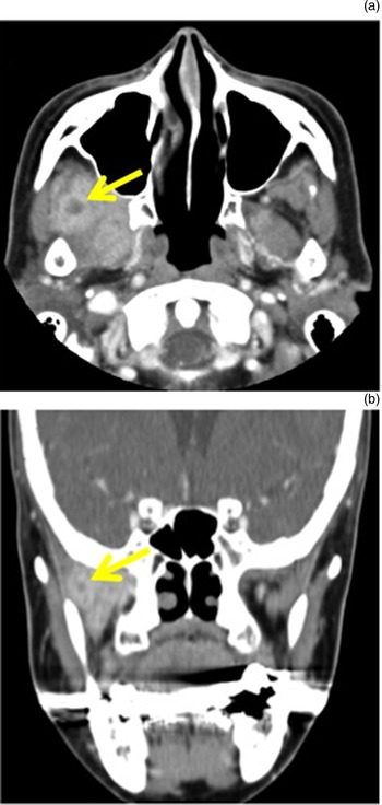
She was admitted to the neurology department of a tertiary care hospital for investigation. She had no history of maxillofacial trauma, diabetes or other immunocompromising disorders. Axial and coronal contrast-enhanced CT showed diffuse enhancement of the lateral and medial pterygoid muscles, and a ring-like region of enhancement in the lateral pterygoid muscle (Figure 1). Magnetic resonance imaging demonstrated a mosaic pattern on T2-weighted images and low intensity on T1-weighted images in the region of the pterygoid muscles (data not shown).
Pre-operative axial (a) and coronal (b) contrast-enhanced computed tomography scans, demonstrating enhanced medial and lateral pterygoid muscles, with a ring-like region of enhancement (arrows) in the lateral pterygoid muscle.

The patient was transferred to our department for surgical intervention. She was taken to the operating theatre and anaesthetised with transnasal intubation because of the trismus.
Surgical technique
All procedures were performed under general anaesthesia, with a 4-mm, 0-degree endoscope (Karl-Storz Endoscopy, Tuttlingen, Germany).
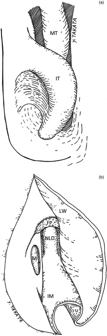
A vertical incision was made in the lateral nasal wall at the mucocutaneous junction, directly behind the piriform aperture, and continued to the nasal floor (Figure 2a).Reference Nakayama, Asaka, Okushi, Yoshikawa, Moriyama and Otori11 The mucosa was elevated from the bone surface, and the inferior nasal concha bone was exposed. Any anterior attachment to the lateral nasal wall was detached with a chisel, and any remaining bone attached to the lateral wall was removed with a diamond burr. Consequently, the entire inferior turbinate could be preserved and displaced medially.
Schema of surgical approach. (a) Incision at the lateral nasal wall. A vertical incision (solid line) is made in the lateral nasal wall at the mucocutaneous junction, directly behind the piriform aperture, and continued to the nasal floor. (b) The nasolacrimal duct, mucosal flap and inferior turbinate are pushed medially and preserved. MT = middle turbinate; IT = inferior turbinate; LW = mucosa of lateral nasal wall; FP = frontal process of the maxilla; NLD = nasolacrimal duct; M = mucosa of the medial wall of the maxillary sinus; IM = mucosa of the lateral wall of inferior meatus

The mucosal flap was elevated posteriorly and the nasolacrimal duct was exposed. The nasolacrimal duct, mucosal flap and inferior turbinate were pushed medially and preserved (Figure 2b). The medial bony wall of the maxillary sinus was drilled with a diamond burr so that the nasolacrimal duct and nasal mucosa could be preserved. The medial bony wall of the maxillary sinus is not extremely thick, so the cutting burr is usually not necessary.
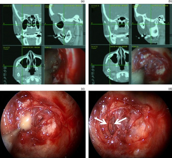
At this stage of the procedure, the posterior wall of the maxillary sinus was in the centre of the field of vision. Under navigation guidance (1-mm thick slices were used), the adjacent mucosa of the posterior wall was scratched and marked (Figure 3a). The posterior wall of the maxillary sinus was thinned and removed; a diamond burr or a blunt elevator can be used. The posterior periosteum was incised. Small veins and arteries were dissected from the fat tissue and muscles. Blunt dissection was needed because bleeding from the internal maxillary artery could be unmanageable with the endoscope. After the pus was drained, the drainage pathway was widened (Figure 3b–d).
Intra-operative photographs obtained with a 0-degree endoscope and screen shots of the navigation guidance. (a) The most appropriate point of the posterior maxillary wall with access to the abscess is determined by navigation guidance. (b) Infratemporal fossa abscess is reached with navigation guidance. (c) Yellow pus drained after removing the navigation probe. (d) Drainage pathway is formed (arrows).

The surface of the exposed pterygoid muscles was covered with Surgicel (Ethicon, Somerville, New Jersey, USA), although the drainage pathway was left without packing material. The inferior turbinate, nasolacrimal duct and lateral mucosal wall of the inferior meatus were replaced. No fistula was made at the inferior meatus. Middle meatal antrostomy was performed for post-operative examination. Calcium alginate dressing (Sorbsan; Alcare, Tokyo, Japan)Reference Okushi, Yoshikawa, Otori, Matsuwaki, Asaka and Nakayama14 was used for soft packing to keep the structures in place.
Post-operative course
Ampicillin sodium 1 g and sulbactam sodium 0.5 g were intravenously administered every 6 hours for 4 days.
The patient's pain remitted the next day. Four days after the operation, antibiotics were changed to meropenem 1 g every 12 hours for 13 days, as trismus and low-grade fever persisted. Trismus had diminished in one week. Abducens nerve palsy gradually recovered over six months. Culture of the abscess showed Propionibacterium acnes.
Discussion
All previous cases of infratemporal fossa abscess except one were treated with the external and/or transoral approach.Reference Akst, Albani and Strome1, Reference Leventhal and Schwartz2, Reference Bradley4–Reference Varshney, Zawawi and Tewfik10 Only one case was treated with the transnasal endoscopic approach.Reference Varshney, Zawawi and Tewfik10 The transnasal endoscopic approach is less invasive and can provide good visualisation. The disadvantage of the normal transnasal endoscopic approach is difficulty in accessing the infratemporal fossa and in controlling unexpected massive bleeding. In the previous case, a trans-septal approach through the contralateral nostril was used to access the infratemporal fossa.Reference Varshney, Zawawi and Tewfik10 To expose the maxillary sinus, middle meatal antrostomy was performed and the inferior half of the middle turbinate was resected.Reference Varshney, Zawawi and Tewfik10
We believe that endoscopic modified medial maxillectomy is the optimal approach to reduce damage to the surrounding structures during surgery, and to avoid using the angled endoscope and curved instruments, which are difficult to handle. Endoscopic modified medial maxillectomy provides access to the anterior wall of the maxillary sinus and is useful for inverted papilloma resection.Reference Nakayama, Asaka, Okushi, Yoshikawa, Moriyama and Otori11 The infratemporal fossa is visualised at the centre of the field of the 0-degree endoscope when performing endoscopic modified medial maxillectomy, and so is accessible with straight instruments. All structures, including the inferior turbinate, lacrimal duct and the membranous lateral wall of the inferior meatus, can be preserved. Epiphora is one of the post-operative complications after medial maxillectomy. Conventional endoscopic medial maxillectomy minimises the incidence of epiphora by identifying and sharply cutting the nasolacrimal duct. We think that endoscopic modified medial maxillectomy is preferable, because the entire nasolacrimal duct and its nasal orifice can be preserved.
As endoscopic modified medial maxillectomy is a new technique, the long-term prognosis remains unclear. Our patient did not have epiphora, but the clinical course will be carefully observed. The conventional transoral approach has the disadvantages of post-operative cheek swelling, cheek pain and facial pain. Most of these symptoms recover within months, but persistent numbness and pain of the cheek is possible. If the surgeon is not familiar with endoscopic modified medial maxillectomy, the transoral approach is a possible option. In such a case, significantly improved vision compared to the unaided eye can be obtained by using an endoscope.Reference Hidaka, Ishida, Suzuki, Matsutani, Kobayashi and Takahashi15 If bleeding occurs at the infratemporal fossa, electrocauterisation or compression with Surgicel is possible. In the worst-case scenario, in which the internal maxillary artery is injured and massive bleeding occurs, radiological intervention with coiling or ligation of the external carotid artery should be considered. Therefore, this technique should only be employed by ENT surgeons with experience in endoscopic skull base surgery who can perform external approaches if required. The endoscopic procedure should be performed with a clear surgical field and blunt dissection to avoid such a situation.
Conclusion
The transnasal approach with endoscopic modified medial maxillectomy is a direct, minimally invasive method that provides a direct field of view for drainage of infratemporal fossa abscess. We recommend this approach for surgeons with experience in endoscopic skull base surgery.