INTRODUCTION
The first Australian specimen in the genus Cirrhigaleus (Squaliformes: Squalidae) was collected off the New South Wales coast (Bass, Reference Bass1979). At that time, the specimen was assigned to the mandarin shark Cirrhigaleus barbifer Tanaka 1912, a species described from Japan to south-western Indonesia, with a south-eastern range to New Zealand and possibly Vanuatu (Garrick & Paul, Reference Garrick and Paul1971). Additional Cirrhigaleus specimens were later collected off the coasts of New South Wales and Tasmania (White et al., Reference White, Last and Stevens2007).
Cirrhigaleus specimens from Australia brought to light differences from the accepted description of C. barbifer, resulting in the description of an Australian/New Zealand endemic, the southern mandarin dogfish Cirrhigaleus australis White, Last & Stevens Reference White, Last and Stevens2007. At the time the species was described, the limited number of specimens suggested that the distribution of C. australis was restricted to south-eastern Australia, between Sydney and Tasmania, although it was also noted that New Zealand specimens might be conspecifics of C. australis (White et al., Reference White, Last and Stevens2007).
Examination of Cirrhigaleus specimens collected off the coast of Western Australia has revealed that these are C. barbifer, giving new information on the distribution and biology of this species. This paper provides a description of the new specimens and presents new data on the reproductive biology and range of C. barbifer.
MATERIALS AND METHODS
Morphometrics
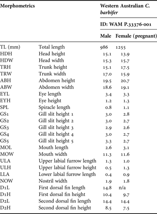
Morphometric measures follow those of White et al. (Reference White, Last and Stevens2007), and are expressed as a percentage of total length (TL) throughout (± standard deviation (SD)), unless otherwise stated (Table 1). Measurements were collected (by Brett Human) from two fresh adult Cirrhigaleus barbifer specimens immediately prior to preservation. Specimens were preserved in 10% formalin (following gut incisions and muscle and cranial injections) for three months followed by a three day soak in water and placed in 75% ethanol for long term storage. Liver tissue samples were taken from both adult specimens prior to preservation, and stored in 100% ethanol. Post-preservation counts were obtained by X-ray for precaudal (monospondylous + diplospondylous to origin of upper lobe of caudal fin) and caudal (centra of the caudal fin) vertebrae; as well as upper and lower tooth row counts. See Table 1 for abbreviation descriptions.
Selected morphometrics expressed as percentages of total length for Western Australian Cirrhigaleus barbifer specimens. Measurements follow the protocol of White et al. (Reference White, Last and Stevens2007). For full morphometric details see online Supplementary Material.

Material examined
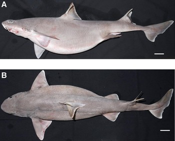
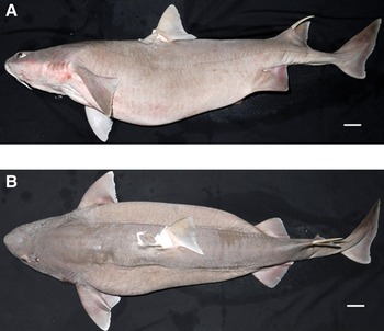
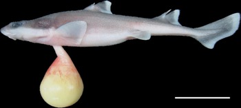
Specimens were collected on 26 February 2011 by a recreational fisherman off the west coast of Rottnest Island in Western Australia (32°03′S 115°21′E; Figure 1) bottom fishing at a depth of 430 m using rod and reel. Specimens were deposited in the Western Australian Museum (WAM) ichthyological reference collection (WAM P.33376.001). Specimens comprised one 986 mm TL mature male (Figure 2) and one 1255 mm TL gravid female (first dorsal spine removed) (Figure 3) with 22 embryos (Figure 4). Embryos were thought to be close to fully developed; ranging in size from 213 to 233 mm.
Map showing the distribution of Cirrhigaleus barbifer ● and C. australis ○.

Cirrhigaleus barbifer Tanaka, 1912, adult male (WAM P.33376.001, 986 mm total length): (A) lateral view; (B) dorsal view. Scale bar = 50 mm.

Cirrhigaleus barbifer Tanaka, 1912, adult female missing first dorsal spine (WAM P.33376.001, 1255 mm total length): (A) lateral view; (B) dorsal view. Scale bar = 50 mm.

Cirrhigaleus barbifer Tanaka, 1912, late stage embryo (WAM P.33376.001, 1 of 22: 225 mm total length): lateral view. Scale bar = 50 mm.

DNA sequencing
DNA isolated from ethanol-preserved liver tissue using DNeasyTM (Qiagen) was used for the polymerase chain reaction (PCR) amplification of a 642 base pairs (bp) fragment of the mitochondrial CO1 gene using the forward (Fish F2) and reverse (Fish R1) primer pair described by Ward et al. (Reference Ward, Zemlak, Innes, Last and Hebert2005). Amplification was carried out using HotStar Taq polymerase (Qiagen) with 35 cycles at an annealing temperature of 48oC for 30 seconds, and an extension temperature of 72oC for 60 seconds and a melting temperature of 94oC for 30 seconds. The resulting fragment was visualized by agarose gel electrophoresis, excised from the gel and isolated with Wizard columns (Promega) using the manufacturer's protocol. After quantification, the amplified DNA was direct sequenced in both directions using the original PCR primers (Fish F2 and Fish R1).
RESULTS
SYSTEMATICS
Order SQUALIFORMES Godrich, 1909
Family SQUALIDAE
Genus Cirrhigaleus Tanaka 1912
Cirrhigaleus barbifer Tanaka 1912
DESCRIPTION
A moderately large dogfish with a fusiform body, a strongly humped nape, a large trunk (TRH: 16.32 %TL ± 1.71 SD; TRW: 16.49 %TL ± 0.78 SD) and abdomen (ABH*: 20.10 %TL ± 0.88 SD; ABW*: 18.84 %TL ± 0.40 SD), and a relatively large head (HDH: 14.49 %TL ± 0.88 SD; HDW: 15.51 %TL ± 0.28 SD). The heterocercal tail has a slight posterior notch to the poorly developed but distinct ventral lobe, and well developed lateral keels, originating slightly posterior to the level of insertion of the second dorsal fin. A depressed head (more flattened dorsally than ventrally) with a dorsally flattened snout, which is narrowly rounded ventrally with a bluntly pointed apex. Small oval eyes (EYL: 3.40 %TL ± 0.07 SD; EYH: 1.25 %TL ± 0.04 SD) and semi-circular spiracles (SPL: 0.95 %TL ± 0.19 SD). Gill openings are long, the first four almost equal in size (GS1–GS4: 2.77–2.89 %TL) followed by a slightly longer fifth (GS5: 3.03 %TL ± 0.45 SD). Large mouth and (MOL: 2.88 %TL ± 0.33 SD; MOW: 11.41 %TL ± 0.21 SD) upper labial furrows much longer (ULA: 1.16 %TL ± 0.16 SD; ULH: 0.89 %TL ± 0.54 SD) than lower (LLA: 0.67 %TL ± 0.36 SD). Small nostrils (NOW: 1.88 %TL ± 0.07 SD) with greatly enlarged barbels extending slightly posterior to the lower jaw margin (anterior to the upper jaw margin in embryos). First dorsal fin origin posterior to pectoral fin insertion; larger than second dorsal fin (D1L: 14.81 %TL ± n/a; D1H: 10.09 %TL ± 0.52 SD; D2L: 14.41 %TL ± 0.01 SD; D2H: 8.01 %TL ± 0.73 SD).
[*Although the female specimen was pregnant the male specimen had a similarly large abdomen.]
TOOTH COUNT
Adult male (left to right): upper jaw—13 + 14 = 27; lower jaw—12 + 12 = 24. Adult female (left to right): upper jaw—15 + 15 = 30; lower jaw—11 + 12 = 23.
VERTEBRAE
113–115 vertebral centra, 49–52 monospondylous, 85–86 precaudal, 28–29 caudal.
COLOUR
Dorsal surface uniformly light grey, ventral surface mostly white, nasal barbels almost uniformly white. Coloration strongly demarcated on head, extending from snout to pectoral-fin base, passing well under eye through the middle of the gill slits. Beyond pectoral fins, coloration becomes less demarcated, the darker dorsal coloration extending more ventrally. Caudal fin, dorsal fins and dorsal surface of the pectoral and pelvic fins light grey with a thin white posterior margin from tip to base. Embryos have much more obvious white posterior margins on all fins. The ventral coloration of the pectoral and pelvic fins slightly darker on posterior half of the fin than on the base.
DNA SEQUENCING
The 642 bp barcode region of the cytochrome oxidase subunit one (CO1) gene from both adult Western Australian Cirrhigaleus specimens was PCR amplified and sequenced (GenBank Accession number KC349854). The sequences are identical to each other and to the GenBank sequence (EU398719 and EU398720) from the two available voucher specimens of C. barbifer (BW-A2280 and BW-A2281), from Indonesia, but show 8 bp differences from the GenBank sequence (DQ108220) of the voucher specimen of C. australis (BW-A137), from Tasmania.
REPRODUCTION
The female specimen was found to exhibit a form of aplacental viviparity (viviparous with a yolk sac), with 22 late stage embryos, 11 in each uterus. Embryos ranged in size from 213–233 mm TL and were not uniformly oriented within the uterus. The embryos appeared to be fully developed, resembling the full adult form, with pigmentation across the entire body and fully developed external features, such as barbels, eyes, and nostrils, although a large amount of yolk still remained (Figure 4). There was no sexual disequilibrium between the embryos with 11 males and 11 females and no size difference found between sexes: males TL (mm): mean 224.91 ± 4.61 SD; females TL (mm): mean 221.73 ± 5.69 SD; t19 = −1.44, P = 0.166.
DISCUSSION
This is the first record of Cirrhigaleus barbifer in Western Australia. The south-eastern Australia species (C. australis) is separated from C. barbifer primarily based on sequence differences in the CO1 gene and also multiple morphological features, including smaller eyes, shorter dorsal–caudal space, and smaller pectoral fins, as outlined by White et al. (Reference White, Last and Stevens2007). It should be noted, however, that only 8 diagnostic base pairs out of 642 bp (1.25%) separate the two species, and so they are very closely related. Specimens collected in Western Australia exhibit an identical CO1 gene sequence to the Indonesian specimens of C. barbifer.
Previously, coloration was highlighted as a characteristic useful in identifying C. barbifer (dark brown dorsally) or C. australis (medium grey dorsally) (White et al., Reference White, Last and Stevens2007). However, the coloration of Western Australian specimens most closely matches C. australis (Figures 2–4). In addition, Western Australian specimens were collected at 430 m depth off the west coast of Rottnest Island (32°S; Figure 1), which corresponds to the depth and upper latitudinal range of C. australis (32°S–48°S).
Given the very close relationship between the two Cirrhigaleus species, it may be challenging to distinguish them based solely on morphological characters. Thus, based on the identical CO1 gene sequences of Indonesian C. barbifer specimens, Western Australian specimens are considered to represent a significant range extension of C. barbifer to Australia. The Western Australian specimens are thought to highlight morphological similarities between the two species.
The female specimen presented here was found to exhibit a form of aplacental viviparity, a reproductive mode common to squaliform sharks. Centrophorus, a genus closely related to Cirrhigaleus, generally exhibit low reproductive fecundity, with a gestation period of one to two years and one to two pups per litter, which attain a maximum total length before birth of up to 361 mm (White & Dharmadi, Reference White2010). Squalus sharks may show a similar gestation period of one to two years, but have larger litters of 2–12 pups, with embryos reaching a smaller maximum total length, up to 300 mm for Squalus acanthias (Yano & Tanaka, Reference Yano and Tanaka1988). Previous records for C. barbifer show a similar litter size to other squalus sharks with a maximum record of 10 pups (Compagno, Reference Compagno1984; Compagno et al., Reference Compagno, Dando and Fowler2004). However, the Western Australian specimens demonstrate that C. barbifer can actually achieve a litter size of at least 22 pups.
ACKNOWLEDGEMENTS
This paper is dedicated to the memory of Brett Human, a good friend and respected chondrichthyan taxonomist who unfortunately passed away during the completion of this work. We thank Steve Downs for collecting, retaining, and presenting the Cirrhigaleus barbifer specimens for scientific investigation. Also thanks to Corey Wakefield (Western Australian Department of Fisheries), for alerting Ryan Kempster to the specimens, Glen Moore (Western Australian Museum) who accessioned and preserved the specimens, and Sue Morrison (Western Australian Museum) for continued help and guidance.
Supplementary materials and methods
The supplementary material referred to in this article can be found online at journals.cambridge.org/mbd.