THE AVERMECTIN/MILBEMYCIN ANTHELMINTICS
The avermectins and milbemycins (A/M), often also referred to as the macrocyclic lactones, are the largest selling anthelmintics in the world. As well as being widely used in veterinary medicine for the treatment of gastro-intestinal nematode infections and ectoparasite infestations (Vercruysse & Rew, 2002), they are used in agriculture for the control of insect pests and in human medicine for the treatment of filarial nematode infections, most notably in the treatment and control of onchocerciasis (Omura & Crump, 2004). This enormous success, which has only very recently been clouded by reports of resistance in veterinary parasites in some parts of the world (Jackson & Coop, 2000; Anziani et al. 2001; Familton, Mason & Coles, 2001; Loveridge et al. 2003), is due to their rapid effects on, and specificity for, the target organisms. Though extremely effective at controlling arthropods as well as nematodes, the A/M are ineffective against flatworms and tapeworms. This article will concentrate on the nematocidal effects of these drugs.
When the A/M anthelmintics are applied to nematodes two main effects, rapid paralysis of movement and of pharyngeal pumping, are observed. As a result, the treated worms are unable to either move or to feed and, for most infections, the paralysed parasites are consequently rapidly removed from the host. The major group of nematodes refractory to A/M treatment, adult macrofilariae, may survive drug treatment because neither movement nor pharyngeal pumping are required for their continued survival in tissues. However, in this case a further effect of the drugs is revealed, a long-lasting reduction in the production of new larvae (Campbell et al. 1983). It is this third effect, coupled with their extreme toxicity to the motile microfilariae, which is responsible for the success of ivermectin treatment in the control of onchocerciasis. These three effects predict that the target of these drugs must have roles in the locomotion, pharyngeal pumping and reproduction of parasitic nematodes.
GLUTAMATE-GATED CHLORIDE CHANNELS
Soon after the introduction of ivermectin, the first A/M to be commercialised, in 1980, electrophysiological experiments showed that the drug caused an increase in the chloride conductance of mammalian neuronal membranes, and that this effect was blocked by picrotoxin (Kass et al. 1980; Supavilai & Karobath, 1981; Graham, Pfeiffer & Betz, 1982; Pong & Wang, 1982). Ivermectin has continued to be the drug used in most published studies on the A/M family, since it is both the largest selling of these compounds and also the least hydrophobic of those readily available. Given the known role of GABA receptors in nematode locomotion (McIntire et al. 1993) and that picrotoxin is a potent GABA-gated chloride channel blocker (Olsen & Tobin, 1990), it was a natural hypothesis that the A/M were acting at GABA receptors, and early data on nematode preparations offered some support for this (Holden-Dye et al. 1988; Martin & Pennington 1989; Holden-Dye & Walker, 1990). However, the concentrations required were rather too high for this effect to be clinically relevant and in some preparations ivermectin inhibited rather than opened GABA receptor channels (Martin & Pennington, 1988; Holden-Dye & Walker, 1990) and, though it is clear that the A/M do have effects on nematode GABA receptors (Feng et al. 2002), attention turned to alternative chloride channels as the likely target.
Experiments in which total mRNA isolated from Caenorhabditis elegans was injected into Xenopus oocytes revealed a glutamate-gated chloride channel (GluCl) that was sensitive to ivermectin (Arena et al. 1991, 1992). Fractionation of this total mRNA into smaller and smaller pools eventually resulted in the identification of two GluCl subunits, designated α and β, from C. elegans (Cully et al. 1994). Expression of the α subunit in the Xenopus oocyte resulted in the appearance of an ivermectin-gated channel, the β subunit produced a glutamate-gated, but ivermectin-insensitive, channel and co-expression of the two subunits resulted in an ivermectin-sensitive, glutamate-gated channel. The properties of this channel were very similar to that produced by the total mRNA preparation, and it was swiftly established that its pharmacology was very similar to that expected for the avermectin target (Arena et al. 1995). The genes encoding these proteins were later designated glc-1 for the GluClα subunit and glc-2 for the GluClβ subunit (glc=glutamate-gated chloride channel).
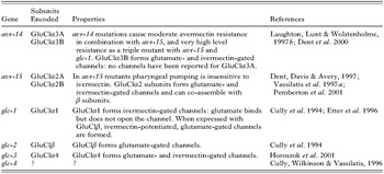
This rather simple picture rapidly became more complex as complementary molecular biological and genetic approaches led to the identification of two further α subunits, GluClα2A and GluClα2B, from C. elegans (Dent, Davis & Avery, 1997; Vassilatis et al. 1997a). These subunits produced channels gated by both glutamate and ivermectin and were products of the alternatively spliced avr-15 (avr=avermectin resistance) gene, providing further evidence that the GluCl were indeed the in vivo avermectin targets. Reporter gene constructs, in which the putative promoter regions of the genes under study are attached to lacZ or GFP (Fire, Harrison & Dixon, 1990), demonstrated that both the GluClα2 and β subunits were expressed in pharyngeal muscle cells and, in the case of the GluClα2 subunits, more widely in the nematode motor nervous system, (Dent et al. 1997; Laughton, Lunt & Wolstenholme, 1997a) consistent with the paralysis of the pharynx and body-wall muscle observed when ivermectin is applied. The genetics of avermectin resistance suggested that this was not the end of the story, since mutations in avr-15 alone did not cause the worms to be drug-resistant and a mutation in a second gene, avr-14 was also necessary for moderate resistance and in a third, glc-1, for high level resistance (Dent et al. 2000). The second gene, avr-14, is identical to the gene also referred to, by us, as gbr-2 (GABA receptor related) (Laughton, Lunt & Wolstenholme, 1997b). It too is alternatively spliced and encodes two subunits, GluClα3A and GluClα3B, which share a common N-terminal half but differ in the C-terminal, channel forming, half of the protein. GluClα3B produces glutamate- and ivermectin-sensitive channels when expressed in Xenopus oocytes, but the GluClα3A subunit has yet to be shown to produce any functional channels (Dent et al. 2000; Rogers & Wolstenholme, unpublished). The complete sequencing of the C. elegans genome allowed bioinformatics to be added to the cDNA cloning and genetic methods previously used. This led to the identification of a fourth α-subunit encoding gene, glc-3: its product, GluClα4, also produced glutamate- and ivermectin-gated chloride channels when expressed in Xenopus oocytes (Horoszok et al. 2001). An additional putative GluCl-encoding gene, glc-4, has also been identified (Cully, Wilkinson & Vassilatis, 1996) that may also be alternatively spliced. The sequence of the predicted glc-4 encoded subunits is rather distant from either the α or β subunits previously described, suggesting that it may belong to a different, γ class. The current list of C. elegans GluCl genes and the subunits they encode is given in Table 1.

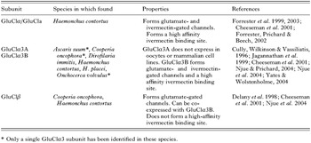
Though the paralytic effects of the A/M on parasitic nematodes had been observed at an early stage, and parasite preparations, especially from Ascaris suum, have been widely used to study the effects of the drugs on non-recombinant targets (Kass et al. 1980; Kass, Stretton & Wang, 1984; Holden-Dye et al. 1988; Martin & Pennington 1989; Holden-Dye & Walker, 1990; Martin, 1996; Adelsberger, Scheur & Dudel, 1997; Brownlee, Holden-Dye & Walker, 1997), progress in the molecular characterization of the GluCl has been much slower in parasites. The sequences of the C. elegans GluCl subunits were used as the basis of a reverse transcriptase-PCR approach using degenerate primers based on highly conserved regions of amino acids. The parasite species most widely studied in this way has been Haemonchus contortus, from which several GluCl cDNAs have been cloned (Delany, Laughton & Wolstenholme, 1998; Forrester et al. 1999; Jagannathan et al. 1999). From this work it is clear that some of these subunits are very similar to those found in C. elegans, including the HcGluClα3A, 3B and HcGluClβ (Hc=Haemonchus contortus) subunits. Others, such as the HcGluCla or α subunit, are less closely related to any particular C. elegans gene product (Yates, Portillo & Wolstenholme, 2003), raising the intriguing possibility that the GluCl gene family varies between nematode species. Of the various subunits described to date, GluClα3 has been found in the largest number of species, with sequences reported from H. placei (Mes, 2004), A. suum (Jagannathan et al. 1999), Cooperia oncophora (Njue et al. 2004; Njue & Prichard, 2004), Dirofilaria immitis (Cully et al. 1996; Yates & Wolstenholme, 2004), and Onchocerca volvulus (Cully et al. 1996). Mutations and polymorphisms in GluClα3 subunits have been associated with avermectin resistance in both C. elegans and C. oncophora (Dent et al. 2000; Njue et al. 2004; Njue & Prichard, 2004) which could be interpreted to suggest that they are important in vivo drug targets. The GluCl subunits currently known in parasitic nematodes are summarised in Table 2.

The sequence of the GluCl subunits clearly revealed them to be members of the ‘Cys-loop’ family of ligand-gated ion channels, closely related to vertebrate GABAA and glycine receptors, and more distantly related to the nicotinic acetylcholine receptors. In fact their closest homologues in vertebrates are probably the glycine-gated chloride channels (Vassilatis et al. 1997b). Each of the subunits has the characteristic topology of 4 membrane-spanning domains with extracellular N- and C-termini. As with other members of this family, the GluCl are predicted to be pentameric structures, with the native channels consisting of more than one subunit type. Table 1 clearly shows that in C. elegans there are more than five predicted subunits and this immediately implies that there must be multiple types of native GluCl, and hence avermectin targets, in this organism, and therefore presumably in other nematodes, each made up of different combinations of subunits and presumably expressed on different tissues. This molecular complexity presents a major challenge to a complete understanding of the A/M's actions, since it is likely that each of these GluCls will have different sensitivities and accessibilities to the drug, and that activation of them will have different effects on the organism. If the potential for variations in the number and expression of the individual subunits are added to the mix, then it is obvious that there is scope for considerable differences in the effects of the A/M between nematode species. The determination of the subunit composition of native GluCl is therefore of major importance, but is not trivial. One advantage may be that nematodes are anatomically rather simple, with relatively few individual cells.
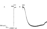
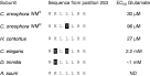
Recombinant nematode GluCl are activated by L-glutamate, but not aspartate, GABA, glycine, histamine or any other amino acid or candidate neurotransmitter. Ibotenate, a conformationally constrained analogue of glutamate, is at least a partial agonist at most GluCl (Cully et al. 1994; Dent et al. 1997; Vassilatis et al. 1997a; Dent et al. 2000; Horoszok et al. 2001; Forrester et al. 2003; Yates & Wolstenholme, 2004). Glutamate activation of most α-subunit- containing receptors produces a rapid opening of the channel that rapidly desensitises (Fig. 1A). If the receptor is composed solely of β subunits, then the receptors do not desensitise, but do close rapidly once agonist is removed (Cully et al. 1994; Njue et al. 2004). The EC50 for activation of recombinant C. elegans GluCl by L-glutamate is usually about 1–2 mM if an α-subunit is present, although the GluClα2B is more sensitive (EC50=0·14 mM) (Cully et al. 1994; Dent et al. 1997; Vassilatis et al. 1997a; Dent et al. 2000; Horoszok et al. 2001), and those composed of β subunits are also more sensitive to the agonist (EC50=0·38 mM) (Cully et al. 1994). There have been fewer quantitative data published for recombinant parasite GluCl: the EC50 for glutamate at the D. immitis GluClα3B receptor is also approximately 1 mM (Yates & Wolstenholme, 2004) but, intriguingly the C. oncophora GluCl subunits are much more sensitive, with the α3 subunit forming a receptor with an EC50 of ~0·03 mM and the β subunit having a EC50 for glutamate of ~0·18 mM (Njue et al. 2004). This increased agonist sensitivity of the GluCl subunits may be conserved in H. contortus, since the HcGluClα subunit forms receptors with an EC50 for glutamate of only 8·4 μM (Forrester et al. 2003) and the HcGluClα3B receptor has a very similar quantitative pharmacology to that from C. oncophora (Rogers & Wolstenholme, unpublished). By analogy with the other members of the ‘cys-loop’ family of ligand-gated ion channels, glutamate would be expected to bind to the extracellular domains of the receptor, possibly at the subunit interface (Cascio, 2004). The agonist-binding site would be made up of six loops, three from each of the adjacent subunits. Consistent with this prediction, a mutation in the extracellular domain of the C. oncophora α3 subunit (L256F) reduced the efficacy of glutamate approximately three-fold and a combination of two polymorphisms in the N-terminal domain (V60A and R100H) abolished the glutamate sensitivity of the β subunit (Njue et al. 2004). The combination of mutations in both subunits reduced the EC50 for glutamate at the α3β heteromeric receptor by 13-fold. It is possible to correlate the amino acid sequence of many GluClα3 subunits around this position with their sensitivity to glutamate (Fig. 2), which may indicate that this part of the subunit either forms part of the binding site or is involved in the efficient coupling of ligand-binding to channel opening.

Fig. 1. Examples of chloride currents produced by activation of recombinant GluCl in Xenopus oocytes. HcGluClα3B cRNA was transcribed in vitro and microinjected into oocytes as described (Yates & Wolstenholme, 2004). Recordings were made 3–5 days later. A) shows the response to 1 mM L-glutamate and B) to 1 μM ivermectin: the horizontal bars above the recordings indicate when agonist was applied. Note the rapid onset and desensitisation of the L-glutamate-induced currents, compared to the slow onset and essentially irreversible current induced by ivermectin.

Fig. 2. Alignment of the partial amino-acid sequence of the GluClα3B subunits from several nematode species, correlated with their sensitivity to activation by glutamate. IVMS and IVMR are ivermectin sensitive and resistant, respectively. Amino-acids that are different from the ivermectin sensitive version of the C. oncophora subunit are shown in white on a black background.
Application of ivermectin to recombinant GluCl produces a markedly different effect from glutamate (Fig. 1B). The channels open very slowly, and the rate of channel opening may be dose-dependent (Rogers & Wolstenholme, unpublished). This channel opening is essentially irreversible, and the channels remain open even when the drug is removed (Cully et al. 1994; Vassilatis et al. 1997a; Horoszok et al. 2001; Forrester et al. 2003; Yates & Wolstenholme, 2004). Ivermectin has no effect on GluClβ subunits (Cully et al. 1994; Njue et al. 2004). Because of the extremely unusual properties of ivermectin and the irreversible channels it opens, it is very difficult to obtain good quantitative data with this drug, but EC50 values in the range of 0·1 to 10 μM have been reported for recombinant GluCl (Arena et al. 1991; Cully et al. 1994; Vassilatis et al. 1997a; Dent et al. 2000; Horoszok et al. 2001; Forrester et al. 2003; Njue et al. 2004). These are rather higher than the concentrations at which the drug shows anthelmintic effects, and also higher than the Kd values (26–100 pM) obtained in radioligand binding experiments to membranes from mammalian cells expressing recombinant H. contortus GluCl (Cheeseman et al. 2001; Forrester, Prichard & Beech, 2002). Very recent experiments have found that the H. contortus GluClα3B subunit can be activated by ivermectin concentrations as low as 0·1 to 1·0 nM (Rogers and Wolstenholme, unpublished), much closer to the concentrations suggested by the binding studies.
One very interesting phenomenon is the interaction between glutamate and ivermectin. The very first paper on the cloning of the GluCl subunits from C. elegans (Cully et al. 1994) showed that concentrations of ivermectin that were too low to directly activate the channels would nonetheless potentiate the effects of simultaneously applied sub-maximal concentrations of glutamate. Similar results were obtained (Martin, 1996) using two-electrode patch clamp recordings from the pharyngeal muscle of A. suum, where milbemycin D caused a dose-dependent potentiation of the observed response to glutamate. A similar result has been obtained using recombinant human glycine receptors: ivermectin acted as an irreversible agonist at higher concentrations (>0·3 μM) but at lower concentrations (30 nM) it allosterically potentiated glycine-gated currents (Shan, Haddrill & Lynch, 2001). Ivermectin has similar allosteric effects at the vertebrate α7 nicotinic receptor (Krause et al. 1998) and here mutations in the TM2 channel domain, believed to change the conformational equilibrium that exist between the active and desensitised states of the receptor, alter ivermectin potentiation. This result was interpreted to suggest that ivermectin binding also changes this equilibrium, leading to an increased probability of channel opening. More recent experiments have also shown the reverse phenomenon, that glutamate potentiates the activity and binding of ivermectin, at least at the HcGluClα receptor (Forrester et al. 2002; Forrester, Beech & Prichard, 2004). Glutamate and ivermectin do not compete for the same binding site and hence have different binding sites on the receptor (Hejmadi et al. 2000) so these data suggest that the two sites exert complementary, and possibly additive, effects on the conformational changes needed for the channels to open. It is possible that interactions between exogenous anthelmintic and endogenous glutamate explain the extraordinary potency of the A/M for killing worms and the much lower concentrations needed to activate native GluCl (Pemberton et al. 2001) than those reported for recombinant channels expressed in Xenopus oocytes (Arena et al. 1991; Cully et al. 1994; Vassilatis et al. 1997a; Dent et al. 2000; Horoszok et al. 2001; Forrester et al. 2003; Njue et al. 2004). Binding of an A/M to a GluCl molecule will both increase the probability of that channel opening but, in addition, greatly stabilise the open state once that opening has taken place, resulting in the essentially irreversible activation observed.
In summary, in vitro molecular cloning and expression of recombinant subunits has provided unequivocal evidence that the GluCl are the nematode targets of the ML anthelmintics. An examination of the in vivo roles of these receptors should explain how the drugs exert their anthelmintic effects, and vice versa.
P-GLYCOPROTEINS AND THE A/M ANTHELMINTICS
It is apparent from the previous section that, in vitro, the A/M do not interact only with the invertebrate GluCl, but also with other invertebrate and vertebrate ligand-gated chloride channels. Is the in vivo selectivity of these compounds for the invertebrate parasites over their vertebrate hosts due solely to a higher affinity at the GluCl over these other channels, or are other factors important? An important finding has been that certain collie dogs are extremely sensitive to treatment with the A/M, and that this sensitivity is associated with mutations in the mdr-1 gene that encodes a P-glycoprotein (Mealey et al. 2001; Roulet et al. 2003). The drugs are extremely good substrates for these pumps in vertebrates and it is likely that their reduced toxicity for host versus parasite is due to their removal from the CNS by non-specific pumps of the blood-brain barrier (Nobmann, Bauer & Fricker, 2001). The A/M are also good substrates for nematode P-glycoproteins (Kerboeuf et al. 2003) and it has been suggested that changes in the expression of these pumps might contribute to drug resistance (Blackhall et al. 1998; Sangster et al. 1999; Drogemuller, Schnieder & von Samson-Himmelstjerna, 2004). However, there are no suggestions that the anthelmintic effects of the A/M are due to their interactions with the P-glycoproteins and it seems this is solely due to their channel-opening properties.
GLUCL IN THE NEMATODE PHARYNX
The nematode pharynx is a muscular organ and its function is to take in and partially process food prior to pumping in into the gut. The structure of this organ varies widely between nematode species and is one of the most characteristic features of their morphology. It contains distinct muscle, nerve and gland cells and is almost a self-contained system (Bird & Bird, 1991). As such, it is amenable to electrophysiological recordings and pharyngeal preparations have been widely used to study the effects of the A/M. It is also easy to observe and measure the rate of pumping. Pumping is a cyclic process and includes a relaxation phase triggered by the glutamatergic inhibitory motor neurone called M3 (Avery, 1993a,b; Raizen, & Avery, 1994; Lee et al. 1999), which implies the presence of inhibitory glutamate receptors on the post-synaptic muscle cells.
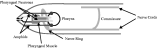
Pharyngeal pumping in nematodes is extremely sensitive to the A/M, and in many species the pharynx is the most sensitive organ to the effects of the drugs, with reported EC50 values of 0·2 to 10 nM (Bottjer & Bone, 1985; Avery & Horvitz, 1990; Geary et al. 1993; Gill et al. 1995; Paiement et al. 1999; Sheriff et al. 2002). This may mean that the primary anthelmintic effect of the A/M is on the pharynx and that its paralysis leads to worm death, either due to starvation or to the loss of internal turgor pressure. The inhibition of pumping is due to the presence of GluCl on pharyngeal muscle cells (Fig. 3) (Martin, 1996; Adelsberger et al. 1997; Pemberton et al. 2001). The irreversible activation of these receptors by ivermectin leads to a depolarisation of the muscle (Pemberton et al. 2001), presumably due to high internal [Cl−], and a cessation of pumping.
In C. elegans, reporter gene experiments have shown that the avr-15 and glc-2 genes, encoding the GluClα2 and β subunits, are indeed expressed in pharyngeal muscle cells (Dent et al. 1997; Laughton et al. 1997a). Pemberton et al. (2001) employed a pharyngeal preparation from wild type and mutant C. elegans to make direct recordings of the effect of glutamate and ivermectin on muscle activity. Some of the properties of these preparations were consistent with the suggestion that the pharyngeal receptor in C. elegans is composed of α2 and β subunits, in particular the loss of ivermectin sensitivity of this preparation in avr-15 mutant worms (Pemberton et al. 2001), but some of the pharmacological details are not. The channels found in avr-15 worms do not possess the picrotoxin sensitivity predicted for a GluClβ receptor and the sensitivity to ivermectin of the preparation is rather higher than that seen for the GluClα2+β combination in the Xenopus oocyte. So is there a third GluCl subunit present in pharyngeal muscle and, if so, which is it? So far the only gene that can be ruled out is avr-14, where neither reporter gene patterns nor the physiological data supports expression in these cells (Dent et al. 2000; Pemberton et al. 2001).
For parasitic species, we have less information. The physiological experiments have been, by and large, carried out on large worms such as A. suum and these clearly indicate the presence of GluCl on pharyngeal muscle, but the only localisation studies have been carried out on H. contortus using antibodies raised against synthetic peptides corresponding to poorly conserved regions of the individual subunits. These studies have not yielded any strong evidence for expression of any H. contortus GluCl subunit in pharyngeal muscle cells, and in particular the HcGluClβ subunit was not found there (Delany et al. 1998), but have suggested the possibility that HcGluClα3B may be expressed in pharyngeal neurones (Portillo, Jagannathan & Wolstenholme, 2003). Irreversible activation of these receptors, which are presumably presynaptic, could explain the inhibition of pumping observed in this species (Geary et al. 1993). However, it may also be that so-far undiscovered GluCl subunits may be present on H. contortus pharyngeal muscle: the absence of the HcGluClβ subunit could well mean that the composition, and hence pharmacology, of any such receptor will differ from that of its C. elegans counterpart.

Fig. 3. Schematic representation of the distribution of GluCl in nematodes. The cuticle is outlined in grey and the pharynx in black. Structures reported to express GluCl are indicated by arrows.
GLUCL AND THE CONTROL OF LOCOMOTION
The second major effect of the A/M on nematodes is an apparent paralysis of body-wall muscle, rendering them immobilised. However, all the evidence points to this being an indirect effect, rather than a direct inhibition of neuromuscular transmission and there are no indications that GluCl are expressed in muscle cells. Locomotion in nematodes is controlled by both excitatory and inhibitory motor neurones, organised into ventral and dorsal nerve cords, each of which innervate body-wall muscle. Waves of reciprocal excitation and inhibition pass down the body, so that the dorsal muscles are relaxed as the ventral muscles are contracted, and vice versa. This results in the characteristic sinusoidal swimming motion. Studies on C. elegans showed that the motor neurones are in turn controlled by command interneurones in the head of the worm that regulate the rate of locomotion and also the frequency at which the worm reverses and moves backwards (White et al. 1986; Zheng et al. 1999).
Early reports showed that, in A. suum, avermectin blocked transmission between interneurones and excitatory motor neurones in the ventral cord, and also ventral inhibitory transmission (Kass et al. 1980, 1984). This would predict that GluCl subunits are expressed on those motor neurones and this prediction has been confirmed in C. elegans, using reporter gene constructs, and H. contortus, using antibodies (Fig. 3). Both avr-14 and avr-15 are expressed in C. elegans motor neurones (Dent 1997, 2000) and, in H. contortus, the HcGluClα, α3A, α3B and β subunits have all been detected on motor neurones (Delany et al. 1998; Jagannathan et al. 1999; Portillo, Jagannathan & Wolstenholme, 2003). All of the H. contortus subunits were detected on motor neurone commissures, structures connecting the dorsal and ventral nerve cords and which form synapses with interneurones on the lateral and sub-lateral nerve cords. The use of anti-GABA antibodies suggested that these were inhibitory motor neurons (Portillo et al. 2003). Application of A/M to channels on these inhibitory motor neurones would therefore presumably result in an irreversible hyperpolarisation of the cells and their consequent inability to produce action potentials. This would prevent inhibitory transmission at the neuromuscular junction and hence the abolition of the waves of muscular relaxation required for movement.
In addition, some GluCl subunits have been detected on interneurones, which might represent the command interneurones. The C. elegans avr-14 gene is expressed in extra pharyngeal head neurones (Dent et al. 2000) and use of an antibody that recognised both GluClα3 subunits produced immunofluorescence in the nerve ring of H. contortus (Jagannathan et al. 1999). Direct confirmation that GluCl are present in at least some command interneurones was provided by the detection of a glutamate-gated chloride current in the AVA interneurone of C. elegans using in vivo recording techniques (Mellem et al. 2002). Mutations in the excitatory glutamate receptors also found in these neurones reduce the frequency at which the worms reverse direction (Brockie et al. 2001). We have examined the effects of mutations in several GluCl genes on this behaviour and have found the opposite phenotype, that is the worms reverse more frequently and the durations of the forward movements are reduced (N. Aptel, A. Cook, L. Holden-Dye & A. Wolstenholme, unpublished). The GluCl thus have a major role in regulating nematode locomotion and it is not surprising that their irreversible activation by A/M anthelmintics causes an effective paralysis.
GLUCL IN OTHER ORGANS
The A/M anthelmintics do not efficiently kill some tissue-dwelling adult worms, especially the macrofilariae. Nonetheless, they are effective for the treatment and prophylaxis of filarial infections such as Onchocerca volvulus due to their microfilariacidal effects and because they dramatically reduce the production of new microfilariae for several months (Klager et al. 1993; Lok et al. 1995). Similar effects on fecundity have been observed in other species (Petersen et al. 1996), implying that the GluCl have a role in reproduction. C. elegans carrying mutations in some GluCl genes do show a reduction in egg production (N. Aptel, V. Portillo & A. Wolstenholme, unpublished), but the nature of that role is currently unknown. No GluCl have been reported to be present on any reproductive tissue.
An intriguing recent suggestion is that GluCl may be present on sensory neurones, or involved in the pathways linking sensory stimulation to behavioural effects. Studies on Brugia pahangi have shown that ivermectin can block physiological responses to chemical stimuli in amphids (Perry, 2001; Rolfe, Barrett & Perry, 2001) and, in H. contortus, the HcGluClα3A subunit was detected in a structure that closely resembled a sensory neurone (Portillo et al. 2003). This is even more intriguing in light of the finding that ivermectin-resistant H. contortus have defects in amphid structure (Freeman et al. 2003): amphids are the sensory organs present in the nematode head that contain the sensory neurones. One of their functions would be expected to be to detect the stimuli that allow adult filaria to locate each other and mate and there are suggestions that ivermectin interferes with this process (Duke et al. 1992). In C. elegans, mechanical stimulation inhibits pharyngeal pumping in adult worms and it was suggested that GluCl containing the avr-14 and avr-15 gene products were expressed in the inhibitory pathway linking mechanosensation to pharyngeal pumping (Keane & Avery, 2003). There is therefore considerable evidence that the GluCl play a role in sensory signalling in nematodes, but as yet it is not clear how important this role is as a target for the A/M anthelmintics. One possibility is that the amphids act as a route of entry for the drugs into the worm.
CONCLUSIONS
It is generally accepted that the molecular targets of the avermectin/milbemycin anthelmintics are the glutamate-gated chloride channels. The number of identified GluCl subunits indicates that nematodes contain multiple forms of these channels, which may differ in their sensitivity to the current drugs, and at least some of these are expressed in the nematode neuromuscular system. The elucidation of the subunit structure of these different forms and its relationship to drug sensitivity remains a major challenge. The GluCl are widely expressed in the nematode nervous system (summarised in Fig. 2) and most of the effects of the A/M, with the possible exception of the reproductive effects, can be explained by this distribution. The implication is that these drugs can kill, or damage, nematodes in a variety of ways and, given that there are multiple forms of GluCl, the most important mechanism may vary between species. This has important implications for the development of resistance, a topic outside the scope of this review, which is becoming a major concern in veterinary parasites (Kaplan, 2004). The mechanisms of A/M resistance are still somewhat controversial (Wolstenholme et al. 2004), but it may be that the multiplicity of GluCl subtypes and functions may be reflected in a similar multiplicity of resistance mechanisms.
ACKNOWLEDGEMENTS
Work on the nematode GluCl in the authors’ laboratory is and has been funded by the BBSRC (awards 86/GAN13134 and BBS/B/07594) and the Wellcome Trust (award 061043).