Introduction
Permanent cardiac pacing is a safe and effective treatment for patients with symptomatic bradyarrhythmias. The vast majority of pacemakers are implanted in older people; the mean age of patients receiving a first pacemaker is 75 years; over 85% of pacemakers are implanted in patients over 65 years old and approximately a third are implanted in patients aged over 80 years, a proportion that continues to increase as the population ages.Reference Birnie, Williams, Guo, Mielniczuk, Davis, Lemery, Green, Gollob and Tang1 A knowledge of the evidence and issues related to pacing therapy in older patients is therefore important for physicians caring for older people. This review aims to provide a clinician's guide to the essential aspects of modern pacemaker therapy in older patients, including bradycardia aetiology, indications for pacemakers, and pacing system function. We will address the equity and under-provision of pacing therapy with particular reference to older patients. We also review clinical outcomes following pacing and ethical and end of life considerations with these devices. PubMed searches using the subject headings cardiac pacing and elderly were used in addition to detailed review of national and international guideline documents.
Pathophysiology of bradyarrhythmias in older people
Diseases that disturb the cardiac conduction system at any point may lead to bradyarrhythmias, for which the only effective long-term treatment is cardiac pacing. The commonest indications for pacing are ‘sinus node disease’ (SND) and ‘atrioventricular (AV) block’, which together account for more than 97% of pacemaker implantations.Reference Birnie, Williams, Guo, Mielniczuk, Davis, Lemery, Green, Gollob and Tang1 These clinical syndromes of heterogeneous aetiology occur with much higher frequency in elderly populations. It is not uncommon for SND and AV block to co-exist in older patients.
Atrioventricular block
AV block may be caused by disturbance of conduction at the level of the AV node, the bundle of His, or the bundle-branch Purkinje system. Acquired AV block in the older patient is characterized by increased collagen fibre deposition around the AV node and His-Purkinje system and abnormalities of apoptosis have been reported.Reference James2 There are a number of secondary causes of AV block, which often occur more frequently in elderly populations, including coronary disease, annular calcification from valvular heart disease, particularly aortic stenosis, infiltrative cardiac disorders, inflammatory disorders, malignancy and trauma (particularly following cardiac valve replacement surgery). Sometimes AV block may be deliberately induced by catheter ablation to control the ventricular rate in refractory atrial tachyarrhythmias with a strategy of permanent pacing to maintain a stable heart rate.
AV block is classified according to the level and degree of the block.Reference Da Costa, Brady and Edhouse3 First-degree AV block occurs when there is delay in conduction from the atria to the ventricles, and is manifest by prolongation of the PR interval on the surface ECG, with maintenance of a 1:1 relationship between the P waves and the QRS complexes. A relationship between P waves and QRS complexes is maintained in second-degree AV block, although it is no longer 1:1. Mobitz type I second-degree AV block describes intermittent failure of AV nodal conduction, with increasing prolongation of the PR interval, until a QRS complex is dropped. This often occurs at the level of the AV node and is also known as the Wenckebach phenomenon. In Mobitz type II block there are also dropped P waves, but the PR interval in conducted beats remains constant. The QRS complex is often wide, and the level of block is usually within the His-Purkinje system. When 2:1 or 3:1 block is observed, it is not usually possible to characterize it further; and it is sometimes called high grade AV block. Third-degree AV block occurs when there is complete dissociation between the P waves and the QRS complexes, and is also known as complete heart block.
Conduction system disease may be fixed, e.g. when there is chronic complete heart block, and it may then be associated with fatigue, exercise intolerance, heart failure, or other consequences of hypoperfusion. High grade and complete AV block may also be intermittent, and may then be associated with dizzy spells, falls, or syncope. Complete heart block may be intermittent in as many as two-thirds.Reference Kojic, Hardarson, Sigfusson and Sigvaldason4, Reference Shaw and Kekwick5 It may also be asymptomatic.
Escape rhythms originating from other parts of the conduction system usually occur in patients with complete AV block, the rate and QRS duration depending on the anatomical site of the escape rhythm. Rhythms arising proximally within the specialized conducting tissue tend to produce a narrow QRS complex typically at a rate of 45–60 per minute, while those arising more distally produce a wide QRS complex at a slower rate.
Sinus node disease
SND (or sick sinus syndrome) occurs when there is a problem with the generation of the action potential within the sinus node, producing a heart rate that is inappropriate for physiological requirements. This may result in resting bradycardia, inability to increase heart rate with exertion (‘chronotropic incompetence’), or pauses due to sinus arrest or exit block. It may be associated with tachycardias, when it forms part of the tachy-brady syndrome.Reference Brignole6 Ageing is associated with a progressive loss of P cells, which are the main pacemaker cells of the sinus node,Reference Shiraishi, Takamatsu, Minamikawa, Onouchi and Fujita7 and varying degrees of fibrosis and fat deposition within the node.Reference Shiraishi, Takamatsu, Minamikawa, Onouchi and Fujita7, Reference Bharati and Lev8 SND may also be secondary to other disease processes that are often more common in elderly people, including hypothyroidism, coronary disease, infiltrative cardiac disorders (haemochromatosis, amyloid), inflammatory disorders (pericarditis, myocarditis, connective tissue diseases), malignancy, and trauma. Older people are also at higher risk for the development of sinus node dysfunction due to extrinsic causes, in particular rate-limiting medications. Drugs such as beta-blockers, calcium channel blockers, and amiodarone are used more frequently in older people for co-morbidities such as hypertension, coronary disease and atrial fibrillation (AF), and older people tend to be more sensitive to the effects of such drugs.
Epidemiology of bradyarrhythmias
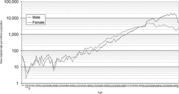
Epidemiological data on the incidence and prevalence of bradycardia, either symptomatic or not, are few. Much of the information available actually comes from series or registers of paced patients. Figure 1 shows the pacemaker implantation rate for each age decade in England and Wales. It is obvious that the need for pacing increases with age, and twice as many men receive pacemakers as do women. The need for pacing is therefore critically age and sex dependent.
Graph illustrating new implant rates (per million population) of permanent pacemakers in the UK according to age and gender. Data are from 2006, courtesy of Dr David Cunningham, Central Cardiac Audit Database, NHS Information Centre.

Studies of older people describe the prevalence of first-degree AV block as 12–17%; second-degree AV block 1–2%, and complete heart block 1–2%.Reference Rajala, Kaltula, Haavisyo and Mattila9–Reference Lakkireddy, Clark and Mohiuddin11 In addition, 5% of the oldest old are already paced.Reference Kelley, Stellingworth, Broyles and Glancy10 The incidence of left bundle branch block is 3.2/10,000/year in men;Reference Kojic, Hardarson, Sigfusson and Sigvaldason4 of these, 2.3% will receive pacemakers during a 5–10 year follow-up period.Reference Miller, Hodge and Hammill12 The prevalence of rhythms associated with sick sinus syndrome is 1–6%.
The only robust epidemiological data in the UK come from the Devon Heart Block and Bradycardia Survey, which was completed over 30 years ago.Reference Shaw and Kekwick5 The incidence of complete heart block was 97/10Reference Brignole6 and of sick sinus syndrome 108/10Reference Brignole6. The number identified potentially requiring pacing therapy was equivalent to 159–174/10Reference Brignole6 (these figures are not age and sex adjusted). The prevalence of AV block in men aged over 80 years exceeded 5000/10Reference Brignole6, and the prevalence of SND was twice as much as that of AV block, implying a total requirement for pacing in the over-80s of 15,000/10Reference Brignole6 (which is actually still more than the pacing rate in this age group now).
The prevalence of bundle branch block increases with age and is estimated at 1% of the population aged 35 and approximately 17% at age 80.Reference Vardas, Auricchio, Blanc, Daubert, Drexler, Ector, Gasparini, Linde, Morgado, Oto, Sutton and Trusz-Gluza13 In patients without syncope, the rate of progression to high-degree AV block is low (0.6–0.8% per year)Reference McAnulty, Rahimtoola and Murphy14, Reference Englund15 and there is no non-invasive technique with a high predictive value.
Equity of provision of pacing
Pacing rates in western Europe, where the age structure of each country's population and economic factors are relatively similar, vary widely.Reference Ector and Vardas16 The reasons for this are not clear. Similar widespread variation is also seen within the United Kingdom, even after correction of implantation rates to take into account the age and sex structure of local populations.Reference Ector and Vardas16, 17 So far there is no evidence that this is related to economic factors or local purchasing decisions.
Potential barriers to access to pacing
The only factors described which might influence pacing rates include concern about co-morbidities,Reference Thomas18 and lack of knowledge of pacing indications and difficulty with ECG interpretation in those to whom these patients present.Reference Cunnington, Plummer and McComb19, Reference Cunnington, Plummer and McComb20
Age
Figure 1 suggests that the pacing rate falls in the oldest old, implying that some of these patients are inappropriately not receiving pacemakers. It is not known whether this is due to failure of referral for pacing, or refusal of pacing. It is increasingly apparent that age itself should never be considered a barrier to pacing which improves quality of life.
Co-morbidity
Similarly, co-morbidities should not necessarily preclude pacing, particularly when not immediately life threatening. Thomas expressed surprise at the ease with which an elderly woman with a clear indication for pacing (complete heart block with symptoms) was accepted for pacing, because she had dementia.Reference Thomas18 Obviously such decisions need to be made on a case-by-case basis, but the default decision should probably be to pace, rather than not to, because of the improvement in quality of life offered by pacing.
Lack of knowledge
Recent work from the UK has suggested that failure to recognize pacing indications may contribute to relatively low pacing rates.Reference Cunnington, Plummer and McComb19, Reference McComb and Plummer21 Roughly a third of patients who received pacemakers had a previous pacing indication which did not trigger a pacing referral, and by inference, not all patients who might benefit from pacing are referred for it. This resulted in significant delays to pacing, often with ongoing symptoms, additional hospitalizations, and unnecessary investigations.Reference Cunnington, Plummer and McComb19 A survey of the knowledge of pacing indications amongst those in hospital to whom potential pacemaker patients present suggested that physicians require further help in determining which patients should be referred for pacing.Reference McComb and Plummer21
Indications for pacing
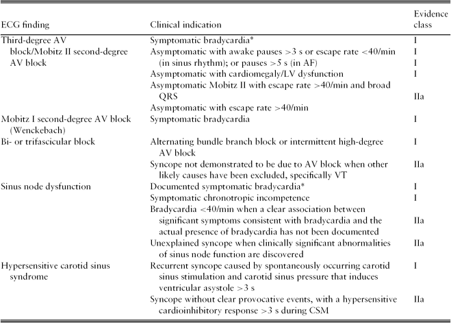
The European Society of Cardiology (ESC) and the American College of Cardiology/American Heart Association/Heart Rhythm Society (ACC/AHA/HRS) have published excellent detailed guidelines for cardiac device based therapy based on the available literature.Reference Vardas, Auricchio, Blanc, Daubert, Drexler, Ector, Gasparini, Linde, Morgado, Oto, Sutton and Trusz-Gluza13Reference Epstein, DiMarco, Ellenbogen, Estes, Freedman, Gettes, Gillinov, Gregoratos, Hammill, Hayes, Hlatky, Newby, Page, Schoenfeld, Silka, Stevenson, Sweeney, Smith, Jacobs, Adams, Anderson, Buller, Creager, Ettinger, Faxon, Halperin, Hiratzka, Hunt, Krumholz, Kushner, Lytle, Nishimura, Ornato, Page, Riegel, Tarkington and Yancy22 A simplified summary of some of the more common pacing indications is presented in Table 1. Essentially, pacing is indicated in all patients with symptomatic bradycardia. There are some conditions, such as advanced AV block, which carry a risk of progression and of life threatening bradyarrhythmias and pacing is indicated regardless of symptoms.
A simplified summary of some of the common pacing indicationsReference Vardas, Auricchio, Blanc, Daubert, Drexler, Ector, Gasparini, Linde, Morgado, Oto, Sutton and Trusz-Gluza13, Reference Epstein, DiMarco, Ellenbogen, Estes, Freedman, Gettes, Gillinov, Gregoratos, Hammill, Hayes, Hlatky, Newby, Page, Schoenfeld, Silka, Stevenson, Sweeney, Smith, Jacobs, Adams, Anderson, Buller, Creager, Ettinger, Faxon, Halperin, Hiratzka, Hunt, Krumholz, Kushner, Lytle, Nishimura, Ornato, Page, Riegel, Tarkington and Yancy22
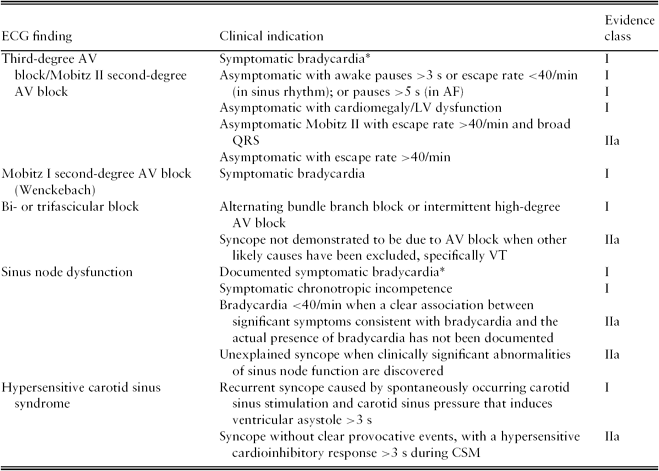
*Bradycardia may be iatrogenic as a consequence of long term drug therapy. Class I: evidence and/or general agreement that pacing is beneficial, useful and effective and should be performed; class IIa: conflicting evidence and/or divergence of opinion about the usefulness of pacing, but weight of evidence/opinion is in favour of benefits outweighing risks and it is reasonable for pacing to be performed. AV block, atrioventricular block; LV, left ventricle; AF, atrial fibrillation; VT, ventricular tachycardia; CSM, carotid sinus massage.
Atrioventricular block
Several studies have shown that permanent pacing improves survival in patients with acquired complete AV block. SiddonsReference Siddons23 showed that patients with complete AV block who received single chamber pacemakers had a survival curve not much different from that of the general population, matched for age and sex, while a similar group with complete AV block, who did not receive pacemakers, had a much higher mortality. Neither the presence of syncope, nor whether the AV block was established or intermittent, affected outcome.Reference Ginks, Leatham and Siddons24
Mobitz type II second-degree AV block has a high incidence of progression to complete heart block (particularly if the QRS complexes are wide, as there is usually advanced disease within the His-Purkinje system, and any escape rhythm may be slow and unreliable) and of symptom development.Reference Dhingra, Denes, Wu, Chuquimia and Rosen25, Reference Shaw, Kekwick, Veale, Gowers and Whistance26 Pacing is therefore indicated for both complete AV block and Mobitz II AV block even in the absence of symptoms. Mobitz type I (Wenckebach) second-degree AV block has a lower rate of progression to advanced AV block and therefore clinical guidelinesReference Vardas, Auricchio, Blanc, Daubert, Drexler, Ector, Gasparini, Linde, Morgado, Oto, Sutton and Trusz-Gluza13, Reference Epstein, DiMarco, Ellenbogen, Estes, Freedman, Gettes, Gillinov, Gregoratos, Hammill, Hayes, Hlatky, Newby, Page, Schoenfeld, Silka, Stevenson, Sweeney, Smith, Jacobs, Adams, Anderson, Buller, Creager, Ettinger, Faxon, Halperin, Hiratzka, Hunt, Krumholz, Kushner, Lytle, Nishimura, Ornato, Page, Riegel, Tarkington and Yancy22 suggest pacing is not routinely indicated (when the level of block has been shown to be above the His-Purkinje system) unless it is associated with symptoms. Wenkebach AV block has, however, been associated with increased mortality, especially in older patients, and benefit from pacing has been demonstrated.Reference Shaw, Kekwick, Veale, Gowers and Whistance26, Reference Shaw, Gowers, Kekwick, New and Whistance27 Pacing should therefore be considered certainly when the patient has symptoms, and perhaps prophylactically in the older patient. Pacing is not generally considered for isolated first-degree AV block, although markedly prolonged PR intervals (>300 ms) sometimes lead to symptoms due to loss of AV synchrony (similar to pacemaker syndrome, where atrial contraction occurs when the AV valves are closed, resulting in cannon waves in the neck, and palpitation, fatigue and exercise intolerance) that may respond to pacing.Reference Barold28 In AF, pauses of at least 5 s in awake, symptom-free patients indicate significant AV conduction defects and are a pacing indication. Shorter pauses in AF associated with symptoms are also a pacing indication.
Fascicular blocks
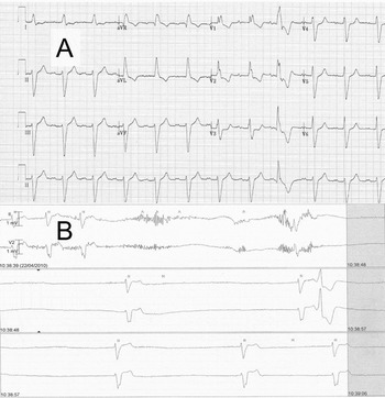
Bifascicular block refers to an electrocardiographic picture of right bundle branch block associated with block in the anterior or posterior fascicles of the left bundle (manifest as an axis deviation), or of left bundle branch block alone.Reference Da Costa, Brady and Edhouse3, Reference Vardas, Auricchio, Blanc, Daubert, Drexler, Ector, Gasparini, Linde, Morgado, Oto, Sutton and Trusz-Gluza13 Trifascicular block is used to describe conduction disease in the right bundle and both fascicles of the left bundle. It may manifest as alternating bundle branch block, right bundle branch block with alternating right and left axis deviation, implying block in both fascicles of the left bundle, or bifasciclar block with first-degree AV block.Reference Da Costa, Brady and Edhouse3 An example is shown in Figure 2. Asymptomatic bi- or trifascicular block is not considered an indication for pacing. However, once syncope has occurred, there is a high rate of progression to high grade AV block (19% per year),Reference Tabrizi, Rosenqvist, Bergfeldt and Englund29 particularly if the QRS is ≥120 ms at presentation (75% within 2 years).Reference Kennebäck, Tabrizi, Lindell and Nordlander30 Pacing is therefore indicated when other likely causes of syncope have been excluded. The other likely cardiac cause of syncope in this situation is ventricular tachycardia, and pragmatic use of echocardiography may be helpful; ventricular tachycardia is uncommon if ventricular function is normal or near normal, and bradycardia may be assumed, if syncope is thought to be cardiac. Studies of contemporary UK pacing suggest low awareness amongst physicians of this pacing indication and found evidence that pacing may be delayed unnecessarily in patients with syncope in the setting of bifascicular block, with consequent additional morbidity.Reference Cunnington, Plummer and McComb19, Reference McComb and Plummer21, Reference Cunnington, Plummer, McDiarmid and McComb31
A, the 12-lead ECG of a 76-year-old patient who presented following an episode of syncope and shows RBBB, left axis and first-degree heart block (trifasicular block). B, the telemetry recording from an episode of dizziness whilst on the cardiology ward showing an episode of complete heart block with a slow broad complex ventricular escape rhythm at a rate less than 30 b.p.m. A dual chamber permanent pacemaker was inserted.

Torsade de pointes
Torsade de pointes (twisting of the points) is an unusual form of polymorphic ventricular tachycardia associated with congenital or acquired lengthening of the QT interval.Reference Viskin32 It may be associated with bradycardia, in particular complete heart block.Reference Kawasaki, Machado, Reinoehl, Fromm, Baga, Steinman and Lehmann33 It may be associated with hypokalaemia or hypocalcaemia, anti-arrhythmic drug therapy or with psychotropic drugs, and is commoner in older people, particularly women.Reference Kawasaki, Machado, Reinoehl, Fromm, Baga, Steinman and Lehmann33 It is an indication for urgent pacing.
Sinus node disease
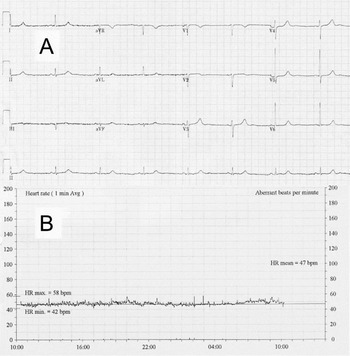
Symptomatic SND is an indication for pacing. Pacing in SND contributes more to relieving symptoms and reducing episodes of AF than to a definite reduction in mortality,Reference Vardas, Auricchio, Blanc, Daubert, Drexler, Ector, Gasparini, Linde, Morgado, Oto, Sutton and Trusz-Gluza13, Reference Shaw, Holman and Gowers34 and hence sinus node dysfunction in the absence of symptoms attributable to bradycardia does not constitute a pacing indication. Symptom-rhythm correlation should be sought, with an ECG recorded during symptoms where possible. Even when a clear association between symptoms and bradycardia has not been documented, it is reasonable to consider pacing when there are other markers of SND, e.g. periods of junctional rhythm, sinus bradycardia (≤40 b.p.m.) or chronotropic incompetence.Reference Vardas, Auricchio, Blanc, Daubert, Drexler, Ector, Gasparini, Linde, Morgado, Oto, Sutton and Trusz-Gluza13, Reference Thomas, Plummer, Turley and McComb35 Exercise intolerance may be associated with chronotropic incompetence. This can be easily demonstrated by encouraging the patient to exercise during ambulatory monitoring (see Figure 3), or by formal exercise stress testing, when the heart rate will fail to rise above 100 b.p.m. during exercise.
A, the 12-lead ECG of an 80-year-old patient presenting with dyspnoea and exercise intolerance. B, the heart rate trend over 24 hours during holter monitoring showing very poor heart rate variability with a maximum rate of 58 b.p.m. despite being active during this period. This is consistent with chronotropic incompetence secondary to sinus node disease and since the patient was not receiving any rate-limiting medications, a dual chamber permanent pacemaker was inserted.

Drug-associated bradycardia
It is important to note that both AV block and sinus bradycardia can occur in patients taking medications which may affect heart rate. In some instances the association is not causal, and pacing is therefore indicated. Patients who experience Mobitz II or complete heart block whilst taking beta-blockers or non-dihydropyridine calcium channel blockers (verapamil and diltiazem) have a high chance of recurrence of AV block even with cessation of the ‘culprit’ medication.Reference Kennebäck, Tabrizi, Lindell and Nordlander30, Reference Zeltser, Justo, Halkin, Rosso, Ish-Shalom, Hochenberg and Viskin36 This suggests that these medications unmask rather than cause AV block and pacing should be considered for high-degree AV block in this setting without a trial of stopping the medication.
When the relationship between drug and bradycardia is probably causal, pacing may be considered to allow continuation of a clinically useful drug. Bradycardias may occur as a consequence of long-term drug therapy required for another indication (such as control of tachyarrhythmias) which cannot easily be stopped. In particular, the use of amiodarone in AF is associated with an increased risk of pacemaker insertion, particularly in women, and when there is a previous history of SND.Reference Essebag, Reynolds, Hadjis, Lemery, Olshansky, Buxton, Josephson and Zimetbaum37 The risk is dose related, and the need for pacing occurs relatively soon after initiation of therapy.Reference Essebag, Hadjis, Platt, Abrahamowicz and Pilote38 Digoxin is also associated with an increase in the need for pacing in these patients.Reference Essebag, Hadjis, Platt and Pilote39 Newer drugs such as donezepil used to treat early Alzheimer's disease may be associated with a risk of sinus node and AV block and again, patients may be considered for pacing to allow continuation of a drug with clinical benefit.Reference Hernandez, Farwell, Cantor and Lawler40, Reference Gill, Anderson, Fischer, Bell, Li, Normand and Rochon41
Neurocardiogenic syndromes
Neurocardiogenic syncope encompasses a spectrum of entities in which triggering of an autonomic neural reflex produces acute and inappropriate vasodilatation and/or bradycardia.Reference Sutton and Petersen42, Reference Grubb43 It includes vasovagal syncope, carotid sinus syndrome, and syncope related to cough, deglutition and micturition. Although neurocardiogenic syncope may be accompanied by bradycardia, there is frequently a substantial vasodepressor element which does not respond to pacing and there has been controversy over the benefit of pacemaker implantation for these conditions.
Vasovagal syncope
Vasovagal syncope is being recognized increasingly commonly in older people.Reference Tan and Parry44 The typical vasovagal prodrome of nausea and diaphoresis is more likely to be absent in older patients.Reference Tan and Parry44 It accounts for up to 50% of all admissions with syncope.Reference Ammirati, Colivicchi and Santini45–Reference Disertori, Brignole and Menozzi47 The role of pacing in vasovagal syndromes is not clear.Reference Sud, Massel, Klein, Leong-Sit, Yee, Skanes, Gula and Krahn48 The early trials, randomizing patients who had vasovagal syncope with bradycardia to receive either pacemaker implantation or no pacemaker implantation, reported lower rates of syncope in the paced groups.Reference Sutton, Brignole, Menozzi, Raviele, Alboni, Giani and Moya49, Reference Connolly, Sheldon, Roberts and Gent50 However, two subsequent double-blind trials in which all patients received a pacemaker which was randomly programmed to pacing on or off showed no reduction in syncope with pacing, suggesting that there is a significant and possibly useful placebo effect of pacemaker implantation.Reference Connolly, Sheldon, Thorpe, Roberts, Ellenbogen, Wilkoff, Morillo and Gent51, Reference Raviele, Giada, Menozzi, Speca, Orazi, Gasparini, Sutton and Brignole52 It should be noted that, although older patients were not specifically excluded from these trials, the mean age of subjects recruited was 55 years,Reference Sud, Massel, Klein, Leong-Sit, Yee, Skanes, Gula and Krahn48 much younger than the average paced patient. Pacing is therefore not indicated as first-line therapy for neurocardiogenic syncope but may be considered in some patients with refractory symptoms and a marked cardioinhibitory component.Reference Vardas, Auricchio, Blanc, Daubert, Drexler, Ector, Gasparini, Linde, Morgado, Oto, Sutton and Trusz-Gluza13
Carotid sinus hypersensitivity
Carotid sinus hypersensitivity (CSH) is characterized by a ventricular pause greater than 3 s and fall in systolic blood pressure of greater than 50 mmHg in response to carotid sinus massage (CSM). Between 25 and 40% of elderly patients with syncope or unexplained falls exhibit CSH,Reference Dey, Stout and Kenny53, Reference Richardson, Bexton, Shaw and Kenny54 but reproduction of syncope with these findings is required to define the carotid sinus syndrome. There is evidence to suggest that patients who have carotid sinus syndrome may have amnesia for loss of consciousness and will therefore present with falls rather than syncope.Reference Parry, Steen, Baptist and Kenny55 Two trials have suggested a reduction in syncope following pacemaker implantation in patients with falls/syncope and an abnormal response to CSM.Reference Brignole, Menozzi, Lolli, Bottoni and Gaggioli56, Reference Kenny, Richardson, Steen, Bexton, Shaw and Bond57 However, both trials were unblinded (a pacemaker was implanted or not) and so did not include a placebo control. Guidelines, based on these data, suggest that permanent pacing is indicated in patients with recurrent syncope whose symptoms are reproduced by CSM associated with ventricular asystole of more than 3 s (during which the patient may be syncopal or pre-syncopal), particularly if usual symptoms appear to be precipitated by inadvertent carotid sinus pressure. However, the only blinded placebo-controlled trial, published recently, has not confirmed benefit from pacing. It included 25 subjects with unexplained falls and an abnormal response to CSM.Reference Parry, Steen, Bexton, Tynan and Kenny58 Patients all received pacemakers which were programmed randomly either to pace or not, with crossover to the other mode after 6 months. In this small study, the pacing intervention had no effect on the number of falls, raising the possibility that much of the positive effect of previous studies may have been placebo. The same group now suggest that CSH may be an age-related epiphenomenon, rather than a cause of syncope.Reference Tan, Newton, Reeve, Murray, Chadwick and Parry59 Further work in this controversial area will be of interest.
Cardiac resynchronization therapy
Cardiac resynchronization therapy (CRT) or biventricular pacing, with pacing leads positioned in the right atrium, right ventricle and left ventricle (usually via the coronary sinus, the branches of which run over the left ventricular epicardium) has also been shown to improve symptoms and survival in patients with advanced heart failure and cardiac dyssynchrony due to intraventricular conduction delay.Reference McAlister, Ezekowitz, Hooton, Vandermeer, Spooner, Dryden, Page, Hlatky and Rowe60 Modelling from trial data has shown that CRT is cost effective in all age groups, including those over 75.Reference Yao, Freemantle, Calvert, Bryan, Daubert and Cleland61 Further discussion of the indications and benefits of CRT is beyond the scope of this review.
Outcome after pacing in older people
Mortality after pacing
Pacing reduces mortality in patients with AV block,Reference Siddons23, Reference Shaw, Kekwick, Veale, Gowers and Whistance26, Reference Shaw, Gowers, Kekwick, New and Whistance27 when compared with patients who did not receive pacemakers, but not in patients with SND.Reference Shaw and Kekwick5 However, despite this lack of reduction in mortality, older patients who receive pacemakers still have a life expectancy of some years. The proportion of older patients over 80 years of age who receive a pacemaker has increased in recent years from 18% in the 1970s, to 25% in the 1980s, to 32% in the 1990s.Reference Schmidt, Brunner, Olschewski, Hummel, Faber, Grom, Giesler, Bode and Zehender62, Reference Birnie, Sambell, Johansen, Williams, Lemery, Green, Gollob, Lee and Tang63 Some are even implanted in centenarians.17, Reference Schmidt, Brunner, Olschewski, Hummel, Faber, Grom, Giesler, Bode and Zehender62 The median survival in patients over the age of 80 who have received pacemakers in the last decade is 8 years,Reference Schmidt, Brunner, Olschewski, Hummel, Faber, Grom, Giesler, Bode and Zehender62 and the annual mortality rate is about 7–15%.Reference Toff, Camm and Skehan64, Reference Lamas, Pashos, Normand and McNeil65 Significant co-morbidities are associated, as might be expected, with reduced survival.Reference Brady, Shen, Neubauer, Hammill, Hodge and Hayes66 Pre-existing cardiac conditions, including coronary artery disease, heart failure and chronic AF, increase risk, as do diabetes, chronic kidney disease and chronic obstructive airways disease.Reference Brady, Shen, Neubauer, Hammill, Hodge and Hayes66 Despite this, about 50% of those with heart failure survive at least 4 years.Reference Brady, Shen, Neubauer, Hammill, Hodge and Hayes66, Reference Sweeney, Hellkamp, Ellenbogen and Lamas67
Quality of life after pacing
Although it is very clear that quality of life improves after pacing,Reference Lamas, Orav, Stambler, Ellenbogen, Sgarbossa, Huang, Marinchak, Estes, Mitchell, Lieberman, Mangione and Goldman68–Reference van Hemel, Holwerda, Slegers, Spierenburg, Timmermans, Meeder, van der Kemp, Kelder and Stofmeel70 it is less clear if dual chamber pacing leads to a greater improvement than single chamber ventricular pacing. This improvement in quality of life persists for at least 2–4 years after pacemaker implantation.Reference Gribbin, Kenny, McCue, Toff, Bexton and McComb69, Reference Fleischmann, Orav, Lamas, Mangione, Schron, Lee and Goldman71 The improvement was significantly less in those over 75 years, but still greater than the decline expected with ageing.Reference Fleischmann, Orav, Lamas, Mangione, Schron, Lee and Goldman71 Co-morbidities, particularly heart failure, may also affect quality of life in these patients. Pacing unfortunately does not prevent further decline in those with pre-existing cognitive impairment.Reference Gribbin, Gallagher, Young, McComb, McCue, Toff, Bexton, Bland and Kenny72
Cost-effectiveness of pacing
There are no data on the cost-effectiveness of pacing for AV block and SND, as the practice was established long before randomized trials were required as evidence. More data are, however, available for the comparison of dual chamber pacing compared with single chamber ventricular pacing. As discussed above, dual chamber pacing is associated with benefits in morbidity and quality of life, particularly in patients with SND, although there is no clear effect on mortality. An economic evaluationReference Castelnuovo, Stein, Pitt, Garside and Payne73 in 2005 estimated the cost of dual chamber pacing over 5 years as about £7400. The overall cost difference between single and dual chamber systems was not large – about £700. The cost-effectiveness of dual chamber compared with single chamber ventricular pacing was estimated to be about £8500 per quality adjusted life year (QALY) in AV block and £9500 in SND over 5 years, and about £5500 per QALY in both populations over 10 years. This is generally considered to be acceptable in health economics terms. Although this analysis was not carried out in an elderly population per se, the four large randomized trials which inform it include large numbers of older patients with an average age between 73 and 80 years.Reference Castelnuovo, Stein, Pitt, Garside and Payne73
Pacemaker principles and nomenclature
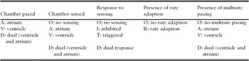
Although the complexity and functionality of pacing systems is advancing rapidly, the fundamental concepts of a modern pacing system are simple. The pulse generator is a roughly circular unit comprising the battery and circuitry, which connects to one or more pacing leads. The leads are composed of electrode(s) positioned within a cardiac chamber and an insulated coiled wire attached to the pulse generator, which provides an electrical connection between the heart and the pulse generator. Electrical signals produced by intrinsic activity of the heart can be detected by the pulse generator (sensing) and electrical output to stimulate cardiac depolarization can be transmitted to the heart (pacing). Pacemakers are classified by a series of five letters (Table 2). The first letter indicates the chamber paced, either ventricle (V), atrium (A) or both (dual, D); the second letter indicates the chamber sensed, the third letter the response to a sensed beat, either inhibited (I), triggered (T) or both (dual, D); the fourth letter indicates the presence of a sensor which can give a rate adaptive response (increasing heart rate with exercise, R). The fifth letter is not widely used but indicates the presence of multisite pacing in one or both chambers.Reference Bernstein, Daubert, Fletcher, Hayes, Lüderitz, Reynolds, Schoenfeld and Sutton74 Multisite pacing in the ventricle is carried out during cardiac resynchronization therapy or biventricular pacing with pacing leads to pace the right atrium, right ventricle and left ventricle.
The five letter classification scheme for cardiac pacemakers

The commonest pacing modes encountered are VVIR and DDD±R. In VVIR pacing, there is a single lead in the right ventricle which senses the intrinsic depolarization and inhibits pacing. If no electrical activity is seen after a preset time, usually 1000 ms, a paced beat is initiated; the pacing rate is adapted according to level of physical activity using an activity sensor incorporated in the system. A DDD system is more complex and has leads in both atrium and ventricle. If an intrinsic beat is not sensed in the atrium, an atrial pacing stimulus will be initiated and given a chance to conduct through to the ventricles. If the ventricular lead does not sense an intrinsic ventricular depolarization in the appropriate time period, a ventricular pacing stimulus will be triggered. Thus an intrinsic stimulus in the atrium may lead to the inhibition of atrial pacing and the triggering of ventricular pacing (justifying the D-dual response in the third letter of the classification). Similarly, if the atrial rate is satisfactory and atrial pacing is inhibited but there is a lack of conduction to the ventricle, failure to sense an ‘expected’ ventricular intrinsic depolarization will also trigger a pacing stimulus. The minimum and maximum pacing rates may vary but often pacemakers are set to pace between 60 and 140 b.p.m. (with rate response, or tracking P waves). If the patient has an intrinsic heart rate greater than the minimum rate then no pacing will occur and no pacing spikes will be evident on the ECG. Various more complex algorithms exist which aim to improve symptoms and minimize inappropriate or unnecessary pacing, discussion of which is beyond the scope of this review.
Pacing modes: dual chamber versus single chamber pacing
The concept of dual chamber cardiac pacing in patients with sinus rhythm is appealing as it seems more physiological, and has the theoretical advantage of maintaining AV and subsequent mechanical synchrony. The potential disadvantages of a dual chamber system include increased implantation and follow-up costs, increased procedural complication rate and decreased battery longevity. Several large multicentre prospective randomized studies have evaluated pacing modes in patients with SND and AV block.Reference Toff, Camm and Skehan64, Reference Lamas, Orav, Stambler, Ellenbogen, Sgarbossa, Huang, Marinchak, Estes, Mitchell, Lieberman, Mangione and Goldman75–Reference Lamas, Lee, Sweeney, Silverman, Leon, Yee, Marinchak, Flaker, Schron, Orav, Hellkamp, Greer, McAnulty, Ellenbogen, Ehlert, Freedman, Estes, Greenspon and Goldman77 Two of these trials specifically excluded younger patients, recruiting only those over 65Reference Lamas, Orav, Stambler, Ellenbogen, Sgarbossa, Huang, Marinchak, Estes, Mitchell, Lieberman, Mangione and Goldman75 or 70.Reference Toff, Camm and Skehan64 Although these trials fail to show any benefit in mortality between the two pacing modes, they do show a reduction in AF with dual chamber pacing, particularly in patients with SND,Reference Lamas, Lee, Sweeney, Silverman, Leon, Yee, Marinchak, Flaker, Schron, Orav, Hellkamp, Greer, McAnulty, Ellenbogen, Ehlert, Freedman, Estes, Greenspon and Goldman77 which, perhaps surprisingly, does not translate into stroke reduction. Additionally, there is a reduction in heart failure symptoms and an increase in quality of life with dual chamber pacing compared with ventricular pacing in patients with SND.Reference Lamas, Lee, Sweeney, Silverman, Leon, Yee, Marinchak, Flaker, Schron, Orav, Hellkamp, Greer, McAnulty, Ellenbogen, Ehlert, Freedman, Estes, Greenspon and Goldman77 It has been suggested that the apparent lack of benefit of dual chamber pacing may, in part, be related to the potentially deleterious effects of perhaps unnecessary right ventricular pacing in this group.Reference Wilkoff, Cook, Epstein, Greene, Hallstrom, Hsia, Kutalek and Sharma78 Pacing the right ventricular apex leads to abnormal left and right ventricular depolarization and dyssynchronous contraction.Reference Wilkoff, Cook, Epstein, Greene, Hallstrom, Hsia, Kutalek and Sharma78 Many patients, particularly with SND, have reliable AV conduction and so normal ventricular activation, and so do not need ventricular pacing. The dual chamber algorithms current at the time these trials were conducted will actually have promoted right ventricular pacing, with possible harm. New algorithms have been developed which promote intrinsic AV conduction and thus minimize pacing, where possible. Disappointingly, a recent randomized trial of one of these algorithms was not designed or powered to detect a change in mortality, although it did show that the incidence of AF was further reduced.Reference Sweeney, Bank, Nsah, Koullick, Zeng, Hettrick, Sheldon and Lamas79 Despite this, dual chamber pacing has been shown to be a cost-effective measure to improve quality of life, and is recommended where possible.Reference Dretzke, Toff, Lip, Raftery, Fry-Smith and Taylor80
Pacemaker implantation: procedure and complications
Pacing may be either temporary, or permanent. Temporary pacing is appropriate if the condition causing symptomatic bradycardia is expected to resolve, or symptoms are so severe that pacing must be achieved as soon as possible. Otherwise, permanent pacing without prior temporary pacing is preferred.Reference Winner and Boon81
Temporary pacing
Temporary cardiac pacing can be achieved either by external pacing,Reference Falk, Zoll and Zoll82, Reference Zoll, Zoll, Falk, Clinton, Eitel and Antman83 using large electrodes placed on the skin of the thorax, or by inserting a temporary electrode transvenously into the heart. External transcutaneous pacing is only suitable for emergency situations, such as cardiac arrest, as it is painful and not always well tolerated by the conscious patient.Reference Zoll, Zoll, Falk, Clinton, Eitel and Antman83 It may not achieve pacing (in over a quarter),Reference Zoll, Zoll, Falk, Clinton, Eitel and Antman83 and its effect may be difficult to assess, as movement and large stimulation artefacts recorded on the ECG may obscure the QRS complex. The equipment required should be available in ambulances, accident and emergency departments, and coronary care units. It cannot be used for prolonged periods, and ideally should be replaced by temporary transvenous or permanent pacing as soon as it can be arranged.
Temporary transvenous pacing is much better tolerated by the patient, but takes longer than transcutaneous pacing to achieve. One or more electrode catheters are inserted, through any large vein, and positioned under fluoroscopic guidance in usually the right ventricle (right atrial pacing may also be performed in the same way). The lead is secured to the skin with sutures, and the electrodes are attached to a temporary pacing box. Temporary transvenous pacing has a high complication rate (32%), particularly in inexperienced hands. Important complications include lead displacement, and infection, including septicaemia, leading to delays in permanent pacing in 23%,Reference Betts84 and more than doubling the risk of infection of the subsequently implanted permanent pacing system.Reference Klug, Balde, Pavin, Hidden-Lucet, Clementy, Sadoul, Rey, Lande, Lazarus, Victor, Barnay, Grandbastien and Kacet85 The majority of patients who receive temporary pacing require permanent pacing (outside the context of acute myocardial infarction),Reference Murphy86 so if possible temporary pacing should be avoided, and a permanent system implanted as a first procedure.
Permanent pacing
Permanent pacemaker insertion is a surgical procedure carried out by cardiologists in most secondary and tertiary centres in the UK. It is usually performed under local anaesthetic and is well tolerated even in older patients with significant co-morbidities. The procedure may be performed electively, as a day case, even in the oldest old,Reference Osman, Krishnamoorthy, Nadir, Mullin, Morley-Davies and Creamer87 but commonly involves an overnight stay in hospital. A skin incision is made in the prepectoral area, usually on the left side, and a ‘pocket’ is created for the generator by blunt dissection in the plane immediately above the pectoral muscle. The leads are inserted into the central venous circulation and are positioned in the heart using X-ray fluoroscopic guidance. Lead fixation within the heart is either passive, where small tines at the end of the lead hold it in place within the cardiac trabeculations, or active, when a retractable screw mechanism is deployed, securing the lead to myocardium. The electrical parameters of the leads are measured, and when satisfactory, they are secured with sutures to the pectoral muscle and then inserted into the pacemaker pulse generator. The generator and any redundant lead loops are placed in the pocket and the wound is closed with sutures. The procedure typically takes less than an hour. The size of the generator can be a concern in thin patients where lack of overlying tissue increases the chance of subsequent erosion, but this can generally be overcome using a smaller device and/or implantation beneath the pectoral muscle.
Early procedural complications may occur but these are very seldom life-threatening. Lead displacement may occur at a rate of 0.7–2% for the ventricular lead and 0.5–4% for the atrial lead.Reference Ellenbogen, Hellkamp, Wilkoff, Camunãs, Love, Hadjis, Lee and Lamas88 This usually requires an early repeat procedure to reposition the lead. When the subclavian approach for venous access is used, inadvertent pleural puncture may occur, leading to a pneumothorax in 0.7–2% of patients.Reference Ellenbogen, Hellkamp, Wilkoff, Camunãs, Love, Hadjis, Lee and Lamas88 Although small asymptomatic pneumothoraces can usually be treated conservatively, insertion of a chest drain may be required. Cardiac perforation by the lead tip may occur at rates of 0.3–1% although this often does not cause any significant compromise.Reference Ellenbogen, Hellkamp, Wilkoff, Camunãs, Love, Hadjis, Lee and Lamas88 Early infection rates of 0.2–1% have been reported and these usually respond to systemic antibiotics.Reference Ellenbogen, Hellkamp, Wilkoff, Camunãs, Love, Hadjis, Lee and Lamas88 Rarely, a complete system extraction may be required. Given the natural history of indications for pacing, most of the patients in the large pacing trials informing these statistics are older. However, there are some data to evaluate the effect of age on complication rates. A recent study,Reference Healey, Armaganijan, Nair, Morillo, Divakaramenon and Connolly89 including >4800 patients, pooled data from three large pacing trialsReference Toff, Camm and Skehan64, Reference Connolly, Kerr, Gent, Roberts, Yusuf, Gillis, Sami, Talajic, Tang, Klein, Lau and Newman76, Reference Andersen, Nielsen, Thomsen, Thuesen, Mortensen, Vesterlund and Pedersen90 and showed an increase in early pacemaker procedural complications from 3.4% in patients under 75 years to 5.1% in those over 75 years, particularly lead problems and pneumothorax. In contrast, late complications, such as lead fracture and need for generator replacement, were less likely among those over 75 years. In summary, the older old may have a slight increase in periprocedural complications, but these are generally not life threatening and are offset by a reduction in longer term complications, probably due to reduced life expectancy.
Pacemaker follow-up
Patients with cardiac pacemakers should remain under regular occasional follow-up to assess clinical status, optimize pacemaker function and assess battery status.Reference Wilkoff, Auricchio, Brugada, Cowie, Ellenbogen, Gillis, Hayes, Howlett, Kautzner, Love, Morgan, Priori, Reynolds, Schoenfeld and Vardas91 The frequency of device follow-up may be driven by clinical circumstances but typically patients are seen about 1 month after implantation to assess early complications such as lead displacement, and to optimize programming of the device. Subsequent follow-up is then at 6- or 12-monthly intervals, and allows evaluation of pacemaker function and identification of any problems with leads or the generator. Reprogramming of device parameters may be done in response to patient symptoms, or changes in status, but are often not required. Battery voltage is checked regularly, and this allows early identification of the need for generator replacement so that this can be planned ahead of the end of the battery life. Pacemaker generator replacement (for battery depletion) for certain pacing indications may be an essential life-sustaining treatment. Generator change should not, however, be undertaken without considering both the continued need for pacing and the patient's wishes. Some patients may not wish to undergo another procedure; however, even the extremely frail and oldest old should be offered generator change if they wish it and if the patient wishes to undergo the procedure, it should not be denied. Another interesting group are those patients with SND who develop permanent AF during follow-up. If AV node conduction is intact, these patients may no longer be at risk of bradycardia, and so may not need continued pacing. Over a third (38%) of patients paced for SND who subsequently developed permanent AF had a minimum intrinsic heart rate of over 60 b.p.m. and 18% had never used ventricular pacing during AF.Reference Huagui, Dhanunjaya, Hema, Karen and Tom92 Although it is unlikely that a pacing system would ever be removed given the risks of lead extraction, a depleted battery may not be replaced in selected patients.
Recurrent syncope after pacing
Patients with pacemakers may experience syncope. This may occur in up to 13% of patients paced for SND over a period of 10 years.Reference Sgarbossa, Pinski, Jaeger, Trohman and Maloney93 It is commonly suspected that pacemaker malfunction is the cause, but in fact this is seldom the case.Reference Sgarbossa, Pinski, Jaeger, Trohman and Maloney93, Reference Pavlovic, Kocovic, Djordjevic, Belkic, Kostic and Velimirovic94 A large series identified pacemaker malfunction as the cause of syncope in 6.5% of patients paced for SND with recurrent syncope. Other causes were vasovagal (18%), orthostatic hypotension (25.5%), rapid atrial tachyarrhythmias (11.5%), ventricular tachycardia (5%), acute myocardial ischaemia (2.5%) and in 29.5% of patients syncope remained unexplained. This reflects the multifactorial nature of syncope and the possible overlap between SND and neurocardiogenic syndromes (in up to 26%).Reference Brignole, Menozzi, Lolli, Oddone, Gianfranchi and Bertulla95 When syncope is attributable to the implanted device, it may occur as a result of lead failure or generator battery depletion or failure.
Atrial fibrillation
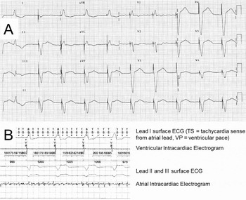
Another frequently encountered clinical problem identified at pacing follow-up is the development of AF. AF is common, particularly in older people. It occurs at a rate of about 3–4% per year in paced patients,Reference Skanes, Krahn, Yee, Klein, Connolly, Kerr, Gent, Thorpe and Roberts96, Reference Toff, Camm and Skehan97 and occurs more commonly in the older old (≥74 years of age), those with a prior history of AF, those paced for SND, and in those who receive VVI (rather than DDD) pacemakers.Reference Skanes, Krahn, Yee, Klein, Connolly, Kerr, Gent, Thorpe and Roberts96 Identification of AF in paced patients at follow-up is important as patients are often asymptomatic, particularly if they have AV block, so that AF is not conducted rapidly to the ventricle. These patients may have a regular and normal (paced) heart rate at pulse check which will not raise suspicion of AF, but importantly will still have an increased risk of stroke associated with the arrhythmia,Reference Glotzer, Hellkamp, Zimmerman, Sweeney, Yee, Marinchak, Cook, Paraschos, Love, Radoslovich, Lee and Lamas98 which is particularly significant in elderly people. If AF is identified at follow-up, either by pacemaker recorded intracardiac electrograms, shown in Figure 3, or on the surface ECG, the responsible physician can assess stroke risk using a risk scoring system such as CHADS2Reference Gage, Waterman, Shannon, Boechler, Rich and Radford99 or one of its refinements, CHA2DS2VAScReference Lip, Nieuwlaat, Pisters, Lane and Crijns100 or HAS BLEDReference Lip, Frison, Halperin and Lane101 to guide decisions regarding anticoagulation.
A, a 12-lead ECG from a patient with persistent atrial fibrillation and complete heart block with a dual chamber permanent pacemaker. The ventricular rhythm is paced and regular at the device base rate of 60 b.p.m. There are no visible P waves. B, the data from interrogation of the pacemaker. TS in the first column refers to pacemaker sensing of rapid atrial activity in AF, which is illustrated in the intracardiac recording from the tip of the atrial lead in the right atrium. The intracardiac recording from the tip of the ventricular lead shows the pacing spike and cardiac depolarization from the tip of the ventricular lead in the right ventricle. Leads II and III are also displayed in the figure and appear similar to the 12-lead ECG.

Ethical and end of life decisions
Pacemaker implantation in older patients has increased rapidly in recent years and this means that the management of these devices in dying patients will be encountered increasingly in clinical practice. The withholding and withdrawal of life-sustaining treatments (e.g. cardiopulmonary resuscitation, mechanical ventilation, or haemodialysis) from terminally ill patients who do not want the treatments is ethical and legal.Reference Mueller, Hook and Hayes102 There is no difference between a competent adult refusing implantation of a pacemaker and requesting its withdrawal.Reference Lampert, Hayes, Annas, Farley, Goldstein, Hamilton, Kay, Kramer, Mueller, Padeletti, Pozuelo, Schoenfeld, Vardas, Wiegand and Zellner103 Withdrawal of pacing can be done easily and non-invasively by programming; it rarely requires surgical removal of the device. If a pacemaker is programmed off in a pacemaker-dependent patient, death may be rapid, if there is no reliable escape rhythm. However, there may be a rhythm that sustains life for some time. As pacing is painless and the patient is rarely aware of it, it is uncommon to be asked to withdraw it. Equally, withdrawal of implantable cardioverter defibrillator (ICD) therapy may be done painlessly by programming, and bradycardia pacing support may be maintained despite disabling tachycardia therapy. Health professionals generally support withdrawal of ICD therapy for tachycardias in the terminally ill, particularly in those for whom a ‘do not resuscitate’ order has been agreed, as shocks delivered by the ICD are painful and unpleasant, and prevent sudden death.
These cases present a difficult dilemma, although with extensive counselling and a detailed informed consent may be considered an option on a case-by-case basis.Reference Mueller, Hook and Hayes102 The key to managing these cases is early communication between the patient, their family and health professionals from both the cardiac team and any health professionals delivering care for a non-cardiac terminal illness. Advanced directives can provide useful guidance to medical teams when a patient's clinical condition deteriorates. Clinicians involved in device education do not commonly have discussions with patients about end of life issues and device deactivationReference Epstein, DiMarco, Ellenbogen, Estes, Freedman, Gettes, Gillinov, Gregoratos, Hammill, Hayes, Hlatky, Newby, Page, Schoenfeld, Silka, Stevenson, Sweeney, Smith, Jacobs, Adams, Anderson, Buller, Creager, Ettinger, Faxon, Halperin, Hiratzka, Hunt, Krumholz, Kushner, Lytle, Nishimura, Ornato, Page, Riegel, Tarkington and Yancy22 and it may be important to address these issues at the time of implantation.
Removal of devices after death
Pacemakers and ICDs should be removed after death if cremation of the body is to be undertaken, as the lithium iodide battery can explode, with possible damage.Reference Gale and Mulley104 Removal of pacemakers poses no electrical risk to the operator. In contrast, handling ICD generators or cutting the leads is an electrical hazard, and shocks can be delivered to the operator if shock therapy has not been disabled.Reference Ghosh, James and Palmer105
Conclusions
Pacing is the only practical method of preventing bradycardia, and should be considered in all patients with symptoms probably related to bradycardia. Symptom rhythm correlation (ECG recording during symptoms) may be helpful, but is not always essential, as there may already be sufficient evidence to support a pacing indication. There is little to gain in stopping ‘culprit’ drugs such as beta-blockers or calcium antagonists, particularly in patients with AV block, as the block is likely to persist or recur. Most recipients of pacemakers are old, and some are among the oldest old. Despite this, pacing is associated with a significant improvement in quality of life, and in some patients increased longevity. The implantation procedure is straightforward, and well tolerated even in the oldest old, despite a small increase in the complication rate in this group. Routine pacemaker follow-up should not be onerous, but is important. Pacing therapy is seldom withdrawn, even at end of life, perhaps because it is painless. After death, the pacemaker generator should be removed prior to cremation.
Conflicts of interest
None of the authors had any conflicts of interest to declare.