Introduction
Acarosporaceae Zahlbr. currently is comprised of Acarospora A. Massal., Glypholecia Nyl., Lithoglypha Brusse, Myriospora Nägeli ex Hue, Pleopsidium Körb., Polysporina Vězda, Sarcogyne Flot., Thelocarpella Nav.-Ros. & Cl. Roux , Timdalia Hafellner and Silobia M. Westberg & Wedin (Tehler & Wedin Reference Tehler, Wedin and Nash2008; Westberg et al. Reference Westberg, Crewe, Purvis and Wedin2011). The family occurs on all continents and, in our estimation, contains over 200 species worldwide, although it definitely has over three times more names than taxa due to a profusion of usually useless infraspecific epithets (CABI Bioscience et al. 2011). As we progress toward more natural species concepts, many old species names continue to be reduced to synonymy (for example see Knudsen & Kocourková Reference Knudsen and Kocourková2008; Westberg et al. Reference Westberg, Crewe, Purvis and Wedin2011; Westberg & Wedin Reference Westberg and Wedin2011). At the same time new taxa are being discovered and regularly described. For instance, in approximately the last year, four new species were described from North America and four new species from Europe (Knudsen Reference Knudsen2010; Knudsen et al. Reference Knudsen, Lendemer and Harris2011; Lumbsch et al. Reference Lumbsch, Ahti, Altermann, Amo de Paz, Aptroot, Arup, Bárcenas Peña, Bawingan, Benatti and Betancourt2011; Westberg et al. Reference Westberg, Crewe, Purvis and Wedin2011).
It is impossible to estimate the real diversity of Acarosporaceae in South America at present. Thirty-nine species of Acarosporaceae (38 Acarospora, and one Sarcogyne) have been reported from South America in the literature, but that number is inaccurate (Magnusson Reference Magnusson1929, Reference Magnusson1935; Knudsen et al. Reference Knudsen, Elix and Reeb2008). With the exception of three yellow Acarospora species and A. lorentzii (Müll. Arg.) Hue, records of most taxa are usually based only on a type or relatively few collections in herbaria. Several widespread polymorphic species such A. chrysops (Tuck.) H. Magn. and A. rhabararina Hue have been described more than once (Knudsen et al. Reference Knudsen, Elix and Reeb2008). At least four Acarospora species described from South America belonged to other families (Magnusson Reference Magnusson1929; Galloway & Quilhot Reference Galloway and Quilhot1998). The specimens documenting some reports are unavailable. A few reports may be projections of species concepts from North America or Europe on related or similar taxa. In every group of collections we studied, there were usually some specimens with taxa probably new to science but which were sterile or depauperate. No robust phylogenetic studies of South American Acarosporaceae have been conducted. The occurrence of cryptic or semi-cryptic species that are best detected using molecular tools combined with taxonomic, ecological, and biogeographical analysis can also be expected (Vondrák et al. Reference Vondrák, Řiha, Arup and Søchting2009; Lendemer & O'Brian Reference Lendemer and O'Brian2011).
This paper is based primarily on the collections of the second and third authors. Recent collections by J. C. Lendemer, Z. Palice and V. Reeb were also examined, as well as the South American collections of Acarosporaceae at GZU.
Materials and Methods
Specimens collected by A. Flakus and M. Kukwa are deposited in KRAM, LPB UGDA, and hb. Flakus, respectively. Other specimens examined are stored in DUKE, FH, GZU, KANU, KEW, MSU, NY, PRA, S, UCR, UPS and W. Hand-cut sections were studied with standard microscopy and measurements were made on material mounted in water; structures were also studied in 10% KOH, and amyloid reactions were studied with IKI 1·5% with or without pretreatment with KOH. Images were photographed using a NIKON ECLIPSE 80i (DIC) compound microscope. Chemical analyses were carried out using thin-layer chromatography (TLC) following Orange et al. (Reference Orange, James and White2001). Extracts were run on aluminium TLC plates in solvent systems A, B and C.
Results
Acarospora dissecta K. Knudsen & Flakus sp. nov
MycoBank No.: MB 561076
Species Acarosporae altoandinae similis sed thallo fisso acidum norsticticum continente, et corona paratheciale absente differt.
Type: Bolivia, Dept. Potosí, Prov. Nor Lípez, Pinturas Rupestres near Mallku Villamar village, S21°46′20″, W67°29′05″, alt. 4038 m, open semi-desert high Andean area, 6 December 2009, A. Flakus 14739 & P. Rodríguez (KRAM!—holotypus).
Paratypes: Bolivia, Dept. Potosí, Prov. Daniel Campos, by Puerto Chuvica village near Salar Uyuni, S20°36′06″, W67°35′50″, alt. 3670 m, open semi-desert high Andean area, 4 December 2009, A. Flakus 14699, 14717 & P. Rodríguez (LPB & hb. Flakus).
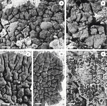
(Fig. 1A & B)
Acarosporaceae, habit of selected species from South America. A & B, Acarospora dissecta (holotype); C & D, Acarospora trachyticola (Flakus 5751, 10021; KRAM); E, Sarcogyne brunnea (holotype). Scales: A–E = 1 mm.

Thallus crustose of dispersed or contiguous gomphate areoles or squamules 1·0–2·0 mm diam., up to c. 0·4 mm thick, light to dark brown, round to angular, with thick mycelial base or stipe, areoles or squamules arising separately from endosubstratic hyphae or dividing vegetatively. Upper and lateral surfaces epruinose, upper surface with abundant cross-hatched fissures, lower surface pale, white to light brown (though could become blackened by substratum interactions). Cortex derived from hyphal bundles penetrating the algal layer, 50–70 µm thick, hyphae differentiating into paraplectenchyma, globose to angular cells, 4–6 µm diam.; upper layer c. 10 µm thick, brown pigmented with a hyaline gelatinized epicortex c. 4–10 µm thick., lower layer hyaline. Algal layer interrupted, to 200 µm thick, the algal cells broken into islands and glomerules, algal cells to 15 µm diam., continuous beneath apothecia. Medulla not distinct, continuous with hyphal bundles penetrating the algal layer and attaching hyphae of the stipe, inspersed with crystals, hyphae c. 2 µm diam., cells mostly 5–9 µm long.
Apothecia usually one per areole or squamule, disc immersed, brown, rugulose, usually epruinose, sometimes surrounded by a narrow thalline collar apparently pushed up by development of apothecium. Exciple narrow, of thin prosoplectenchyma, 10–15 µm wide, not expanding near surface of the disc, not forming a distinct parathecial crown. Hymenium hyaline 170–200 µm tall, IKI+ blue, yellow-green or red; epihymenium reddish brown, conglutinated, 10–15 µm tall; paraphyses mostly 2 µm diam. at mid-level, septate, cells mostly 5–8 µm long with oil drops, apices mostly unexpanded or barely expanded, embedded in pigmented gel and sometimes with pigmented caps, not branching. Asci cylindrical-clavate, 130–140 × 12–25 µm, c. 200 ascospores per ascus. Ascospores simple, hyaline, 4–6 × 2·0–2·5 µm, mostly ellipsoid. Subhymenium 30–40 µm thick, remaining IKI+ deep blue. Hypothecium narrow, continuous with exciple.
Conidiomata not seen.
Chemistry. Norstictic acid was detected in all specimens. Thallus medulla reacts K+ yellow then red, forming crystals, C−.
Etymology. The name refers to the characteristic fissuring of the cortex in mature squamules that often initiates vegetative division.
Distribution and ecology. Acarospora dissecta grows on volcanic rock at a high arid elevation in the Andes in an exposed location. Currently known only from two localities in Bolivia, at an elevation near 4000 m. We do not expect this species to be rare on the Altiplano plateau. It may also occur in North America (Colorado; see comments below).
Discussion. Acarospora dissecta appears closely related to A. altoandina H. Magn. in general appearance and structure of the squamules (Magnusson Reference Magnusson1956; Knudsen et al. Reference Knudsen, Elix and Reeb2008). In both species the cortex is formed from anticlinal hyphae that interrupt a thick and jagged algal layer as well as form an indistinct medulla undifferentiated from the hyphal bundles penetrating the algal layer and the attaching hyphae forming the stipe. They differ in secondary chemistry (A. dissecta produces norstictic acid, A. altoandina lacks secondary metabolites) and the height of hymenium (170–200 µm in A. dissecta, 130–160 µm in A. altoandina). Additionally, A. dissecta develops a narrow, unexpanded exciple not forming a parathecial crown concolorous with the thallus, while A. altoandina usually forms a parathecial crown. However, the two species can still look similar because the apothecia of A. dissecta sometimes appear to push up a thalline collar around the disc during the expansion of the apothecium. The fissuring may be variable and it is possible there are specimens with a relatively smooth upper surface, as such variation in cross-hatch fissuring is known in Acarospora strigata (Knudsen Reference Knudsen, Nash, Gries and Bungartz2007a) as well as A. bullata Anzi in Europe.
Acarospora dissecta has asci overlapping in size with those of A. altoandina, but they contain 200 or more ascospores instead of usually c. 100 ascospores present in the asci of the latter species. Acarospora dissecta also has ellipsoid ascospores, which are usually narrower than those of A. altoandina (2·0–2·5 µm vs. 2·0–3·5 µm), but ascospore size can vary in both. Both species occur in the Andes at high elevations.
Four other species of Acarospora reported from South America produce norstictic acid. Acarospora rouxii is a rugulose yellow species of South America (Bolivia, Chile and Peru), as well as North America (Mexico, Arizona). It differs from A. dissecta in having rhizocarpic acid and epanorin, as well as often an effigurate thallus (Knudsen Reference Knudsen, Nash, Gries and Bungartz2007a; Knudsen et al. Reference Knudsen, Elix and Reeb2008). The other three species of Acarospora with norstictic acid are brown: A. lorentzii, A. basiliensis Zahlbr., and Silobia smaragadula (Wahlenb.) M. Westb. & Wedin. The most common species of these three is the South American endemic A. lorentzii (Magnusson Reference Magnusson1929; Knudsen et al. Reference Knudsen, Elix and Reeb2008), which differs from A. dissecta especially in having a usually smooth, broadly attached, areolate thallus, which can be quite flat in some specimens, often forming an effigurate margin. It also has a shorter hymenium (usually 100–120 µm, rarely up to 140 µm). A second brown species, A. brasiliensis, was reported with norstictic acid (Zahlbruckner Reference Zahlbruckner and Schiffner1909; Magnusson Reference Magnusson1929), but it is a synonym of A. lorentzii (see below). Silobia smaragdula, which is verified from the coast of Chile (see below), differs from both in general appearance (Westberg et al. Reference Westberg, Crewe, Purvis and Wedin2011) as well as having thin branching paraphyses, mostly 1 µm diam.
During this study we examined specimens collected in Colorado by C. A. Morse (KANU). Based on general thallus structure and the presence of norstictic acid, we can assume that A. dissecta or a closely related taxon occurs in Colorado. Since the Colorado material is mostly sterile, however, we refrain from reporting it as new to North America at this time.
Acarospora lorentzii (Müll. Arg.) Hue
Morph. et Anat. 162 (1909)—Placodium lorentzii Müll. Arg., Lichen. Argent. in Flora, 510 (1889); type: Argentina, Concepción, Lorentz s. n. (P & W! syntypes).
Syn. nov. Acarospora brasiliensis Zahlbr., Ergebn. Botan. Exped. Kaiserl. Akad. Wissensch. Südbrail 1901, 54 (1909); type: Brazil, Rio de Janeiro, at the top of Mount Italiaya, about 2750 m, September 1901, V. Schiffner s. n. (W!—holotype; FH!—isotype).
Note. For the description and images of the species see Knudsen et al. (Reference Knudsen and Kocourková2008). The species is common and occurs in Argentina, Brazil, Chile, and Uruguay (Magnusson Reference Magnusson1929; Knudsen et al. Reference Knudsen, Elix and Reeb2008), and is reported here as new from Peru. Acarospora brasiliensis was described from Brazil at 2750 m above Rio de Janeiro (Zahlbruckner Reference Zahlbruckner and Schiffner1909; Magnusson Reference Magnusson1929). Though Magnusson recognized it as a separate taxon (Magnusson Reference Magnusson1929), we found A. brasiliensis to be a synonym of A. lorentzii, a species with great variation in the appearance of the thallus in the over thirty specimens we have examined. In several effigurate species in South America [e.g., A. trachyiticola (Müll. Arg.) Hue, A. subcastanea (Nyl.) Hue, and A. rouxii], marginal lobes are not always produced, and this is also the case in A. lorentzii. In the type of A. brasiliensis, lobes are lacking and the thallus is more reduced than in many specimens of well-developed A. lorentzii. Therefore, we synonymize A. brasiliensis with A. lorentzii.
Selected specimens examined. Peru: Dept. Arequipa: Prov. Caylloma, Cañón del Colca, Quebrada Sutanay, 15°53′06″S, 72°12′22″W, alt. 3320 m, A. Flakus 10050 & M. Kukwa 6059 (KRAM, UGDA); ibid., near Paso del Cruz , 15°53′07″S, 72°13′00″W, alt. 3250 m, A. Flakus 10048 & M. Kukwa (KRAM); ibid., Llahuar village, 15°34′52″S, 72°00′37″W, alt. 2085 m, M. Kukwa 6130 (UGDA); ibid., near Cabanaconde village, 15°38′18″S, 71°57′43″W, alt. 3480 m, A. Flakus 9640 & B. Cykowska (KRAM); ibid., near Soccoro village, 15°38′32″S, 71°43′22″W, alt. 3349 m, A. Flakus 9506 & B. Cykowska (KRAM); ibid., near Achoma village, 15°39′26″S, 71°43′38″W, alt. 3500 m, A. Flakus 9382 & B. Cykowska (KRAM).
Acarospora trachyticola (Müll. Arg.) Hue
Nouv. Arch. Mus. Hist. Nat., Paris, 5 Sér. 1: 163 (1909).—Placodium trachyticolum Müll. Arg., Hedwigia 31: 279 (1892); type: Peru, in Andibus, collo Inter Lampa et Arequipa, Lechler 3316 (KEW!—holotype).
(Fig. 1C & D)
Thallus areolate, usually determinate with a lobed margin, areoles plane to convex, broadly attached, of different shapes and sizes, 0·2–1·5 mm diam., <1 mm tall. Upper surface epruinose, reddish brown to blackish brown. Cortex 40–50 µm (–100 µm) thick; epicortex lacking or layer of gelatinized hyphae less than 5 µm thick, upper layer brown, c. 10 µm thick, lower layer hyaline, hyphae paraplectenchymatous 4–7 µm diam. The upper region of the algal layer is uneven, sometimes with deep clefts filled with hyaline cortical cells, occasionally penetrated by hyphal bands. Algal layer to 140 µm thick, algal cells c. 10 µm diam., not continuous below the apothecium, uninterrupted by hyphal bundles. Medulla continuous with attaching hyphae, obscure in water, hyphae 3–5 µm wide, cells 4–7 µm long, branching, often encrusted with small crystals.
Apothecia at first punctiform, less than 0·2 µm wide, up to 8 per areole, but often only 1 per areole, dilating to up to 1 mm diam. Disc dark, immersed, epruinose. Exciple narrow, of thin prosoplectenchyma or expanded to form a parathecial crown to 70 µm wide. Hymenium usually 140–180 µm tall; epihymenium conglutinated with yellowish brown pigment, 15 µm tall; paraphyses 2–3 µm wide, sometimes branching, especially at mid-height, septate, rarely constricted at the septum, cells 5–10 µm long, apices rarely expanded but infrequently encased in the pigmented hood to 5 µm wide. Asci mostly 70–120 × 16–20 µm, c. 200 ascospores per ascus. Ascospores hyaline, simple, mostly narrowly ellipsoid, 4·0–5·5 × 1·5–2·0 µm. Subhymenium 50–60 µm thick. Hypothecium narrow, continuous with exciple.
Conidiomata not seen.
Chemistry. Secondary metabolites lacking.
Distribution and ecology. Acarospora trachyticola grows on volcanic rock at high arid elevations in the Andes in exposed locations. It is common in Peru. It is reported new to Argentina, Bolivia, and Venezuela.
Discussion. Although A. trachyticola is a common species in Peru, it was impossible to identify using Magnusson (Reference Magnusson1929), because the description and classification are based only on the type. The species is usually effigurate, but the type is predominately non-effigurate (with just a few outer lobes forming) and was not included by Magnusson among the effigurate species in his key, though he mentions some lobes. The type also has punctiform apothecia, but in most specimens they commonly become dilated.
Acarospora trachyticola is similar to A. macrocyclos Vain. from Antarctica and A. molybdina (Wahlenb.) Trevis from the north Atlantic coast of North America and Europe (Magnusson Reference Magnusson1929). All lack secondary metabolites and are effigurate. Acarospora macrocyclos definitely has a different habit: long narrow lobes and a well-developed, thick, rugulose, usually distinctly reddish brown thallus, with sessile apothecia having thalline margins. Acarospora molybdina differs in having longer lobes and usually sessile apothecia, but is more polymorphic than the specimens of A. macrocyclos we examined.
Specimens examined. Argentina: Prov. Tucumán: Valle de Tafí, Carapunco-Infiernillo, alt. 2800 ft., on schistose rock, I. M. Lamb 5170, 5190 & 5443 (FH).—Bolivia: Dept. La Paz: Prov. Murillo, near Cumbre pass, 16°20′37″S, 68°02′21″W, alt. 4670 m, A. Flakus 5751 (KRAM, LPB); ibid. 16°21′59″S, 68°02′37″W, alt. 4604 m, leg. A. Flakus 16307 & P. Rodríguez (hb. Flakus). Dept. Potosí: Prov. Nor López, near Volcín Ollagüe, 21°17′13″S, 68°05′32″W, alt. 4095 m, A. Flakus 14729 & P. Rodríguez (LPB); Pinturas Rupestres near Mallku Villamar village, 21°46′20″S, 67°29′05″W, alt. 4038 m, A. Flakus 14743 & P. Rodríguez (hb. Flakus).—Peru: Dept. Arequipa: Prov. Caylloma, by Cañón del Colca, between Gloria Huasi colony and Paso del Cruz, 15°52′43″S, 72°14′22″W, alt. 2926 m, A. Flakus 10028, 10021 & M. Kukwa 6027 (KRAM, UGDA, hb. Flakus); ibid., by Cañón del Colca, Quebrada Sutanay, 15°54′05″S, 72°11′07″W, alt. 3760 m, A. Flakus 10083 & M. Kukwa (KRAM).—Venezuela: Estado Merida: Paramo de Mucuchies, Rücken an der Strase von Apartaderos nach Pinango, alt. 4000–4200 m., B. & F. Oberwinkler & J. Poelt (GZU).
Selected specimens examined of A. molybdina. Norway: Finnmark: Varager Peninsula, Nesseby Parish, Fugleberget 2–3 km E. of Mortensnes, on rocks 15 m from shore, Tibell 2876 (FH, UPS) .—Sweden (as var. confusa): Bohuslän: Lysekil, Stångehuvud, on sun-exposed rocks, R. Santesson s. n. (FH). Ödesmål: Grötåns holme, A. H. Magnusson, Malme, Lich. Suec. Exs. No. 716 (FH).—USA: Maine: Mt. Desert, Tuckerman s. n. (FH); Rockland, on ledges near the shore, (annotated as f. ereutica by Magnusson), G. K. Merrill s. n. (FH); Washington Co., Petit Manan Wildlife Refuge, Hollingsworth Trail, 44·424°N, 67·890°W, side of granitic rock in littoral zone, A. M. Fryday 9227 et al. (MSU).
Selected specimens of A. macrocyclos examined. Antarctica: Graham Land: Trinity Peninsula, Hope Bay, alt. 200 ft., I. M. Lamb 2356 (FH); Island in Maguerita Bay, D. C. Nutt 205 (FH).—South Shetlands: Deception Island: S.E. side of the harbour, alt. 50 ft., on flat stones in scree slope, I. M. Lamb 1116 (FH).
Polysporina urceolata (Anzi) Brodo
Mycotaxon 28: 95 (1987).—Sarcogyne urceolata Anzi, Comment. Soc. Crittog. Ital. 1(3): 157 (1862); type: Italy, Monte della Parete, ad rapes marmoreas supra terminum arboreum [Anzi, Lich. Rar. Langob. Exs. No. 285] (BM—syntypes, n .v.).
Notes. For the description of the species see Knudsen (Reference Knudsen, Nash, Gries and Bungartz2007b). Polysporina urceolata occurs in North America and Europe. The species is reported as new for South America and Bolivia, where it was found on calciferous brick. This is also the first member of the genus recorded in South America.
The apothecia in the specimen studied were superficial, but can occur also in pits. It can be confused only with the lichenicolous and algicolous fungus P. pusilla (Anzi) M. Steiner ex Kantvilas, which is widespread like P. urceolata, and may occur on calcareous substrata in South America too. For the description of P. pusilla see Knudsen & Kocourková (Reference Knudsen and Kocourková2008).
Specimen examined. Bolivia: Dept. La Paz: Prov. Bautista Saavedra, Area Natural de Manejo Integrado Nacional Apolobamba, near Taypi Cañuma village, 15°03′20″S, 69°09′07″W, alt. 4506 m, A. Flakus 17494 & P. Rodríguez (LPB).
Sarcogyne brunnea K. Knudsen & Flakus sp. nov
MycoBank No.: MB 561077
Species Sarcogyne crustaceae similis sed conidiis 3–4 × 2–3 µm et thallo brunneo, albo-pruinoso differt.
Typus: Bolivia, Dept. Potosí, Prov. Nor Lípez, Pinturas Rupestres near Mallku Villamar village, S21°46′20″, W67°29′05″, alt. 4038 m, open semi-desert high Andean area, 6 December 2009, A. Flakus 14792 & P. Rodríguez (LPB—holotypus).
Paratype: Ecuador, Prov. Cotopaxi, N.P. Cotopaxi, NNE slope of the volcano Cotopaxi in a grass páramo, 00° 36′ 50″S, 78° 24′ 15″W, 3800 m, on soil on a steep eroding slope above a brook, 23 June 1999, Z. Palice 3338 (PRA).
(Fig. 1E)
Thallus of contiguous angular areoles, broadly attached, up to 1·5 mm diam., mostly less than <1 mm wide, separated by deep fissures, vegetatively dividing, or arising from a endostratic hypothallus of anastomosing thin-walled hyphae, hyaline, 3–4 µm diam., indistinctly septate, or distinct cells of to 5 µm long, continuous with medullary hyphae. Upper surface white or mottled white and brown, heavily pruinose with large flakes in the holotype, or more lightly pruinose in the paratype, but brown when wetted, fissured. Cortex mostly 20–60 µm thick; the upper layer dark reddish brown above to lighter brown below, with or without a thin epicortex of gelatinized hyphae, the lower layer hyaline, paraplectenchymatous, globose or angular, to 7 µm diam. differentiating from hyphae 3–4 µm wide, with cells 5–7 µm long. Algal layer interrupted by hyphal bundles, up to 200 µm thick, thinner but continuous below apothecia, algal cells (6–)10–15 µm diam. Medulla of hyphae 2–4 µm wide, with cells mostly 5–7 µm long, inspersed with crystals and gelatinized, to100 µm thick, continuous with attaching hyphae.
Apothecia < 0·6 mm diam., 1–6 per areole, immersed. Disc black, epruinose, rough with distinct narrow dark brown margin raised above the disc, black when wet. Exciple of hyaline radiating hyphae, continuous with the hypothecium, 10–15 µm wide, expanding to 80 µm wide at the surface of the disc, forming a dark brown parathecial crown elevated above the disc. Hymenium (100–)140–200 µm tall, hyaline, IKI+ blue, yellowish green, or with blue streaks turning to red; epihymenium conglutinated in reddish brown gel, 10–25 µm tall; paraphyses 1·5–2·5 µm diam., filled with oil drops, septate, apices unexpanded, some branching. Asci cylindrical to broadly clavate (50–)100–150 × 15–25(–40) µm, approximately 100–200 per ascus. Ascospores hyaline, simple, various, mostly 4–6 × 2–3 µm. Subhymenium 20–40 µm thick, IKI+ blue. Hypothecium thin, continuous with exciple.
Conidiomata pycnidia, common, 200–300 µm diam., conidia simple, hyaline, 3–4 × 2–3 µm.
Chemistry. No substances detected by TLC.
Etymology. The name refers to the corticated and often pruinose brown areoles that distinguish it from several species known from Europe, North America and Asia, with a white ecorticate thallus.
Distribution and ecology. Sarcogyne brunnea grows on volcanic rock and soil at high arid elevations in the Andes, in exposed sunny locations. Currently known from the type locality in Bolivia at an elevation of over 4000 m, and the slope of the volcano Cotopaxi in Ecuador at 3800 m. We do not expect this species to be rare.
Discussion. While many species of Sarcogyne have endolithic thalli, several produce chasmolithic or epilithic thalli of varying thickness. In some species the thallus is chasmolithic (mixed with uppermost layer of rock particles) and occurs especially on decaying rock, sometimes appearing as a ‘white margin’ below or surrounding the exciple, as in Sarcogyne arenosa (Herre) K. Knudsen & Standley from North America (Knudsen & Standley Reference Knudsen, Standley, Nash, Gries and Bungartz2007; Lendemer et al. Reference Lendemer, Kocourková and Knudsen2009). In other species the thallus is epilithic, visible as a thin white coating, as in S. magnispora K. Knudsen & Halici from Turkey (Knudsen et al. Reference Knudsen, Halici and Kocakaya2009), or as a thick white crust up to 5 mm thick in S. cretacea Poelt on limestone in the Alps (Clauzade & Roux Reference Clauzade and Roux1985). In all these species the thallus is white or grey and always ecorticate.
Recently we revised Biatorella terrena Hasse from southern California as Sarcogyne crustacea K. Knudsen & Kocourk., changing the name because it would have been a homonym of S. terrena H. Magn. (Knudsen & Kocourková Reference Knudsen and Kocourková2010). This was the first species with a corticated epilithic thallus. The epruinose thallus was not distinctly areolate or determinate and the surface was a blackened blue-grey colour and grew on soil. The new species, S. brunnea, differs in having a pruinose areolate thallus that is definitely brown when wetted. Unlike S. crustacea, the algal layer of S. brunnea is interrupted by hyphal bundles, though not always distinctly. The conidia of S. crustacea are longer and narrower than the conidia of S. brunnea [4·0–5·0(–5·5) × 0·5–1·0 µm vs. 3–4 × 2–3 µm], while the ascospores of S. crustacea are usually narrower than the ascospores of S. brunnea (mostly 4–5 × 1·5–2·0 µm vs. 4–6 × 2–3 µm).
The well-developed hypothallus of S. brunnea can stabilize eroding soil, acting as rhizohyphae, and it is expected on both volcanic rock and in biological soil crusts. The occurrence on soil and rock is not unusual in the family. For instance, Acarospora obpallens (Nyl. ex Hasse) Zahlbr. in western North America and South America is a saxicolous species that is common on soil in biological soil crusts, especially in southern California (Knudsen Reference Knudsen, Nash, Gries and Bungartz2007a).
Sarcogyne terrena H. Magn
Ann. Cryptog. Exot. 7: 132.—Biatorella terrena H. Magn. (illegit. nom.), Meddelelser fran Göteborgs BotaniskaTrädgard 3: 13 (1927); non Biatorella terrena Hasse, Bryologist 14(1): 3 (1911); type: Brazil, Rio Grande do Sul: Pelotas, 9 February 1892, G. O. Malme s. n. (S L2740!—holotype).
Epilithic thallus lacking.
Apothecia dispersed to contiguous, broadly attached to continuous endosubstratic hypothallus of branching and anatomosing rhizohyphae binding the soil. Vegetative hyphae hyaline, thin-walled, to 3 µm diam., cells 3–7 µm long, branching, continuous with medullary hyphae, becoming gelatinized. Algal layer scattered, thin, not continuous, below apothecia. Non-pruinose apothecia immersed to sessile, < 0·3 mm wide, <0·2 mm tall, dull pale brown, colour darkening red when wetted. Non-thalline margin entire, non-crenulate, non-carbonized, elevated above immersed punctiform disc. Disc non-pruinose, rough, often with accretions on the disc surface, < 0·1 mm wide. Exciple up to 40–70 µm wide of radiating hyphae mostly 1 µm diam., septate, cells 3–5 µm long, hyaline; outer layer formed of melanized hyphae, reddish brown, up to 10 µm; inner layer reddish yellow to hyaline. Hymenium 100–150 µm tall, I+ red; conglutinated epihymenium reddish brown to black to 20 µm tall; paraphyses mostly 1·5–2·0 um wide, septate, cells to 7 µm long, moderately branching, apices barely expanded. Asci 70–90 × 25–35 µm, broadly clavate, c. 100–200 ascospores per ascus. Ascospores narrow to broadly ellipsoid, various, mostly 4–5 × 1·5 µm, but most asci immature in type. Subhymenium < 20 µm thick, I+ red or dirty blue. Hypothecium thin beneath apothecium, 10 µm tall, continuous with the exciple.
Conidiomata not seen.
Chemistry. No secondary metabolites with spot tests. Not examined with TLC due to small size of the holotype.
Distribution and ecology. Known only from the historical type collection from Pelotas in the state of Rio Grande do Sul in southern coastal Brazil. It was growing on the fine sandy soil of the coastal plain of Pelotas. This species may be extirpated from the type locality or may be extremely rare or even extinct.
Discussion. Based on its endosubstratic thallus of dense branching rhizohyphae and its occurrence in fine sandy soil, Sarcogyne terrena is probably an exclusively terricolous species. The apothecia are the smallest we have seen in the genus and the crust of clustered apothecia on soil that it forms is quite distinctive in the genus.
Terricolous lichen species can be regional endemics (either relics or newly evolved species) that occur in biological soil crusts in areas such as valleys, prairie, or deltas that are suitable for agriculture, ranching or urbanization. In southern California, for instance, these endemic terricolous species rapidly became extirpated from most of their range and several are extremely rare or extinct (Knudsen & Kocourková Reference Knudsen and Kocourková2010; Knudsen Reference Knudsen2010). In South America, the terricolous Acarospora terrigena K. Knudsen, Elix & Reeb was collected in 1902 by G. O. Malme, in the state of Rio Grande do Sul in Brazil (Knudsen et al. Reference Knudsen, Elix and Reeb2008). The type locality was on the rich delta of Porto Alegre that has been heavily developed in the last hundred years. Acarospora terrigena was probably extirpated from the type locality. If it was a regional endemic of southern Brazil, A. terrigena may even be extremely rare or extinct. Sarcogyne terrena was also collected in 1902 by Malme in Rio Grande do Sul, but on the coastal plain of Pelotas, another area that has become heavily developed in the last hundred years. Like A. terrigena, S. terrena has probably been extirpated from the type locality and may be extremely rare or extinct.
Silobia smaragdula (Wahlenb.) M. Westb. & Wedin
Lichenologist 43: 21 (2011).—Endocarpon smaragdulum Wahlenb., in Acharius, Method. Lich. Suppl.: 29 (1803); type: Finnmarkiae Norvegicae in petra insula sinus Altenis, 10 May 1802, G. Wahlenberg s. n. (UPS L-137960, n. v.—lectotype, designated in Westberg et al. Reference Westberg, Crewe, Purvis and Wedin2011).
Note. For description of the new genus and the species see Westberg et. al (Reference Westberg and Wedin2011). North American lichenologist J. C. Lendemer (NY) recently collected Silobia smaragdula, syn. A. smaragdula (Wahlenb.) A. Massal., on the coast of Chile. The species was previously reported from Chile but we could not verify the report (Galloway & Quilhot Reference Galloway and Quilhot1998). Silobia smargadula differs from A. dissecta especially with its thin, branching paraphyses (mostly 1 µm diam.) and its distinctive areoles, with several apothecia and a usually distinctive pale yellowish brown colour. As with most collections in North America (Knudsen Reference Knudsen2007c), S. smaragdula was collected along the coast.
Specimen examined. Chile: Chiloé: Isla Grande de Chiloe, Duhatao, 22·5 km S of Quetelmahue, J. C. Lendemer 16290 w/ A. Moroz (NY).
Conclusion
In this continuing study of South American Acarosporaceae, we only recognize species that we have verified ourselves. An on-going checklist of the Acarosporaceae of South America is available online at Lichens and Lichenicolous Fungi of Bolivia http://botan.botany.pl/lichens-bolivia and will be regularly updated. A partial key is available (Knudsen et al. Reference Knudsen, Elix and Reeb2008) and a revised one will be included in a future instalment of this series. We recognize six more species of Acarosporaceae occurring in South America. We currently recognize 21 species and five genera of Acarosporaceae in South America (16 Acarospora, two Sarcogyne, one Pleopsidium, one Polysporina and one Silobia) (Knudsen Reference Knudsen, Nash, Gries and Bungartz2007a; Knudsen et al. Reference Knudsen, Elix and Reeb2008; Knudsen & Flakus Reference Knudsen and Flakus2009; Knudsen et al. Reference Knudsen, Reeb, Westberg, Srikantha and Bhattacharya2010).
We thank our reviewers, the curators of DUKE, FH, GZU, KANU, KEW, KRAM, MSU, NY, PRA, S, UCR, UGDA and UPS, Harrie Sipman (Berlin) for kind help with checking the Latin diagnosis, as well as the Director of Herbario Nacional de Bolivia and Mrs Rosa Isela Moneses Q. (Instituto de Ecología, Universidad Mayor de San Andrés, La Paz) for their generous cooperation. This research received support from the National Centre for Research and Development (NCBiR) in Poland under the LIDER Programme (2010–2013).