The vertebrate retina contains two synaptic layers. At the first synapse, between the photoreceptors and bipolar cells, lateral inhibition from horizontal cells removes the background light signal and generates a spatially and temporally contrast-enhanced representation of the visual input that is passed, via the bipolar cells, to the ganglion cells and amacrine cells in the inner retina (Srinivasan et al., Reference Srinivasan, Laughlin and Dubs1982; Attwell & Wilson, Reference Attwell and Wilson1983; Lipin et al., Reference Lipin, Smith and Taylor2010). The bipolar cells provide the feed-forward excitatory signals that drive the ganglion cells and amacrine cells. Lateral inhibition within the second synaptic layer is mediated by arrays of 30–40 different amacrine cell types (Vaney, Reference Vaney, Osborne and Chader1990; MacNeil et al., Reference MacNeil, Heussy, Dacheux, Raviola and Masland1999). The amacrine cells make contacts with bipolar cell terminals, ganglion cell dendrites, and other amacrine cells. The mechanisms of signal processing within the inner plexiform layer and the roles of the various amacrine cells remain poorly understood. In this review, we will consider the properties of a single type of amacrine cell, the so-called starburst amacrine cell (SBAC) that has been the subject of a considerable body of research reaching back to the early 1980s. SBACs perform at least two distinct functions; during development, they underlie spontaneous waves of excitatory activity that sweep across the retina, and which are thought to be instrumental in the formation of visual circuits (Feller et al., Reference Feller, Wellis, Stellwagen, Werblin and Shatz1996; Zhou, Reference Zhou1998). This developmental role is the topic of another review in this issue and will not be considered further here. After eye opening, the SBACs are key elements in generating directional signals that underpin the performance of the direction-selective ganglion cells. We will review this role of SBACs in the adult retina and evaluate the current models that have been proposed to account for the physiological performance.
Anatomical studies
The SBACs were the first amacrine cells distinguished with somas “displaced” to the ganglion cell layer (Hayden et al., Reference Hayden, Mills and Masland1980; Hughes & Vaney, Reference Hughes and Vaney1980). Displaced amacrine cells account for 32 and 12% of the somas in the ganglion cell layers of rabbit and mouse, respectively (Vaney et al., Reference Vaney, Peichi and Boycott1981; Jeon et al., Reference Jeon, Strettoi and Masland1998). There is a second population of SBACs with conventionally placed somas at the outer margin of the inner plexiform layer (Perry & Walker, Reference Perry and Walker1980; Vaney et al., Reference Vaney, Peichi and Boycott1981; Kao & Sterling, Reference Kao and Sterling2006). The dendritic arborizations of these two populations were shown to be sharply stratified within the ON and OFF sublaminae of the inner plexiform layer (IPL) (Famiglietti, Reference Famiglietti1983; Tauchi & Masland, Reference Tauchi and Masland1984). The displaced population is excited by light (ON cells) and stratify within the ON sublamina of the IPL at about 30% in rabbit (a vertical stratification level of 0% corresponds to the edge of the IPL at the ganglion cell layer and 100% corresponds to the edge of the IPL at the inner nuclear layer), while the second population with somas in the inner nuclear layer are excited by decreased illumination (OFF cells) and stratify within the OFF sublamina of the IPL at about 70% (Brandon, Reference Brandon1987a; Famiglietti & Tumosa, Reference Famiglietti and Tumosa1987). Images of fluorescently filled cells revealed a strikingly symmetric morphology, comprising a small soma ∼10 μm in diameter with three to five radial dendrites, each with three to five bifurcations, and studded with numerous terminal varicosities, in the outer third, reminiscent of starburst fireworks. However, the synaptic connections of the starburst cells show polarization with respect to the radial distance from the soma. The cell’s inputs are made across the entire dendritic arbor, but its outputs are restricted to the varicosities of outer dendrites (Famiglietti, Reference Famiglietti1991). This anatomical polarization has important functional implications, as outlined below.
Efficient targeting of individual SBACs for microinjection with fluorescent dye in living tissue was possible because these cells selectively accumulate the nuclear stain 4′,6-diamidino-2-phenylindole (DAPI), when injected into the vitreous of rabbits 1 or 2 days prior, which made possible quantitative analysis of their distribution and density. The SBACs, both ON and OFF, were found to be present at a high density, relative to the size of their dendritic arbor, so that each point on the retina is covered by the dendrites of 25–70 SBACs, the highest known coverage factor of any neuron in the retina (Tauchi & Masland, Reference Tauchi and Masland1984; Vaney, Reference Vaney1984). The overlapping dendrites do not crisscross randomly but rather run in tight fascicles together with the dendrites of the ON–OFF and ON DSGCs (Vaney et al., Reference Vaney, Collin, Young, Weiler and Osborne1989; Famiglietti, Reference Famiglietti1991; Vaney & Pow, Reference Vaney and Pow2000), consistent with extensive synaptic connections between the SBACs and DSGCs (Vaney et al., Reference Vaney, Collin, Young, Weiler and Osborne1989).
SBACs are rare among neurons in supporting the release of two neurotransmitters, one excitatory, acetylcholine, and the other inhibitory, GABA (Hayden et al., Reference Hayden, Mills and Masland1980; Brecha et al., Reference Brecha, Johnson, Peichl and Wässle1988; Vaney & Young, Reference Vaney and Young1988; O’Malley & Masland, Reference O’Malley and Masland1989). SBACs are the only cholinergic neurons in the retina (but see Vaney, Reference Vaney, Osborne and Chader1990), and due to strong expression of choline-acetyltransferase (ChAT), the synthesizing enzyme for acetylcholine, immunohistochemical staining for ChAT is widely used as a reference for establishing the vertical lamination of processes within the inner plexiform layer and for comparison of lamination between species. While it seems likely that the cholinergic output could target other ganglion cell types (Baldridge, Reference Baldridge1996), the inhibitory output is believed to target cells within the direction-selective (DS) circuitry, and physiological analyses have established SBACs as crucial elements in this circuitry.
SBACs are the source of directional signals in the retina
Neurons that respond selectively to the direction of image motion are widely distributed in the visual systems of all phyla. In the vertebrate visual system, the first directional neurons encountered are the direction-selective ganglion cells (DSGCs) (Barlow et al., Reference Barlow, Hill and Levick1964; Barlow & Levick, Reference Barlow and Levick1965). Direction selectivity arises through interactions at the second synapse in the visual system. Similarly, in invertebrates, and in particular flies, directional signals are generated after only two synaptic stages (Borst et al., Reference Borst, Haag and Reiff2010). Directional signals are also generated de novo in cortical structures from nondirectional inputs (reviewed in Priebe & Ferster, Reference Priebe and Ferster2008). In each case, the directional neurons must combine a spatiotemporal correlation of contrast across the visual field with a nonlinearity, to compute the direction of motion. For the vertebrate retina, it is believed that the SBACs are the critical neural elements in the presynaptic circuit that mediate the necessary asymmetric spatiotemporal correlation found in the DSGC.
GABAergic transmission is absolutely required for generating directional signals (Caldwell et al., Reference Caldwell, Daw and Wyatt1978), and the tight cofasciculation of the GABAergic SBAC dendrites with those of the DSGCs (Famiglietti, Reference Famiglietti1987; Vaney et al., Reference Vaney, Collin, Young, Weiler and Osborne1989) suggested that SBACs might provide the critical inhibitory inputs to the DSGCs. The central role for SBACs in generating directional signals was demonstrated by complementary approaches that succeeded in selectively ablating SBACs and thereby also abolishing DS signaling in the retina (Yoshida et al., Reference Yoshida, Watanabe, Ishikane, Tachibana, Pastan and Nakanishi2001; Amthor et al., Reference Amthor, Keyser and Dmitrieva2002). While these results indicated that SBACs are essential for generating DS signals, it remained unclear whether the critical asymmetry is due to anatomical connections, the physiological properties of the SBACs, or a combination of both.
A correlation model for directional signaling mapped onto the retinal circuitry
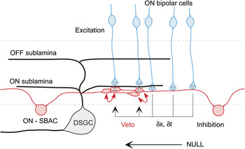
Barlow and Levick (Reference Barlow and Levick1965) originally proposed that spatially asymmetric inhibition fed forward through a delay line to veto excitation during null but not preferred direction motion. During preferred direction motion, the inhibition lagged behind the stimulus and thus arrived too late to veto excitation. Their model corresponds to a correlation-type detector that uses a delay line to compare the intensity at two spatially offset locations and veto the output if the delayed signal coincides with the local input (Fig. 1).
Inhibitory connections from SBACs to DSGCs and between SBACs. Null-side cells (right side) preferentially connect to the DSGC and make reciprocal inhibitory connections with oppositely oriented SBAC dendrites. A spatiotemporal offset arises due to locations of the bipolar cell inputs to the SBAC relative to its GABA release points, allowing inhibition to veto excitation of the DSGC in the null direction. This arrangement is repeated many times across the DSGC dendritic field. This circuit is presumably recapitulated in the OFF sublamina.

Analyses of the starburst connectivity to the DSGC had failed to reveal any anatomical asymmetry that would support such a model (Famiglietti, Reference Famiglietti1991; Dacheux et al., Reference Dacheux, Chimento and Amthor2003; Wei et al., Reference Wei, Hamby, Zhou and Feller2011), but physiological studies consistently showed that GABAergic connections from SBACs to DSGCs were stronger from the null side (Fried et al., Reference Fried, Münch and Werblin2002, Reference Fried, Münch and Werblin2005; Lee & Zhou, Reference Lee and Zhou2006; Zhou & Lee, Reference Zhou and Lee2008; Wei et al., Reference Wei, Hamby, Zhou and Feller2011; Yonehara et al., Reference Yonehara, Balint, Noda, Nagel, Bamberg and Roska2011). Most recently, however, a large-scale serial section electron microscopic reconstruction of DSGCs and their presynaptic SBACs has provided quantitative evidence for a marked anatomical asymmetry, in which SBACs on the null side of a DSGC make many more connections than those of the preferred side (Briggman et al., Reference Briggman, Helmstaedter and Denk2011), essentially as proposed some 20 years earlier (Vaney, Reference Vaney, Osborne and Chader1990).
This anatomical arrangement would appear to be sufficient to make the spatiotemporal correlation required to generate a DS signal in the DSGCs; for motion from the null side, the connected SBACs with their larger dendritic arbors, and output restricted to the peripheral dendrites, would inhibit underlying DSGCs ahead of a stimulus edge, while for motion in the opposite direction, the inhibition would trail excitation and thus have little effect. Such a model would predict that the magnitude of the inhibition observed within the DSGC should be independent of the direction of motion with the relative timing of inhibition and excitation being the deciding factor. Physiological evidence, however, shows that the magnitude of excitation and inhibition does change with direction, in a way consistent with generating a directional signal in the DSGC (Taylor & Vaney, Reference Taylor and Vaney2002; Fried et al., Reference Fried, Münch and Werblin2005). These observations support the notion that presynaptic mechanisms also contribute to DS signaling, and the discovery of directional calcium transients in the dendritic tips of SBACs provided indirect evidence that is at least consistent with directional release of neurotransmitter from these cells (Euler et al., Reference Euler, Detwiler and Denk2002; Lee & Zhou, Reference Lee and Zhou2006). While the source for directional inhibitory inputs to the DSGCs seems certain, the mechanisms generating directional excitation and the role of SBACs in this process remain uncertain. An obvious suggestion is that the GABAergic output from the SBACs is directed both to the DSGCs and also back onto the bipolar cell synaptic terminals that make input to the DSGCs, which would establish a push–pull arrangement in the null direction: increased GABAergic inhibition and suppressed glutamatergic excitation. Contrary to this hypothesis is evidence showing that SBACs do not make contact with bipolar cells (Brandon, Reference Brandon1987b; Famiglietti, Reference Famiglietti1991; Dacheux et al., Reference Dacheux, Chimento and Amthor2003); on the other hand, such connections are seen in primate (Yamada et al., Reference Yamada, Dmitrieva, Keyser, Lindstrom, Hersh and Marshak2003). Further work will be required to settle this issue.
In summary, SBACs contribute to generating directional signals at two levels in the retina, by direct inhibition of the DSGCs and presynaptically by generating directional release of GABA from individual dendrites. These two circuits will be discussed separately below.
Correlation model: postsynaptic
In considering how the inhibition generated in SBACs modulates the activity of the DSGCs, one needs to consider briefly how the DSGCs integrate the synaptic inputs and convert postsynaptic potentials into DS spiking. The veto operation proposed by Barlow and Levick (Reference Barlow and Levick1965) could simply comprise a linear inhibitory interaction preceding a threshold, for example, spike generation. In their initial characterization, Barlow and Levick showed that directional signals could be evoked by motion that covers a small fraction of the dendritic arbor, and they proposed that the directional circuitry was repeated numerous times across the receptive field. This distributed nature of the circuit precludes linear summation of synaptic inputs, with a threshold at the soma, because such a system would fail if numerous subunits were activated simultaneously with different temporal delays. In light of such considerations, Torre and Poggio (Reference Torre and Poggio1978) developed a specific model, in which shunting inhibition produced nonlinear independent subunits, and which required that each inhibitory synapse be located within about one tenth of an electrotonic space constant from the excitatory synapse. Such a model would predict that excitatory and inhibitory inputs should be clustered across the dendritic arbor, but contrary to this prediction, a detailed analysis of synaptic locations on DSGCs failed to show any such systematic spatial correlation between excitatory and inhibitory synapses (Jeon et al., Reference Jeon, Kong, Strettoi, Rockhill, Stasheff and Masland2002).
Observation of dendritic spikes in the DSGCs (Oesch et al., Reference Oesch, Euler and Taylor2005) suggested an alternative possibility, in which local linear inhibitory interactions within the dendritic arbor could be combined with a local dendritic spike threshold to generate directional dendritic spikes that propagated to the soma. A subsequent modeling analysis demonstrated the feasibility of such a scheme and showed that directional signals could be generated independently within numerous subunits across the dendritic arbor of the DSGCs (Schachter et al., Reference Schachter, Oesch, Smith and Taylor2010). These functional subunits provide the neural substrate for the summing point in the correlation model where the veto operation is performed. The modeling suggested that the veto works to suppress the initiation of dendritic action potentials because realistic magnitudes of inhibition were insufficient to cancel a dendritic action potential once initiated (Schachter et al., Reference Schachter, Oesch, Smith and Taylor2010).
The existence of multiple computational subunits within the DSGC arbors may provide some rationale for the very high density of SBACs, which have dendritic fields showing a 25- to 70-fold overlap (Tauchi & Masland, Reference Tauchi and Masland1984; Vaney, Reference Vaney1984); each dendritic subunit will require input from at least one SBAC dendritic branch of the appropriate orientation, thus requiring several coordinated SBACs to provide input across the dendritic arbor of each DSGC (Vaney, Reference Vaney, Osborne and Chader1990). Since the SBAC dendritic fields are larger than the DSGCs, they will overlap considerably. Given a subunit size of ∼40 μm, then each DSGC will require at least ∼10 SBACs within the dendritic field, which is within an order of magnitude of the observed SBAC coverage. Because each SBAC has dendrites emanating in all four directions, each of which acts independently, the same SBAC array can accommodate the inputs to all four populations of DSGC.
Correlation model: presynaptic
As noted above, the release of GABA from the terminals of SBACs is DS, which represents a second level of spatiotemporal correlation within the DS circuit. The spatial offset for the correlation is the same for postsynaptic and presynaptic mechanisms since the length of the SBAC dendrites determines it. One might expect that such a system would suppress excitation during null motion most efficiently at a velocity defined by the ratio of the spatial offset and a temporal delay defined by the circuit. In other words, for optimal DS responses, there should be an inverse relationship between the spatial frequency of a grating stimulus and the velocity, such that the temporal frequency matches the time constant of the delay (Borst et al., Reference Borst, Haag and Reiff2010). Such temporal tuning is evident in the fly visual system, where the optimal temporal frequency is ∼1 Hz, and the spatial offset matches the angle between adjacent ommatidia in the compound eye. Indeed, the DSGC shows speed tuning during preferred direction motion (Wyatt & Daw, Reference Wyatt and Daw1975) that presumably reflects the spatial summation and kinetics of the excitatory bipolar cell inputs. However, the strength of the directional inhibitory response generated by the null-direction veto operation is relatively insensitive to velocity (Grzywacz & Amthor, Reference Grzywacz and Amthor2007). The absence of a fixed temporal frequency for this inhibitory veto is perhaps expected because the excitatory drive to the SBACS is arrayed along the length of the dendrites, and the GABAergic output synapses are distributed over the outer third of the dendritic arbor. Therefore, a range of spatial offsets and temporal delays are possible between input and output. However, the nature of the temporal delay and the mechanisms for the asymmetric response of SBACs remain uncertain.
Mechanism for intrinsic SBAC DS
Numerous studies over recent years have generated a plethora of observations regarding the functional properties of SBACs and have led to three proposals for the origin of directional signals. One proposal is that DS is intrinsic to the dendrites of the SBAC (Euler et al., Reference Euler, Detwiler and Denk2002; Tukker et al., Reference Tukker, Taylor and Smith2004; Hausselt et al., Reference Hausselt, Euler, Detwiler and Denk2007; Oesch & Taylor, Reference Oesch and Taylor2010), the second idea invokes network interactions between connected SBACs (Lee & Zhou, Reference Lee and Zhou2006; Enciso et al., Reference Enciso, Rempe, Dmitriev, Gavrikov, Terman and Mangel2010), and a third suggests a unique role for intracellular chloride gradients (Gavrikov et al., Reference Gavrikov, Dmitriev, Keyser and Mangel2003). The common goal of these studies has been to explain the DS release of neurotransmitter from the SBAC.
Early modeling studies of the SBAC hypothesized that the dendritic branches, attached to the soma through thin initial segments, could be electrotonically isolated and therefore may represent independent processing subunits (Miller & Bloomfield, Reference Miller and Bloomfield1983; Velte & Miller, Reference Velte and Miller1997). A more recent modeling study showed that the dendritic morphology, without asymmetric inhibition, could generate DS postsynaptic potentials (PSPs) at the tips of the dendrites where the output terminals are located (Tukker et al., Reference Tukker, Taylor and Smith2004). This study provided an analysis of the morphological and electrotonic parameters that were important for this property and showed that a centrifugal stimulus (soma outwards) along a dendrite will generate direction selectivity purely by virtue of morphological asymmetries. However, it was also evident that the somatic preference was centripetal (towards the soma), as required for a linear model. The directional signals at the dendritic tips resulted from a phase shift of the voltage at the dendritic tips relative to the soma that was most effective at high stimulus velocities (∼1 mm/s ≈ 3 deg/s in rabbit). In this model, the delay line originally envisaged by Barlow and Levick (Reference Barlow and Levick1965) comprises conduction delays during passive spread of signals within the dendrites of the SBACs.
A later study of calcium signals in the distal dendrites of the SBAC showed a significant nonlinearity in the dendritic tips but the same directional preference at the tips and the soma (Hausselt et al., Reference Hausselt, Euler, Detwiler and Denk2007). Hausselt et al. (Reference Hausselt, Euler, Detwiler and Denk2007) proposed that the standing glutamatergic input that SBACs are known to receive (Taylor & Wässle, Reference Taylor and Wässle1995; Peters & Masland, Reference Peters and Masland1996) produces a voltage gradient within each SBAC dendrite, with the terminals being more depolarized than the soma, and that this couples with voltage-gated calcium channels to produce an asymmetric release of GABA. A subsequent study indicated that tetrodotoxin-resistant sodium channels could amplify a preexisting directional component of PSPs in the dendritic tips of SBACs (Oesch & Taylor, Reference Oesch and Taylor2010). Together, these studies suggest that voltage-gated sodium and calcium channels may represent a nonlinearity in the dendritic tips that amplifies small directional signals intrinsic to the morphology of the dendrites.
Reciprocal connections observed between SBACs (Millar & Morgan, Reference Millar and Morgan1987) and evidence for tonic GABAergic inhibition of SBACs (Massey & Redburn, Reference Massey and Redburn1982) laid the groundwork for the suggestion that directional signals might arise through network interactions involving a plexus starburst cells (Dacheux et al., Reference Dacheux, Chimento and Amthor2003; Lee & Zhou, Reference Lee and Zhou2006; Münch & Werblin, Reference Münch and Werblin2006; Enciso et al., Reference Enciso, Rempe, Dmitriev, Gavrikov, Terman and Mangel2010; Poznanski, Reference Poznanski2010). The central idea is that reciprocal GABAergic connections between opposing dendrites of SBACs produce a positive feedback network that can enhance the asymmetric DS voltage response in the peripheral dendrites of the SBACs. For motion in the null direction, the SBAC connected to the DSGC hyperpolarizes the opposing SBAC, resulting in a positive feedback loop, since the reciprocal inhibition from the opposing SBAC is suppressed. For motion in the preferred direction, the SBAC connected to the DSGC is hyperpolarized by the same mechanism. Thus, it has been argued that network interactions could produce or at least strongly enhance DS GABA release from the SBAC terminals (Lee & Zhou, Reference Lee and Zhou2006; Münch & Werblin, Reference Münch and Werblin2006). However, such positive feedback does not completely account for directional GABA release from SBACs since blocking GABA receptors does not abolish directional calcium transients in the SBACs (Lee & Zhou, Reference Lee and Zhou2006; Hausselt et al., Reference Hausselt, Euler, Detwiler and Denk2007) or directional voltage responses in SBACs (Oesch & Taylor, Reference Oesch and Taylor2010).
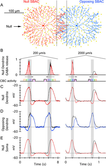
In order to assess how effective reciprocal inhibition might be in generating directional responses, we implemented a realistic model of two opposing SBACs with reciprocal GABAergic connections (Fig. 2A) and simulated a light bar moving across the dendritic arbors of the two cells from left to right and back. The black traces in Fig. 2B–2E show the model simulations without reciprocal inhibition. During motion directed towards the dendritic tips, which represents null-direction motion for the underlying DSGC, the peak voltages in the dendrites show directional differences (Fig. 2C), which are stronger at the higher velocity. These directional differences, seen in the absence of reciprocal inhibition, are intrinsic to the morphology of the SBACs, as shown previously (Tukker et al., Reference Tukker, Taylor and Smith2004). In this simple model, the voltage in the dendrite is converted into a GABA release rate (Fig. 2B) by inclusion of voltage-gated calcium channels with an activation threshold at −45 mV. The voltage-gated calcium channels introduce a threshold nonlinearity during the release of GABA. At the high velocity, the release in the preferred direction was ∼48% of that in the null. The addition of reciprocal inhibition to the model suppressed preferred direction GABA release to ∼11% of that in the null (compare the red shaded areas in Fig. 2B). At the low velocity, the corresponding values were 92% (no inhibition) and 55% (reciprocal inhibition). Thus, reciprocal inhibition enhanced directional GABA release, an effect that was particularly important at low velocities where the intrinsic morphological mechanism was relatively weak. For this model, the presence or absence of reciprocal inhibition had very little effect on GABA release in the null direction. However, in the opposite preferred direction, the null-direction SBAC was initially hyperpolarized by the opposing SBAC, which suppressed subsequent GABA release and thus enhanced directional signaling. The model predicts that for a bar moving across the entire dendritic arbor, the voltage at the soma should comprise a central depolarizing phase flanked by hyperpolarizations, which is very similar to observed responses (see fig. 5b, Enciso et al., Reference Enciso, Rempe, Dmitriev, Gavrikov, Terman and Mangel2010). This simple model does not include all nonlinear properties of SBACs that have been proposed to contribute to directional GABA release (Gavrikov et al., Reference Gavrikov, Dmitriev, Keyser and Mangel2003, Reference Gavrikov, Nilson, Dmitriev, Zucker and Mangel2006; Hausselt et al., Reference Hausselt, Euler, Detwiler and Denk2007; Oesch & Taylor, Reference Oesch and Taylor2010).
Reciprocal inhibition between opposing SBAC dendrites amplifies DS. (A) Morphology of SBAC used for the model simulations. This cell was used in our previous study (Tukker et al., Reference Tukker, Taylor and Smith2004). The orange spots show bipolar cell inputs. The red SBAC is connected to a DSGC having the indicated null direction. (B–E) Simulation of a bar moving forward and backwards along the axis connecting the somas of the SBACs. The shaded bars show the region of dendritic overlap. (B) Predicted GABA input to the DSGC from the null dendrite, with reciprocal inhibition (red) and without (black). Note that at the high velocity, the morphology alone produces marked directional release (black traces), as shown previously (Tukker et al., Reference Tukker, Taylor and Smith2004). The three lower panels show the voltage within the terminal dendrites (C, D) and soma (E) of the SBACs, with and without reciprocal inhibition.

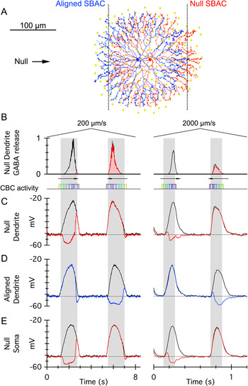
SBACs are present at high density, with a mean soma spacing of ∼45–50 μm, about one fourth the diameter of the dendritic field. Therefore, adjacent somas will lie within the dendritic extent of their neighbors, and the overlapping dendrites will tend to be aligned, not opposing (Fig. 3). Paired recordings have shown that the strength of reciprocal inhibitory connections is similar between closely spaced SBACs (as in Fig. 3) and widely spaced cells (as in Fig. 2. See fig. 1 in Lee & Zhou, Reference Lee and Zhou2006). Simulation of reciprocal inhibition between closely spaced cells with similarly oriented dendrites suggests that DS GABA release would be attenuated rather than amplified (Fig. 3). The problem arises because the blue SBAC is activated ahead of the red one, and therefore inhibits it. Similarly spaced SBACs located orthogonal to the null direction (i.e., the blue soma rotated 90 degrees around the red one), may also contribute to reciprocal inhibition, but since they would be activated synchronously with the null-direction SBAC, they will not produce an opposing directional signal. These observations suggest that reciprocal connections between SBACs that are aligned along the preferred-null axis are counterproductive. A possible solution to this dilemma is suggested by a preliminary finding that GABA receptors are preferentially located on the varicosities in outer third of the SBAC dendrites (Auferkorte et al., Reference Auferkorte, Kaushalya, Reddy, Euler and Haverkamp2011). Such an arrangement would tend to preclude reciprocal interactions along the preferred-null axis but would allow connections between overlapping orthogonally directed dendrites. These orthogonal connections could account for the recriprocal signals observed between closely spaced SBACs (Lee & Zhou, Reference Lee and Zhou2006). It should be noted that these simple models, employing two isolated cells, may not fully capture the behavior of SBAC dendrites when embedded within a continuous plexus that extends many dendritic field diameters across the retina. Additional modeling, using a wide array of SBACs, could be used to determine the key properties of a reciprocally connected network that would produce robust DS signals in SBACs.
Reciprocal inhibition between aligned SBAC dendrites attenuates direction selectivity. (A) SBACs from Fig. 2, with a soma separation of 50 μm, which is similar to that observed in rabbit. (B–E) Simulation of a bar as for Fig. 2. The aligned (blue) SBAC inhibits the null SBAC during null-direction motion, resulting in larger GABA release for the opposite preferred direction (shaded red). This directional signal opposes and thus attenuates that shown in Fig. 2.

Finally, a combination of network and intrinsic models has been proposed, which emphasizes a unique role for an asymmetric distribution of chloride channel transporters, and a resulting chloride gradient within the dendrites of SBACs (Gavrikov et al., Reference Gavrikov, Dmitriev, Keyser and Mangel2003, Reference Gavrikov, Nilson, Dmitriev, Zucker and Mangel2006; Enciso et al., Reference Enciso, Rempe, Dmitriev, Gavrikov, Terman and Mangel2010). Further analysis is required to determine the relative importance of intrinsic and network mechanisms in generating the directional output of GABA from SBACs.
The role of nicotinic excitation
SBACs are the major and most likely the only cholinergic neurons in the retina. Paired recordings have demonstrated fast nicotinic synapses between SBACs and DSGCs (Lee et al., Reference Lee, Kim and Zhou2010); however, nicotinic receptors appear to be present on a number of other ganglion cells types that do not necessarily costratify with the SBACs (Masland & Ames, Reference Masland and Ames1976; Ariel & Daw, Reference Ariel and Daw1982). The role of nicotinic transmission in these ganglion cells, and the DSGCs, remains unclear. Blocking nicotinic receptors attenuates spiking by 50% in rabbit DSGCs (Kittila & Massey, Reference Kittila and Massey1997), an effect that appeared to apply equally to ON and OFF responses. However, analysis of synaptic currents in rabbit DSGCs indicated a reduction in the OFF response, with no discernible effect on the ON response (Fried et al., Reference Fried, Münch and Werblin2005), while a subsequent study found that nicotinic inputs contributed to both ON and OFF responses (Lee et al., Reference Lee, Kim and Zhou2010). These differences echo our own unpublished observations, in which we have been unable to demonstrate clear and reproducible effects of nicotinic blockade on the ON and OFF excitatory synaptic currents in rabbit DSGCs. Perhaps these diverse results arise in part due to the use of stimuli that differentially engage cholinergic mechanisms (Grzywacz et al., Reference Grzywacz, Amthor and Merwine1998). Blocking GABAergic inhibition can produce upregulation of the magnitude of cholinergic inputs to DSGCs that appear to be spatially symmetric (Chiao & Masland, Reference Chiao and Masland2002; Fried et al., Reference Fried, Münch and Werblin2005). Such symmetry of cholinergic input is unexpected given the strong spatial asymmetry in synaptic connections reported at the electron microscopic level (Briggman et al., Reference Briggman, Helmstaedter and Denk2011). One possible explanation for the variable results concerning nicotinic transmission is offered by recent results showing that acetylcholine release from SBACs may require higher intracellular calcium concentrations and thus requires stronger calcium influx than for GABA release (Lee et al., Reference Lee, Kim and Zhou2010). Thus, for example, the large nicotinic EPSCs observed in DSGCs during GABA block may result because the removal of inhibition allowed stronger and nonphysiological depolarization of SBACs. In summary, the anatomical and physiological specificity of nicotinic transmission from SBACs to DSGCs and other types of retinal ganglion cells remains unclear.
OFF and ON pathways
The SBACs are central components to the DS circuit, and given the mirror symmetry in the ON and OFF SBAC populations, it is tempting to assume that the DS synaptic mechanisms are similarly recapitulated. However, it is important to note that apart from a single study showing voltage responses (Bloomfield, Reference Bloomfield1992), the physiological properties of OFF SBACs remain largely unexplored. To date, all the imaging of SBACs and physiological studies of SBAC/DSGC pairs have targeted the ON cells since they are easily accessible, with somas displaced to the ganglion cell layer. One of the first studies to quantitatively examine the synaptic mechanisms in DSGCs documented differences between the ON and OFF synaptic inputs (Taylor & Vaney, Reference Taylor and Vaney2002). For example, the OFF inhibition but not the ON inhibition displayed a temporal offset relative to the excitation, which could result from a difference in the spatial offset within the synaptic circuitry. However, given the morphological similarities between the SBAC populations, it is not obvious how such a difference might arise. The same study showed that on average the directional difference in the total inhibitory input was larger for ON than for OFF responses, suggesting corresponding differences in the two SBAC populations. ON–OFF differences have been observed in other studies. For example, the OFF excitation but not the ON excitation was suppressed by nicotinic antagonists (Fried et al., Reference Fried, Münch and Werblin2005), while the ON inhibition was actually enhanced by these same antagonists and the OFF inhibition was unaffected. Moreover, the original anatomical work noted quantitative differences in the density (OFF SBAC < ON SBAC), receptive field size (OFF SBAC > ON SBAC, ∼10%), and somatic arrays (ON SBAC less regular than OFF SBAC) of the ON and OFF SBACs (Vaney, Reference Vaney, Osborne and Chader1990). Lastly, functional asymmetries are evident between retinal ON and OFF pathways, with the ON pathway displaying more linear response properties than the OFF pathway, likely due to crossover inhibition from the ON pathway that rectifies the output of OFF bipolar cells (Zaghloul et al., Reference Zaghloul, Boahen and Demb2003; Liang & Freed, Reference Liang and Freed2010). If similar mechanisms are active within the DSGC circuits, then the ON and OFF SBACs may display differences in the synaptic mechanisms generating ON and OFF directional responses.
Conclusions
In summary, much progress has been made in elucidating the physiological role of SBACs in the DS circuitry. The SBACs generate directional signals at two levels, by asymmetric release of GABA and by asymmetries in morphology and connectivity with the DSGCs. However, the mechanisms intrinsic to the SBAC plexus that generate DS release of GABA remain unclear. The role of reciprocal inhibition between SBACs, and the contributions from voltage-gated channels, and chloride transporters remain to be clearly established. Finally, the role of cholinergic mechanisms in directional signaling and a comparison of the synaptic mechanisms in OFF versus ON SBACs merit further investigation.