Background
Blindness has a significant impact on the quality of a person’s life, often resulting in depression, social isolation, and premature death. This poses a major burden to society due to lost productivity and earnings as well as the cost of treatment, rehabilitation, education of the visually impaired and provision of visual aids. Recent estimates indicate that the global number of people with sight loss is 285 million, of whom 39 million are classified blind (Pascolini & Mariotti, Reference Pascolini and Mariotti2012). Visual impairment is not evenly distributed across age groups, with more than 82% of blindness occurring beyond 50 years of age. Although the prevalence of blindness in children is approximately 10 times lower than in adults, childhood blindness remains a high priority because the predicted duration of their suffering is protracted (Pascolini & Mariotti, Reference Pascolini and Mariotti2012). A conservative analysis estimated that the loss of productivity of individuals with visual impairment in 2003 bore a $42 billion impact on society with a projected rise to $110 billion by 2020; hence, the return on investment is potentially high compared with the research costs involved in the development of new treatment modalities.
Diseases affecting the outer retina including age related macular degeneration (AMD) and hereditary retinal dystrophies (HRDs) account for approximately 26% of blindness worldwide. The incidence is expected to double by 2020 and to increase to 75% by 2040 as the world population steadily ages (Pascolini & Mariotti, Reference Pascolini and Mariotti2012). Although in some cases the primary pathological event originates in the retinal pigmented epithelium (RPE, a supportive monolayer of pigmented cells forming part of the blood-retinal barrier), the final impact of both AMD and HRDs is the loss of photoreceptors. While there are a number of agents (including high dose antioxidants, neuronal survival agents, and vascular endothelial growth factor [VEGF] inhibitors in patients with neovascular AMD: Krishnadev et al., Reference Krishnadev, Meleth and Chew2010; Arias, Reference Arias2010; Kaiser et al., Reference Kaiser, Brown, Zhang, Hudson, Holz, Shapiro, Schneider and Acharya2007; Rosenfeld et al., Reference Rosenfeld, Brown, Heier, Boyer, Kaiser, Chung and Kim2006) that have been shown to slow outer retinal disease progression, there are no treatments to restore photoreceptors that have already been lost; hence, there is a pressing need for research into the replacement and/or reactivation of dysfunctional photoreceptors as well as RPE in order to restore visual function in these cases. The eye is well suited for the development of cell transplantation therapies as it is easily accessible, allowing the accurate delivery of cells to the retina with minimal risk of systemic effects. To date, gene therapy has dominated the majority of clinical trials for the treatment of eye disease (Otani et al., Reference Otani, Dorrell, Kinder, Moreno, Nusinowitz, Banin, Heckenlively and Friedlander2004; Bainbridge et al., Reference Bainbridge, Smith, Barker, Robbie, Henderson, Balaggan, Viswanathan, Holder, Stockman, Tyler, Petersen-Jones, Bhattacharya, Thrasher, Fitzke, Carter, Rubin, Moore and Ali2008; Maguire et al., Reference Maguire, Simonelli, Pierce, Pugh, Mingozzi, Bennicelli, Banfi, Marshall, Testa, Surace, Rossi, Lyubarsky, Arruda, Konkle, Stone, Sun, Jacobs, Dell’Osso, Hertle, Ma, Redmond, Zhu, Hauck, Zelenaia, Shindler, Maguire, Wright, Volpe, McDonnell, Auricchio, High and Bennett2008; Smith et al., Reference Smith, Bainbridge and Ali2009; Cideciyan et al., Reference Cideciyan, Jacobson, Beltran, Sumaroka, Swider, Iwabe, Roman, Olivares, Schwartz, Komáromy, Hauswirth and Aguirre2013); however, this approach is applicable only to HRDs that are well characterized genetically, show early onset symptoms and slow degeneration. Early treatment of such forms of HRD by gene therapy is likely to succeed in both improved visual function and photoreceptor protection; however, at later stages of the disease this method is unlikely to be effective and will necessitate additional approaches combined with gene therapy (Cideciyan et al., Reference Cideciyan, Jacobson, Beltran, Sumaroka, Swider, Iwabe, Roman, Olivares, Schwartz, Komáromy, Hauswirth and Aguirre2013). In such cases, cell replacement therapies offer an attractive complementary approach.
Pluripotent is best: Why and how?
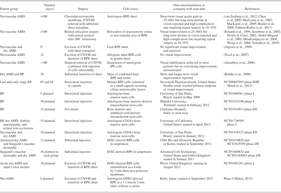
A key reason for using stem cell–based therapies to treat retinal disorders is the prospect of generating unlimited quantities of desired cell types for transplantation. A variety of cell types have been tested in animal models and human clinical trials (Table 1) for their ability to repopulate the degenerate retina or rescue retinal neurons from further degeneration, in particular the RPE. In most cases, these sources include autologous cells from the eye itself (iris pigmented epithelial cells [IPE] and RPE). As can be seen from Table 1, both autologous RPE transplantation and retinal relocation surgery to a healthy area of RPE can result in increase in visual acuity or stabilization of vision in a proportion of patients; however, postoperative complications and the likelihood of continued deterioration of visual acuity secondary to disease recurrence are also quite high, making these procedures unlikely to succeed in a large number of patients with AMD or HRD. Nevertheless, they provide proof of concept that diseased RPE replacement, probably most optimally performed in a sheet configuration, can result in visual improvement and photoreceptor rescue in some cases.
A summary of clinical trials focusing on stem cell transplantation in AMD and HRD patients

Neurosensory retinal regeneration is more complex. Transplants of hematopoietic and mesenchymal stem cells isolated from adult bone marrow are being tested in patients with retinitis pigmentosa (RP), Stargardt’s disease and AMD, and the clinical outcomes are eagerly awaited (Table 1). Notwithstanding this, studies conducted in animal models have demonstrated that these cells are unable to repopulate the degenerate retina and give rise to functional photoreceptors, but act to prevent further retinal degeneration via the production of neurotrophic factors. Therefore, although this approach may be useful for retinal protection during the early degenerative phase, it is unlikely to work in cases of advanced degeneration where substantial retinal remodeling has already taken place. Other studies have investigated the possibility of deriving photoreceptor cells by reprogramming cell sources found within the eye, either using genetic manipulation or coaxing cell plasticity by manipulating culture conditions. To this end, various ocular tissues have been tested including RPE (Li et al., 2010), IPE (Haruta et al., 2001), ciliary body (MacNeil et al., 2007) and limbal epithelium (Zhao et al., 2008). Of these, RPE cells appear the most promising to date with experimental findings showing that reprogramming of chick RPE with neurogenin can give rise to cell layers expressing photoreceptor and phototransduction genes (Li et al., 2010). Functional assays such as the response to light and 9-cis-retinal were however not performed, nor transplantation tests, thus leaving open the question whether full reprogramming of RPE to functional photoreceptors had taken place.
More recently, pluripotent stem cells have been investigated for their ability to contribute to retinal restoration. The term pluripotent stem cell is commonly used to describe two types of stem cells characterized by their indefinite self-renewal ability and the capacity to give rise to any somatic cell type in the adult, namely embryonic stem cells (ESCs) and induced pluripotent stem cells (iPSCs) (Mellough et al., Reference Mellough, Steel and Lako2009). Human ESCs (hESCs) are derived from surplus in vitro fertilized embryos and have been widely used in the last decade as a research tool to understand the mechanisms behind the maintenance of pluripotency, human embryonic development and congenital disease. The use of human embryos for research purposes is surrounded by a number of ethical issues, prohibiting hESC derivation and application in several countries. In addition, a major issue related to their biological application is the evidence that their differentiated progeny expresses human leukocyte antigens (HLAs) that could result in graft rejection. This could be overcome by the creation of HLA-typed hESC banks, from which a best match can be selected for each transplant recipient (Taylor et al., Reference Taylor, Bolton, Pocock, Sharples, Pedersen and Bradley2005). However, human iPSCs (hiPSCs) bypass both of these issues as they are generated by reprogramming somatic cells back to a pluripotent-like state akin to ESCs (Lako et al., Reference Lako, Armstrong and Stojkovic2010; Rashid & Vallier, Reference Rashid and Vallier2010). As such, these cells share many key characteristics of hESCs including the ability to proliferate indefinitely and differentiate into different cell types, but also represent a source of autologous stem cells given that they can be derived from the patients themselves. Such patient-derived cells present a unique opportunity to create in vitro disease models, which can be exploited to understand disease pathology and drug discovery (see Fig. 1). This becomes extremely important for degenerative diseases such as those affecting the retina, where availability of patient-specific cells (i.e., photoreceptors and RPE) is only possible with invasive surgery or post mortem. New tools developed in the gene therapy field including improved and safer viral vectors as well as the possibility of correcting endogenous mutations through the application of site-specific restriction endonucleases, such as Zinc Finger Nucleases (ZFNs) and Transcription Activator-Like Effector Nucleases (TALENs), also mean that functional cells for transplantation can be produced from hiPSCs that, without genetic modification techniques, would otherwise harbor typical disease characteristics and therefore be less suitable for transplantation (Sundaram et al., Reference Sundaram, Moore, Ali and Bainbridge2012; Collin & Lako, Reference Collin and Lako2011).
Schematic summary of hiPSC derivation and its applications in disease modeling, drug discovery, and cell replacement therapies.
The choice of cell type for transplantation into the neurosensory retina is heavily dictated by the ability of grafted cells to integrate with high efficiency within the correct retinal layer and contribute to improved vision by restoring retinal circuitry. Studies performed by Robin Ali’s group in murine retina have shown that postmitotic photoreceptor precursors isolated at a very specific time point during mouse embryonic development (MacLaren et al., Reference MacLaren, Pearson, MacNeil, Douglas, Salt, Akimoto, Swaroop, Sowden and Ali2006; Lakowski et al., Reference Lakowski, Baron, Bainbridge, Barber, Pearson, Ali and Sowden2010) demonstrate the highest levels of engraftment within host retinae and acquire the specialized morphological features of mature photoreceptors. For the transplants to be successful, other events have to occur in addition to photoreceptor cell engraftment into the correct retinal layer. An important step is the ability of newly integrated cells to make the appropriate synaptic connections with the host retina and to restore functional retinal circuitry to a measurable degree that can be demonstrated by different functional tests. A recent study (Gonzalez-Cordero et al., Reference Gonzalez-Cordero, West, Pearson, Duran, Carvalho, Chu, Naeem, Blackford, Georgiadis, Lakowski, Hubank, Smith, Bainbridge, Sowden and Ali2013) has shown that newly transplanted rod photoreceptor precursors isolated from postnatal mice form typical triad synaptic connections with second-order bipolar and horizontal cells of the recipient retina and generate visual signals which are transmitted to higher visual areas in the brain. Improved vision was shown in transplanted mice, as assessed by optomotor head-tracking responses and a visually guided water-maze test, thus demonstrating the feasibility of photoreceptor transplantation as a therapeutic strategy. Notwithstanding these remarkable results, one has to be aware of the multiple challenges on the route to restoring functional retinal circuitry by transplanted photoreceptors, not the least of which is the ability to achieve a sufficient critical mass of engrafted photoreceptors (Gonzalez-Cordero et al., Reference Gonzalez-Cordero, West, Pearson, Duran, Carvalho, Chu, Naeem, Blackford, Georgiadis, Lakowski, Hubank, Smith, Bainbridge, Sowden and Ali2013). In addition, changes in the host retinal environment such as glial scarring and the integrity of the outer limiting membrane (which acts as a barrier to integration) differ with the type and stage of disease and, as shown in a recent paper, bear a heavy impact on photoreceptor transplantation (Barber et al., Reference Barber, Hippert and Duran2013), thus suggesting that both need to be fully assessed and manipulated to achieve the best photoreceptor transplantation across a wide range of retinal diseases at both early and late stages of degeneration.
All the above studies have been achieved in mouse and for this success to be translated to humans, equivalent cells would need to be isolated from second trimester fetuses, an issue which raises obvious ethical concerns. Multiple reports have shown that in vitro differentiation of hESCs and hiPSCs can in fact recapitulate many aspects of human embryonic development (Lako et al., Reference Lako, Armstrong and Stojkovic2010; Rashid & Vallier, Reference Rashid and Vallier2010). Therefore, if human embryonic retinal precursors represent the optimal transplantable tissue for retinal restoration, then in vitro differentiation of hESCs and hiPSCs provides the best currently available platform to achieve this goal.
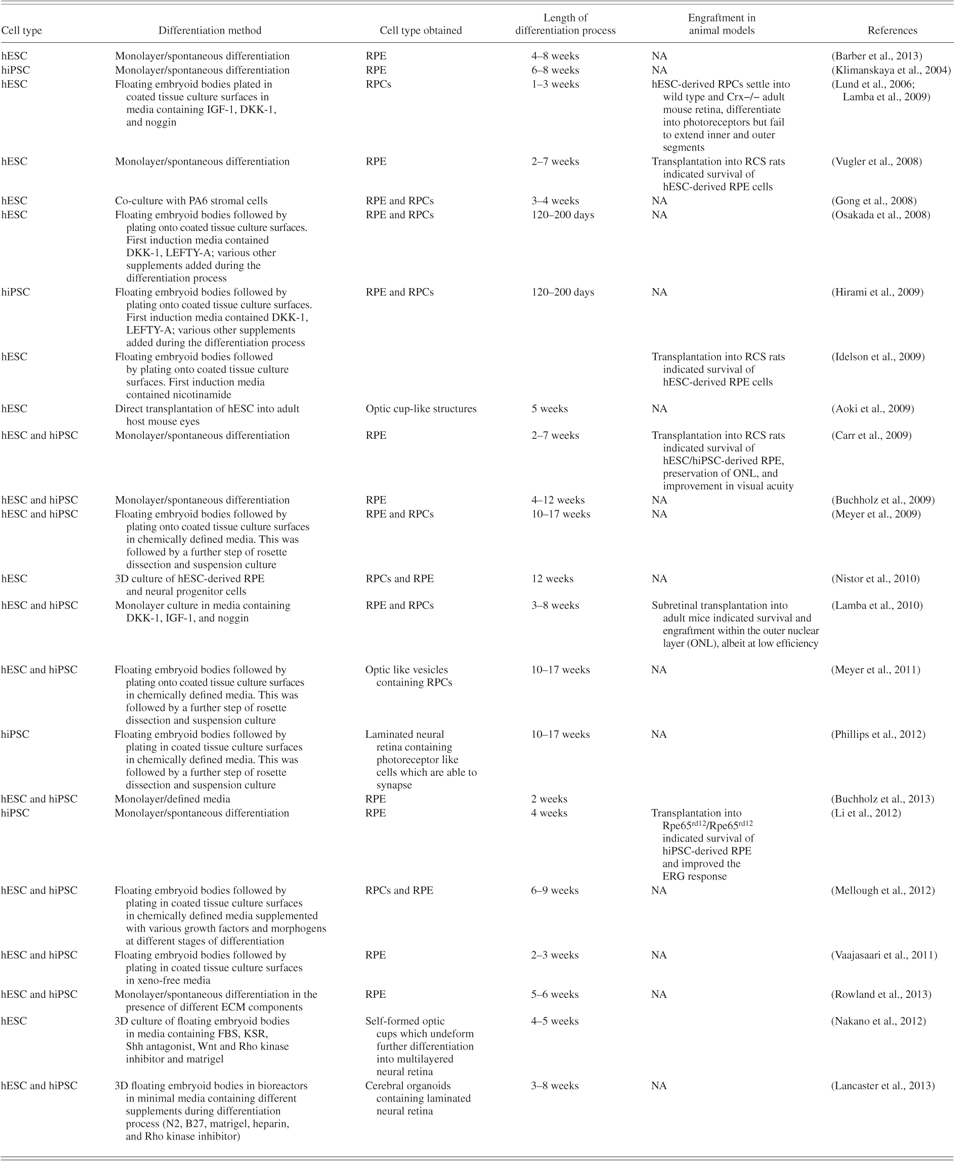
The earliest indication of the possibility of replicating the murine data using human-derived cells came from the first report of the production of human RPE during the spontaneous differentiation of hESCs in 2004 (Table 2; Aoki et al., Reference Aoki, Hara, Niwa, Yamada and Kunisada2009; Barber et al., Reference Barber, Hippert and Duran2013) and the presence of optic cup-like structures containing primitive neural retina and RPE in teratomas formed from pluripotent stem cells. With an aim to enhance the differentiation process and produce functional photoreceptors and RPE from hESCs and hiPSCs, several researchers in the field have used a number of growth factors and morphogens that are known to enhance anterior neural specification (bone morphogenetic protein [BMP] and Wnt inhibitors: Osakada et al., Reference Osakada, Ikeda, Mandai, Wataya, Watanabe, Yoshimura, Akaike, Sasai and Takahashi2008; Hirami et al., Reference Hirami, Osakada, Takahashi, Okita, Yamanaka, Ikeda, Yoshimura and Takahashi2009; Mellough et al., Reference Mellough, Sernagor, Moreno-Gimeno, Steel and Lako2012; Lund et al., Reference Lund, Wang, Klimanskaya, Holmes, Ramos-Kelsey, Lu, Girman, Bischoff, Sauvé and Lanza2006), retinal precursor cell (RPC) emergence (insulin-like growth factor 1 [IGF-1]: Lund et al., Reference Lund, Wang, Klimanskaya, Holmes, Ramos-Kelsey, Lu, Girman, Bischoff, Sauvé and Lanza2006; Lamba et al., Reference Lamba, Karl, Ware and Reh2006; Mellough et al., Reference Mellough, Sernagor, Moreno-Gimeno, Steel and Lako2012), photoreceptor maturation (activin A, taurine, sonic hedgehog [Shh]: Mellough et al., Reference Mellough, Sernagor, Moreno-Gimeno, Steel and Lako2012), or RPE differentiation (nicotinamide, activin A: Idelson et al., Reference Idelson, Alper, Obolensky, Ben-Shushan, Hemo, Yachimovich-Cohen, Khaner, Smith, Wiser, Gropp, Cohen, Even-Ram, Berman-Zaken, Matzrafi, Rechavi, Banin and Reubinoff2009; refer to Table 2). Each of these studies was performed by differentiating cells as a monolayer under two-dimensional (2D) conditions and, despite being able to generate photoreceptor-like cells with high efficiency (up to 80% (Mellough et al., Reference Mellough, Sernagor, Moreno-Gimeno, Steel and Lako2012), upon transplantation into animal models of retinal degeneration, such hESC-derived photoreceptors engrafted with low efficiency (an average of 3000 cells Nrl+ human cells found in the Crx−/− retina after transplantation) failed to develop photoreceptor outer segments (an essential component for the phototransduction process) and only in very rare cases acquired the expression of photoreceptor markers (Lamba et al., Reference Lamba, Karl, Ware and Reh2009), suggesting that 2D culture conditions may not be the ideal route for generating fully mature functional photoreceptors. What is fascinating is that although unable to elaborate outer segments following subretinal transplantation into Crx−/− mice, in the same study, hESC-derived retinal cells grafted into normal adult mouse retina were capable of outer segment formation, indicating that existing host photoreceptors or interphotoreceptor matrix may provide vital support for the final functional maturation of this cell type upon transplantation (Lamba et al., Reference Lamba, Karl, Ware and Reh2006).
Summary of main protocols used to date to cue differentiation of hESC and hiPSC to RPE and photoreceptor cells
Recent ground-breaking work from Yoshiki Sasai’s group has shown that both murine ESCs (mESCs) and hESCs are able to generate self-organizing optic cups when cultured under three-dimensional (3D) minimal culture conditions (Eiraku et al., Reference Eiraku, Takata, Ishibashi, Kawada, Sakakura, Okuda, Sekiguchi, Adachi and Sasai2011; Nakano et al., Reference Nakano, Ando, Takata, Kawada, Muguruma, Sekiguchi, Saito, Yonemura, Eiraku and Sasai2012). Most importantly, mESC- and hESC-derived optic cups can generate fully laminated neural retina containing all classes of retinal cells including the light sensitive photoreceptors, following the normal sequence of retinal development (Fig. 2). A surprising finding of this study was that optic cups formed independently of any interaction with neuroepithelial cells, surface ectoderm or mesenchymal tissues that ordinarily surround them in the developing embryo, challenging our traditional understanding of this developmental process. Importantly, murine rod precursors arising from the 3D differentiation system described above have the ability to integrate within the degenerate retina in adult mice and mature into photoreceptors showing outer segment formation (Gonzalez-Cordero et al., Reference Gonzalez-Cordero, West, Pearson, Duran, Carvalho, Chu, Naeem, Blackford, Georgiadis, Lakowski, Hubank, Smith, Bainbridge, Sowden and Ali2013), while no outer segments were observed in optic cups derived from hESCs.
Schematic representation of retinal ontogenesis.
Human transplantation studies have yet to be attempted with hESC and hiPSC 3D-derived photoreceptors; nevertheless, the above findings highlight an important facet of ESC and iPSC biology that is essential for the field of retinal regeneration: a latent intrinsic ability to self-organize giving rise to a multilayered neural retina which can be exploited for drug discovery purposes, disease modeling, and retinal regeneration. Such in vitro grown retina has profound potential for determining the molecular and inductive interactions that are essential for eye development which have not yet been elucidated from embryological studies due to the scarcity of human embryonic material. Furthermore, the ability to produce laminated neural retina containing functional photoreceptors with fully formed outer segments is invaluable for producing in vitro models of retinal disease that are sufficiently close to in situ retina in order to obtain clinically useful results. The current cost of bringing a new drug to the market has been evaluated in the range of 4 to 11 billion USD. This has been associated with a high failure rate due to the lack of appropriate biological models that reflect patient populations. The reproducible generation of fully functional neural retina from a large number of hESC and hiPSC lines offers an amazing prospect for drug discovery and disease modeling using hiPSCs generated from patient tissues. The generation of photoreceptor precursors within a 3D in vitro microenvironment which closely resembles that of the developing human retina may also be our best chance of producing photoreceptor precursors that have received the relevant “priming” signals during their development for them to act in an appropriate functional manner following transplantation in humans. It is also possible that transplantation of combined RPE/retinal sheets derived from pluripotent stem cells may be a successful strategy as carried out in pilot studies with human fetal tissue (Radtke et al., Reference Radtke, Aramant, Petry, Green, Pidwell and Seiler2008).
Given the incredible promise of pluripotent stem cell differentiation, it is not surprising that clinical safety studies focused on the more straightforward challenge of transplanting hESC and hiPSC-derived RPE have already started (Table 1). The first trial was approved by the U.S. Food and Drugs Administration in January 2011 to enable Advanced Cell Technology (ACT) to perform a phase I/II multicenter clinical trial to treat dry AMD patients with RPE cells derived from hESCs. Japan has already approved the second clinical trial, scheduled to start in September 2013, and is intended to treat six patients with wet AMD using autologous RPE cells obtained from iPSCs. Three additional trials (Table 1) are also imminent in the United Kingdom, United States, and Korea. Although the long-term outcome of these trials is eagerly awaited, early results from the ACT study have underlined the safety and tolerability of these cells for clinical trials and have set the scene for pioneering new therapies for retinal disease (Schwartz et al., Reference Schwartz, Hubschman, Heilwell, Franco-Cardenas, Pan, Ostrick, Mickunas, Gay, Klimanskaya and Lanza2012).
What does the future hold?
It is clear that the generation of laboratory grown synthetic retina from hESCs and hiPSCs provides one of the best tools to date for modeling human retinal disease, large-scale drug screening, and disease reversion by transplantation of patient-specific photoreceptors and/or RPE following the correction of disease-linked gene mutations. To realize these goals, we need to optimize current differentiation protocols to achieve the efficient and reproducible generation of synthetic retina from a large number of hESC and hiPSC lines in defined media conditions and within time periods that are amenable to human cell replacement therapies. Current hESC-based studies suggest that it takes up to 126 days to generate a fully laminated neural retina (Nakano et al., Reference Nakano, Ando, Takata, Kawada, Muguruma, Sekiguchi, Saito, Yonemura, Eiraku and Sasai2012). Moreover, culture media commonly include fetal bovine serum (FBS) which is of animal origin and can show batch-to-batch variability and hence is not suitable for clinical applications (Nakano et al., Reference Nakano, Ando, Takata, Kawada, Muguruma, Sekiguchi, Saito, Yonemura, Eiraku and Sasai2012). Under these conditions, around 58% of hESC-derived aggregates contain optic-like vesicles. However, the optic vesicles that undergo further differentiation to stratified neural retina contain photoreceptors lacking outer segments and therefore unable to respond to light. In conclusion, there remains clear scope for improvement and below we have highlighted several methods that may enhance our ability to generate functional photoreceptors:
The provision of a microenvironment which allows endogenous signaling to guide retinal formation
Most cell culture–based studies tend to rely on the application of fetal bovine serum or similar substituents (for example knock-out serum [KSR]) to achieve either cell expansion or differentiation toward desired lineages. This has proven to be counterproductive in protocols designed to generate synthetic retina, as knock-out serum caudalises hESC-derived neural progenitors, contributing to low yields of ESC-derived optic cups which ordinarily emerge from anterior forebrain (diencephalon) during embryonic development (Eiraku et al., Reference Eiraku, Takata, Ishibashi, Kawada, Sakakura, Okuda, Sekiguchi, Adachi and Sasai2011; Nakano et al., Reference Nakano, Ando, Takata, Kawada, Muguruma, Sekiguchi, Saito, Yonemura, Eiraku and Sasai2012). Indeed, lowering the knock-out serum concentration results in greater efficiency of optic cup formation from both hESCs and mESCs (Eiraku et al., Reference Eiraku, Takata, Ishibashi, Kawada, Sakakura, Okuda, Sekiguchi, Adachi and Sasai2011; Nakano et al., Reference Nakano, Ando, Takata, Kawada, Muguruma, Sekiguchi, Saito, Yonemura, Eiraku and Sasai2012). Our work and that of others have shown that this is due to the ability of differentiating hESCs to endogenously upregulate the expression of a range of factors important for retinal cell type specification (Mellough et al., Reference Mellough, Sernagor, Moreno-Gimeno, Steel and Lako2012). For example, embryoid bodies obtained from hESCs are able to upregulate the expression of EGF, DKK1, NGF, NODAL, and SHH when allowed to differentiate in the absence of any added morphogens or growth factors. This suggests that, in the absence of external cues, a proportion of hESCs follow an intrinsic neural and retinal default differentiation pathway. However, when these conditions are maintained long term, we have also noticed a delay in the onset of expression of mature photoreceptor markers, which can be rescued by the addition of two culture supplements, N2 and B27, at specific stages of the differentiation process (Mellough et al., Reference Mellough, Sernagor, Moreno-Gimeno, Steel and Lako2012). Combining these two nutrients with matrigel, a gelatinous protein mixture mimicking the complex extracellular environment under bioreactor culture conditions (allowing more efficient exchange of gas and nutrients), has facilitated the generation of cerebral organoids containing an immature neural retina within 3 weeks of culture, although the efficiency, reproducibility, and functional maturity of this system remain to be investigated further (Lancaster et al., Reference Lancaster, Renner, Martin, Wenzel, Bicknell, Hurles, Homfray, Penninger, Jackson and Knoblich2013).
Modulation of extracellular signaling
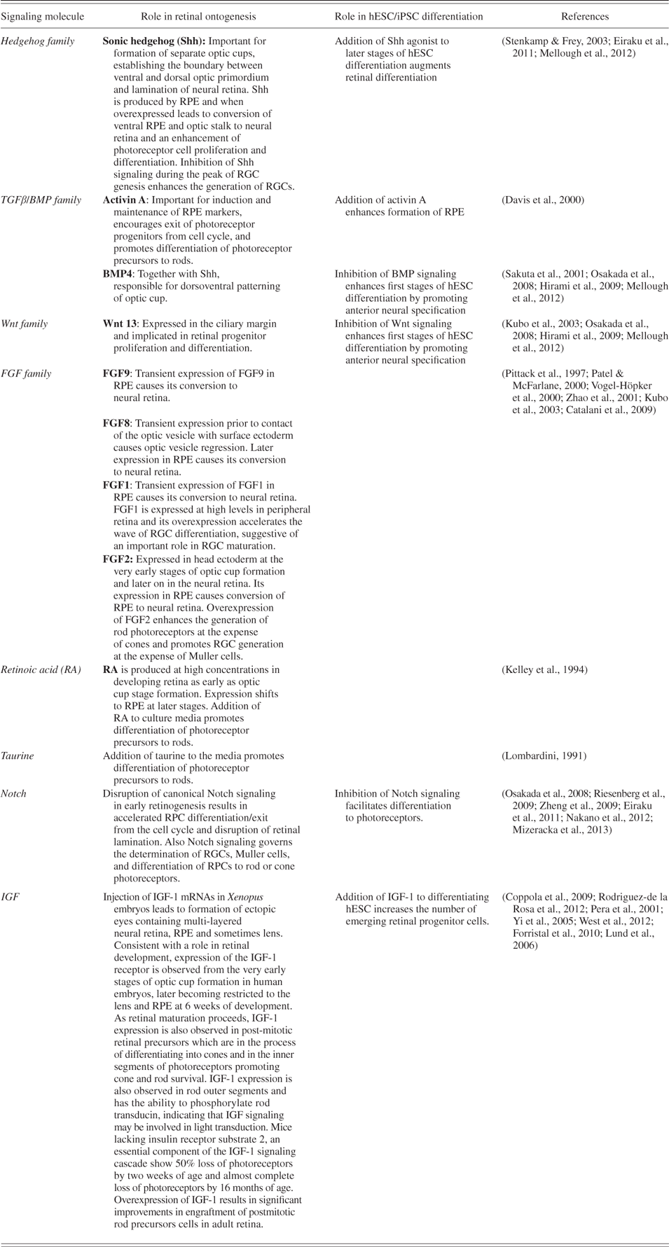
The retina is derived from RPCs, a partially restricted neuroepithelial cell population that undergoes rapid and dramatic expansion in size by proliferation and differentiation to give rise to retinal neurons in a temporally ordered sequential fashion (Swaroop et al., Reference Swaroop, Kim and Forrest2010). Photoreceptors have a complex, highly unique and specific phenotype, many aspects of which arise from their interaction with other retinal cell types as well as with the vitreous and outer limiting membrane of the retina. In the last twenty years, several studies have demonstrated that the competence of RPCs to generate each of the specific retinal cell types can be altered by manipulating both intrinsic and extrinsic factors (Swaroop et al., Reference Swaroop, Kim and Forrest2010). While these studies have been performed in various animal models (chick, frog, mouse), there is a general consensus that six major families of signaling molecules, namely Shh, transforming growth factor beta (TGFβ), BMP, Wnt, fibroblast growth factor (FGF), Notch, and IGF-1 govern key events during retinogenesis. The process begins with the establishment of bilateral apically concave optic vesicles and their subsequent invagination to form the optic cup, a bilayered apically convex structure, determining cell fate commitment to RPE (the outer layer of the optic cup) or neural retina (the inner layer), influencing RPC proliferation, exit from cell cycle and the decision to become rod/cone photoreceptors, retinal ganglion cells (RGCs), Muller cells, and each of the retinal interneurons. This developmental knowledge has been incorporated into hESC differentiation protocols (Table 3). For example, the application of Shh agonists and Notch inhibitor to cultures during the latter stages of differentiation (Nakano et al., Reference Nakano, Ando, Takata, Kawada, Muguruma, Sekiguchi, Saito, Yonemura, Eiraku and Sasai2012; Eiraku et al., Reference Eiraku, Takata, Ishibashi, Kawada, Sakakura, Okuda, Sekiguchi, Adachi and Sasai2011; Lund et al., Reference Lund, Wang, Klimanskaya, Holmes, Ramos-Kelsey, Lu, Girman, Bischoff, Sauvé and Lanza2006) enhances the generation of photoreceptors, as expected from developmental studies in animal models (Table 3). Similarly, the addition of IGF-1 to the culture media enhances the generation of RPCs from hESCs (Lamba et al., Reference Lund, Wang, Klimanskaya, Holmes, Ramos-Kelsey, Lu, Girman, Bischoff, Sauvé and Lanza2006), in accord with the multiple roles played by IGF-1 during eye development (Table 3). While application of some of those factors has already been tested in the most recent 3D hESC differentiation protocol (Eiraku et al., Reference Eiraku, Takata, Ishibashi, Kawada, Sakakura, Okuda, Sekiguchi, Adachi and Sasai2011), these experiments were undertaken in the presence of FBS which, by its undefined composition and containment of multiple signaling molecules, may hide or mask the true effects of the signaling molecules being tested. It is imperative now to apply those extracellular signaling molecules to define minimal media at particular stages of differentiation to assess their impact on optic cup emergence, neural retina and RPE formation, emergence of mature photoreceptors, and formation of fully formed neural retina. Of equal interest is to determine whether manipulation of the hESC differentiation cultures with FGFs (for example FGF8 or 9) upon the emergence of the bilayered optic cup is able to alter the balance between neural retina and RPE formation. This is important for designing directed differentiation strategies for the production of desired cell types for use in cell replacement therapies or drug discovery studies. From a scientific and biological point of view, it is also of tremendous interest to investigate the expression of these signaling molecules, their receptors and downstream effectors in hESC/hiPSC-derived synthetic retina and to compare and contrast those to native developing human retina. This will provide the answer to whether we are able to replicate the formation of retinal cell types using pluripotent stem cells as it happens in vivo.
Summary of key signaling molecules involved in retinal ontogenesis
Modulation of oxygen concentration
A number of studies have revealed that oxygen (O2) is a potent regulator of stem cell maintenance and differentiation, thereby controlling the provision of O2 to the ESC or iPSC differentiation cultures could provide a powerful tool for enhancing differentiation regimes (Forristal et al., Reference Forristal, Wright, Hanley, Oreffo and Houghton2010; Mondragon-Teran et al., 2011a). Oxygen tension in the uterus of mammalian species ranges from 1.5 to 8.7%. In humans, the mean O2 tension at the uterine interface during weeks 7–10 of gestation is 2.4% and after 11 weeks rises to 7.8% as a result of maternal blood flow to the fetus (Rodesch et al., Reference Rodesch, Simon, Donner and Jauniaux1992; Fischer & Bavister, Reference Fischer and Bavister1993). Although the O2 tension of the human diencephalon from which the optic cup emerges has never been directly measured, there is an understanding from work done in other species that a low O2 tension (between 0.8% and 4%) is prevalent across different locations of the developing brain. In accordance with these findings, it has been shown that lowering oxygen levels to 2% during the differentiation of murine and hESC results in a significant increase in the expression of key retinal markers and the yield of photoreceptors (Garita-Hernández et al., Reference Garita-Hernández, Diaz-Corrales, Lukovic, González-Guede, Diez-Lloret, Valdés-Sánchez, Massalini, Erceg and Bhattacharya2013; Bae et al., Reference Bae, Mondragon-Teran, Hernandez, Ruban, Mason, Bhattacharya and Veraitch2012). Although the exact mechanisms by which hypoxia improves differentiation to retinal photoreceptors are not yet known, it is envisaged that this is orchestrated by stabilization of hypoxia-inducible factor 1-alpha (Hif1α) and subsequent activation of VEGF, both known to have neuroprotective and proliferation inducing abilities in RPCs (Grimm et al., Reference Grimm, Wenzel, Groszer, Mayser, Seeliger, Samardzija, Bauer, Gassmann and Remé2002). Mammalian embryos are however exposed to increasing O2 as development proceeds, and in the very early stages of postnatal development (postnatal day 10–20) hypoxia has counterproductive effects resulting in photoreceptor death (Maslim et al., Reference Maslim, Valter, Egensperger, Holländer and Stone1997; Mervin & Stone, Reference Mervin and Stone2002); hence, increasing the O2 concentration in a stepwise fashion as suggested by Mondragon-Teran et al., 2011b may yield improved differentiation results. This, combined with controlled and reproducible generation of floating 3D structures using custom-made tissue culture wells (for example aggrewells) subsequently expanded in bioreactors, which enable more efficient oxygen distribution within differentiating cultures, could provide the support that is needed for more efficient generation of synthetic retinae from pluripotent stem cells.
Modulation of the extracellular matrix
The extracellular matrix (ECM) plays an important role in regulating stem cell proliferation and providing inductive cues for differentiation (Kazanis et al., Reference Kazanis, Lathia, Vadakkan, Raborn, Wan, Mughal, Eckley, Sasaki, Patton, Mattson, Hirschi, Dickinson and ffrench-Constant2010; Keung et al., Reference Keung, de Juan-Pardo, Schaffer and Kumar2011; Keung et al., Reference Keung, Asuri, Kumar and Schaffer2012). ECM can immobilize secreted proteins and alter their biochemical activities so that they are able to exert highly specific effects upon the cells in their vicinity. Moreover, changes in ECM characteristics can influence signal transduction systems and regulate the movement and migration of stem cells; hence, the ECM is a critical component in establishing and maintaining the stem cell microenvironment. Both aspects have been shown to be important for pluripotent stem cell differentiation, for example the presence of soft ECM promotes the early neural differentiation of iPSCs (Keung et al., Reference Keung, Asuri, Kumar and Schaffer2012; Boucherie et al., Reference Boucherie, Mukherjee, Henckaerts, Thrasher, Sowden and Ali2013), while addition of Matrigel to the media enhances human and mESC self-organization into optic cup-like structures under three-dimensional culture conditions (Eiraku et al., Reference Eiraku, Takata, Ishibashi, Kawada, Sakakura, Okuda, Sekiguchi, Adachi and Sasai2011; Nakano et al., Reference Nakano, Ando, Takata, Kawada, Muguruma, Sekiguchi, Saito, Yonemura, Eiraku and Sasai2012). Together these data suggest that the manipulation of ECM characteristics is a promising method that could influence pluripotent stem cell differentiation toward synthetic retina. Defining the optimal ECM components that are physiologically relevant during human retinal development and which may enhance the hESC/iPSC differentiation system to synthetic retina will therefore be of great importance. Multiple lines of published evidence suggest that laminins and their receptors are key candidates that deserve attention when designing new differentiation regimes. Laminins are a family of heterotrimeric glycoproteins, each composed of an α, β, and γ chain that combine to form at least 15 different laminin isoforms. Several members of this family (α3, α4, α5, β2, β3, γ2, and γ3 chains likely to organize into Laminin 5, 14, and 15) are expressed in the interphotoreceptor matrix (IPM) during murine retinal development, prior to and during photoreceptor differentiation (Libby et al., Reference Libby, Champliaud, Claudepierre, Xu, Gibbons, Koch, Burgeson, Hunter and Brunken2000). Single and double knockdown studies of β2 and β2/γ3 chains have respectively shown their importance in the maintenance of rod photoreceptors (Hunter et al., Reference Hunter, Murphy, Olsson and Brunken1992; Libby et al., Reference Libby, Lavallee, Balkema, Brunken and Hunter1999), formation of synaptic ribbons and photoreceptor outer segments, the integrity of the inner limiting membrane, Müller cell attachment and retinal organization (Pinzón-Duarte et al., Reference Pinzón-Duarte, Daly, Li, Koch and Brunken2010). Furthermore, disruption of integrin α6 that functions together with integrin β1 as a major laminin receptor leads to abnormalities in retinal laminar organization in mice (Georges-Labouesse et al., Reference Georges-Labouesse, Mark, Messaddeq and Gansmüller1998), suggesting that laminins and their receptors are key players in the development of several retinal cell types as well as in the maintenance of retinal cyto-architecture. Indeed, the replacement of matrigel with laminin during the differentiation of murine ESCs under 3D conditions allows the successful generation of optic cups and their further differentiation to a fully stratified neural retina, although this remains to be tested in human tissue (Eiraku et al., Reference Eiraku, Takata, Ishibashi, Kawada, Sakakura, Okuda, Sekiguchi, Adachi and Sasai2011). Furthermore, disruption of integrin signaling results in the inhibition of apical nuclear deviation, giving rise to an unusually thin hESC-derived neural retina that fails to evert suggesting that, at least in the human scenario, ECM driven integrin signaling is critical for the formation of the neural retina (Nakano et al., Reference Nakano, Ando, Takata, Kawada, Muguruma, Sekiguchi, Saito, Yonemura, Eiraku and Sasai2012).
Recapitulation of the interphotoreceptor matrix microenvironment
The IPM surrounds the photoreceptors inner and outer segments and plays an important role in the trafficking of retinoids and metabolites, maintenance of the photoreceptor-specific microenvironment, photoreceptor alignment and cellular interactions between the outer segments and RPE (Hollyfield, Reference Hollyfield1999). It is tempting to speculate that recreation of the embryonic IPM microenvironment during hESC and hiPSC differentiation may provide the critical microenvironmental cues needed for the formation of photoreceptor outer segments in vitro. A major component of the IPM in human adult retina is hyaluronic acid (HA), a large non-sulfated linear polysaccharide of (1-β-4)D-glucuronic acid and (1-β-3)N-acetyl-D-glucosamine, which is able to bind a number of secreted proteins of IPM (for example SPACR and SPACRCAN) and also forms a scaffold that fills the IPM (Hollyfield, Reference Hollyfield1999; Keenan et al., Reference Keenan, Clark, Unwin, Ridge, Day and Bishop2012). Formation of this scaffold is enabled by link proteins (for example HAPLN1, shown to be present in the IPM), which form a ternary complex with HA and proteoglycans (for example aggrecan, also expressed in the IPM) (Keenan et al., Reference Keenan, Clark, Unwin, Ridge, Day and Bishop2012). Whether this is the case in developing human retina is unknown, but an important aspect of future investigation. Identification of proteoglycans that are expressed during distinct stages of human retinal ontogenesis is likely to greatly facilitate improvements in existing differentiation regimes. With current improvements in tissue engineering and polymer scaffolds, one can easily envisage the encapsulation of hESC and hiPSC into HA hydrogels. The advantage of this system is that other ECM components and specific growth factors can be added and immobilized to create a more efficient environment for hESC and hiPSC differentiation toward synthetic retinas in vitro. Recent findings indicate that HA hydrogels can be successfully used to deliver donor cells to the retina and help minimize cell aggregation and degradation in the first week following transplantation (Gerecht et al., 2007). Optimization of 3D differentiation regimes using HA hydrogels in longer term may not only be beneficial for improving in vitro differentiation, but also for delivering the hESC- and hiPSC-derived retinal structures in vivo.
The power of iPSC-based approach for disease modeling, drug discovery, and cellular therapies
In retinal disease with monogenic inheritance where the gene defect is known, genetically modified animals produced by targeting specific gene functions have been the mainstay of modeling and investigating disease mechanisms. This is not the case for complex retinal disease (such as AMD) or inherited gene disorders where the gene defect is unknown, for which no truly representative disease models have yet been created. In addition, several key differences exist between animal models and humans in terms of lifespan, tissue composition, anatomy and physiology. Mice typically live for 1–2 years and have no fovea (the centrally positioned cone-enriched region of the retina that is essential for fine visual acuity in humans). Furthermore, it is known that even single gene defect disorders can be affected by non-pathological variations in a large number of other genes in addition to as yet poorly understood epigenetic differences. This is particularly important in complex polygenic diseases (such as AMD) where a large number of identified genetic associations are thought to be disease modifying rather than disease causing, as well as single gene defects with varying penetrance (such as RP caused by mutations in the splicing factor PRPF31). In the same way that studying disease mechanisms is limited by representative tissue, the assessment of any therapeutic effect of novel agents and understanding variable pharmacodynamics is limited by the lack of suitable disease models. Together these studies highlight an imminent need for the generation of human disease-specific models, which can be used for drug screening and investigating disease pathology. Postmortem eyes can be used to investigate the pathophysiology of disease, but have limited availability, and such samples often represent end stage disease that has been altered by secondary disease processes rather than evolving disease. Although some complex diseases such as AMD are relatively common, the acquisition of tissue samples from all disease variants would be difficult and time consuming. In addition, studies that focus on the correlation between cellular function and clinical phenotype/molecular genotype are difficult to perform with high accuracy using postmortem material.
The advent of iPSC technology and the relative ease with which patient somatic cells can be reprogrammed to pluripotency have opened new and exciting avenues for in vitro disease modeling. hiPSCs can self-renew indefinitely in culture, endogenously express pluripotency genes at normal levels, and bear the genetic profile of the patient, thus increasing the likelihood of recapitulating important disease mechanisms. In the case of retina, this is particularly beneficial, since retinal tissue is not amenable to routine tissue biopsy, and methods already exist to coax hiPSCs toward photoreceptors and RPE cells (refer to Table 2). The key question is whether hiPSC-based modeling can provide a platform for all retinal degenerative diseases or be limited to some forms only. This is very dependent on disease complexity, the retinal cell type being affected and the stage of disease onset; for example, age-related diseases may be harder to model. It is unlikely that many diseases affecting multiple cell types and organs and triggered or influenced by environmental factors that are difficult to control (e.g. lifestyle or diet) can be easily modeled with the iPSC approach. However, RPE-based disorders are ideal for this type of modeling, given the ease of RPE generation from hiPSCs using simple protocols and the ability of hiPSC-RPE cells to display key morphological, physiological, and functional features akin to primary human RPE. Furthermore, proposed environmental effects can be mimicked. Indeed, Singh et al. have used this approach to model Best Vitelline Macular Dystrophy (BVMD), an autosomal dominant disorder with highly variable age of onset, which often begins in childhood or adolescence and is caused by over 100 mutations in the BEST1 gene, manifesting in the appearance of a yellow “egg yolk” lesion in the subretinal space (Singh et al., 2013). Although BVMD is known to arise from malfunction of the RPE leading to secondary photoreceptor degeneration, there are no clear conclusions as to how mutations in BEST1 cause the RPE to malfunction. Derivation of BVMD patient iPSCs and their differentiation into RPE indicates that RPE derived from BVMD patients and unaffected controls show similar molecular and physical characteristics under steady state conditions. Upon exposure to physiological stress (such as long-term feeding with photoreceptor outer segments or the addition of exogenous ATP to the culture media), reduced fluid transport in patient-derived RPE was observed, thus recapitulating the clinical features of BVMD. Further investigation into the role of BEST1 using hiPSC-derived RPE also revealed an important role for BEST1 in endoplasmic reticulum-mediated calcium transients, thus providing for the first time a conclusive mechanism that links BEST1 function to BVMD. In a similar study, this time using photoreceptors generated from patients with sporadic RP, Tucker et al. were able to uncover new insights into the consequences of Alu insertion discovered by exome sequencing studies (Tucker et al., Reference Tucker, Scheetz, Mullins, DeLuca, Hoffmann, Johnston, Jacobson, Sheffield and Stone2011). Using elegant molecular biology analysis, the authors were able to show that Alu insertions into the exon 9 of a patient’s MAK gene prevented the expression of a specific retinal isoform which is expressed in adult human retina, thus uncovering new mechanisms that link the disease causing mutation to the transcriptome of photoreceptor cells (Tucker et al., Reference Tucker, Scheetz, Mullins, DeLuca, Hoffmann, Johnston, Jacobson, Sheffield and Stone2011). In addition to uncovering new insights for RP modeling, this study makes a compelling case for the usefulness of an iPSC modeling approach that is able to provide access to patient-specific photoreceptor cells that can be used to address explicit questions about the function and expression of particular candidate genes in retinal cells. hESCs can also be used to model retinal disease caused by single gene mutations, as these can now be easily introduced in the genomes of cells using engineered nucleases (such as ZFNs, TALENs, or CRISPR/CAS9 based methods). However, diseases that are influenced by extrinsic or epigenetic factors, or show varying penetrance such as PRPF31 RP cannot be accurately modeled this way, highlighting once more the value of patient-specific hiPSC-based modeling.
In addition to gaining insights into disease onset and pathology, hiPSC-derived retinal cells from both patients and unaffected controls can be used as a platform to discover new drugs and test the pharmacological effects of existing drugs. To date there have been only two studies investigating the effects of existing drugs on iPSC-derived retinal cells. In the first study, Jin et al. generated iPSCs from patients with RP caused by mutations in RP1, PRPH2, RHO, and RP9 and observed a significant degeneration of iPSC-derived rod photoreceptors under in vitro conditions between days 120 and 150 of differentiation. Treatment with α-tocopherol (acting as an antioxidant) rescued rod photoreceptor death in cell lines from patients with RP9 mutations, but not in experiments using lines harboring other forms of RP causing mutations, suggesting that this approach can be used to quickly screen new drugs and narrow down the number of candidates that go forward to clinical trials to treat particular forms of retinal disease, making the process safer and more effective (Jin et al., Reference Jin, Okamoto, Osakada, Homma, Assawachananont, Hirami, Iwata and Takahashi2011). In a second study, Meyer et al. were able to derive iPSCs from a patient with gyrate atrophy, a rare autosomal recessive retinal disorder that primarily affects the RPE, leading to secondary photoreceptor loss and degeneration (Meyer et al., Reference Meyer, Howden, Wallace, Verhoeven, Wright, Capowski, Pinilla, Martin, Tian, Stewart, Pattnaik, Thomson and Gamm2011). Supplementation of culture media with vitamin B6 resulted in the restoration of ornithine transferase activity in RPE cells derived from this patient, but not the fibroblasts used for hiPSC derivation, underscoring once more the necessity to screen drugs on the affected retinal cell type.
To date, neither 2D nor 3D culture approaches have resulted in the generation of mature photoreceptors bearing outer segments, which are capable of transducing light in vitro. This may pose some limitations when trying to understand the functional consequences of gene mutations using patient-derived retinal cells. The possibility of generating fully laminated neural retina under 3D culture conditions (Eiraku et al., Reference Eiraku, Takata, Ishibashi, Kawada, Sakakura, Okuda, Sekiguchi, Adachi and Sasai2011; Nakano et al., Reference Nakano, Ando, Takata, Kawada, Muguruma, Sekiguchi, Saito, Yonemura, Eiraku and Sasai2012) has however opened new horizons for investigations of this nature, but still relies on some fine tuning of the retinal production process for it to be of greatest use. Nonetheless, 3D generated neural retina will be particularly important for investigating the genesis of specific photoreceptor types (rods vs. cones) in order to study congenital disorders such as enhanced S-cone syndrome, which manifests as a gain in the number and function of S cones (short wave length, blue cones), night blindness and varying degrees of L (long red) and M (middle green)-cone vision due to mutations in NR2E3 gene. It is speculated that this disorder is caused by a fault in the process determining photoreceptor identity, but how this develops in humans remains unclear. In vitro 3D generated neural retina presents an unprecedented opportunity to address these questions and to assess photoreceptor production during human development (Haider et al., Reference Haider, Jacobson, Cideciyan, Swiderski, Streb, Searby, Beck, Hockey, Hanna, Gorman, Duhl, Carmi, Bennett, Weleber, Fishman, Wright, Stone and Sheffield2000).
Recent developments in targeted genetic engineering through the use of ZFN, TALENs, and CRISPR/CAS9 technologies have made the correction of mutations at endogenous loci in hESC and hiPSC possible with much greater ease and efficiency. Although some questions still remain regarding potential harmful consequences of off-target effects in the genome, the prospect of combining stem cell and gene therapy holds great promise for restorative therapies in the eye. To date, there has been only one successful report of the functional correction of a retinal disease causing mutation in patient-specific hiPSC using bacterial artificial chromosome mediated homologous recombination (Meyer et al., Reference Meyer, Howden, Wallace, Verhoeven, Wright, Capowski, Pinilla, Martin, Tian, Stewart, Pattnaik, Thomson and Gamm2011), and no doubt additional examples will emerge as the technology moves forward. The correction of a gene defect in hiPSCs coupled with 3D differentiation would not only provide a powerful tool with which to generate fully functional patient-specific retinal cells, but also an appropriate control that encompasses the genetic background and environmental influences when investigating patient-specific hiPSC for disease insights and drug testing.
The first proof of principle studies highlighted in this review and encompassing literature has clearly demonstrated the utility of hiPSC for disease modeling, drug screening, and future cell replacement therapies. However, one needs to be aware of a number of issues related to reprogramming strategies (genetic and epigenetic differences between hiPSC clones derived from the same patient and between patients, incomplete reprogramming, imprinting, repeat instability, copy number variations, and mutations that may arise during reprogramming etc.), banking of hiPSC (availability of GMP compatible hiPSC derivation protocols, scalability, numbers of hiPSC lines that need to be banked in each country), safety (lack of tumor formation upon transplantation), development of robust differentiation protocols for generating mature photoreceptors (scalability, GMP compliance, timing), and identification of disease relevant and interesting phenotypes that can benefit from the benefits that the hiPSC system offers. All these issues have been well addressed in a recent review to which the reader is encouraged to refer (Wright et al., Reference Wright, Phillips, Pinnilla, Hei and Gamm2014) and for this reason will not be expanded herein.
Conclusions and future directions
While the progress of pluripotent stem cells for retinal disease modeling and treatment has surpassed most expectations, a number of hurdles in the clinical, biological, and regulatory field need to be addressed before the promise of lab-made synthetic retina becomes a reality. At the moment, it is not clear whether gene corrected patient-specific iPSC will make its way to the clinic and pave the path toward personalized medicine; or will generic retinal cells created from carefully selected hiPSC and hESC banks on the basis of homozygosity for HLA-A, B, and DR ride the fast track to the clinic? A series of careful considerations must be made before these decisions can be reached and will of course also be influenced by cost, the time it takes to prepare the required retinal cells before individual treatments can commence, ease of distribution to the clinics, and legislation that governs health and safety across countries. As is often the case, one approach is unlikely to provide an all-encompassing solution; instead we will likely witness the tailored application of both hESCs and hiPSCs across various aspects of disease modeling, drug screening, drug discovery, and cell-based replacement therapy. Indeed, ongoing clinical safety trials using hESC-derived RPE in the United States and hiPSC-derived RPE in Japan are an important test of current approaches and provide an excellent indication of how far and fast we have come toward developing new therapies for blinding diseases from the first derivation of hESC in 1998 and hiPSC in 2007. If we can achieve such progress in just 15 years, no doubt the next 15 years will be an extremely exciting period for visual research and is anticipated to reveal new knowledge about disease mechanisms and the advent of therapies to restore vision in patients affected by currently untreatable retinal disease.
Acknowledgments
The authors are grateful to Fight for Sight UK (1456/1457), Macular Disease Society UK, RP Fighting Blindness UK (GR5840), BBSRC UK (BB/I02333X/1, European Research Council (614620) and Sunderland Eye Infirmary for funding this work and Simon Foster for help with the design of figures included in this review.