Introduction
Paediatric cardiomyopathies represent a heterogeneous group of diseases associated with high morbidity and mortality. Despite their low prevalence, they are the leading indication for heart transplantation beyond the first year of life. Reference Lipshultz, Sleeper and Towbin1–Reference Lipshultz, Law and Asante-Korang4
Understanding of paediatric cardiomyopathies has expanded since the first registries in the late 1990s and early 2000s. Reference Lipshultz, Sleeper and Towbin1–Reference Nugent, Daubeney and Chondros3 However, most studies on cardiomyopathy are derived from adult populations in high-income countries, Reference Ware, Bhatnagar and Dexheimer5–Reference Rath and Weintraub9 leaving significant gaps in paediatric data, particularly in developing nations, where genetic diversity, socio-economic conditions, and healthcare infrastructure may influence disease presentation, management, and outcomes.
This study presents data from the first regional registry of paediatric cardiomyopathies in Brazil, aiming to characterise the clinical and epidemiological profiles.
Methods
The CHARISMA Registry (CHildren and Adolescent Registry in Myocarditis and CArdiomyopathy) is a prospective observational study (2019–2024) including children and adolescents with cardiomyopathy.
Participants were recruited through the Brazilian Unified Health System (SUS) at a university-affiliated quaternary hospital and a municipal paediatric hospital in the metropolitan region of Rio de Janeiro, Brazil. This region is composed of seven cities with distinct characteristics that vary from large urban centres to rural regions, with a total of 1,908,751 inhabitants, of which 429,512 are children and adolescents, according to data from the Brazilian Institute of Geography and Statistics. 10
Inclusion criteria were echocardiographic confirmation of the phenotype and onset of symptoms before 18 years of age. The definitions of the European Society of Cardiology and American Society of Cardiology were used to confirm the diagnosis. Reference Elliott, Andersson and Arbustini11,Reference Maron, Towbin and Thiene12
Echocardiographic parameters were used based on the z-score relative to body surface area, age, and sex. Hypertrophic cardiomyopathy was considered a z-score greater than + 2 for measurements of the interventricular septum and/or posterior wall during diastole. While dilated cardiomyopathy was defined by the left ventricle end-diastolic diameter above z-score + 2, systolic dysfunction was defined as a left ventricular ejection fraction below 55%. Reference Lipshultz, Law and Asante-Korang4,Reference Elliott, Andersson and Arbustini11,Reference Maron, Towbin and Thiene12
Noncompaction cardiomyopathy was confirmed by cardiac MRI or by Jenni criteria on echocardiography (ratio of noncompaction to compaction layer greater than 2 during diastole). Reference Rohde, Muslem and Kaya13,Reference Petersen, Jensen and Aung14
Arrhythmogenic cardiomyopathy was based on the 2019 Heart Rhythm Society criteria. Reference Towbin, McKenna and Abrams15
Secondary causes such as arterial hypertension, valve disease, children of a diabetic mother, or those who had the symptoms onset after 18 years of age were excluded.
The study was approved by the local Research Ethics Committee. After signing the informed consent, the data were obtained and applied “the five steps of investigation of paediatric cardiomyopathy” published in a previous study by the group of researchers. Reference Torbey, Couto and Grippa16 Each participant was assessed for: phenotype, definition of aetiology, classification proposed by the World Heart Federation- MOGE(S), Reference Arbustini, Narula and Dec17 definition of prognosis, and availability of specific therapies.
The MOGE(S) Reference Arbustini, Narula and Dec17 classification analysis includes five criteria: morphofunctional phenotype (M), involvement of organs other than the heart (O), genetic or family inheritance pattern (G), aetiology (E), heart failure stage proposed by the AHA, and functional class (S).
To assess functional class, the Ross classification was used for children up to six years of age and the NYHA classification for participants aged six or over. The presence of a history of difficulty or fatigue during breastfeeding, difficulty in gaining weight, sweating during breastfeeding, and fatigue during exertion were considered signs of heart failure.
After 2022, with the implementation of the RENOMICA Study Reference Kasai-Brunswick, Campos and Braga18 (National Network of Cardiovascular Genomics), it was possible to expand the genetic investigation of CHARISMA participants by building a collaborative network between research projects.
The variables studied were reason for diagnosis of cardiomyopathy, age at onset of symptoms, age at diagnosis of cardiomyopathy, personal and family history, available laboratory tests, such as N-terminal pro-B-type natriuretic peptide, electrocardiogram data, 24-hour Holter monitoring, echocardiogram, and other imaging tests, such as cardiac magnetic resonance imaging, and results of genetic tests. Ejection fraction was considered using the Teicholz technique, since few patients underwent the Simpson technique.
Participants were grouped according to phenotypes. The outcomes were death, indication for heart transplantation, and the use of an implantable cardioverter-defibrillator.
Statistical analysis was performed using RStudio 2024.12. Variables were described as absolute numbers, percentages, means, medians, and standard deviations. Categorical variables were compared using Fisher’s exact test. One-way analysis of variance assessed differences in continuous variables among phenotypes, with Bonferroni-corrected post hoc analysis. The t-test compared means between two groups. Survival was analysed using the Kaplan–Meier method, with group comparisons by log-rank test. A 95% confidence interval and a significance level of p < 0.05 were adopted.
Results
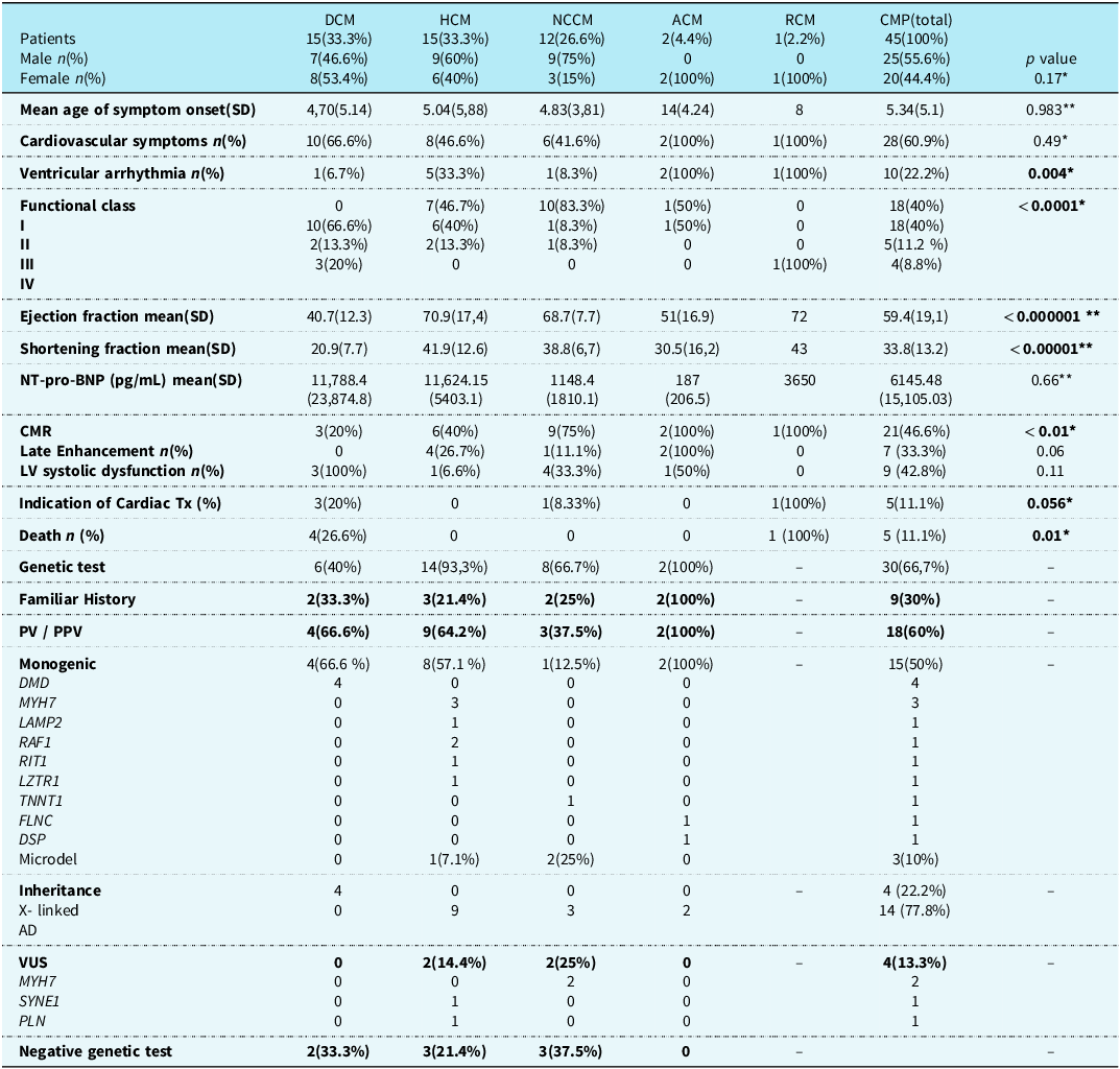
During the study, 45 cases of cardiomyopathy were identified. There was a similar distribution between dilated and hypertrophic cardiomyopathies (33.3% each), which were the two predominant phenotypes. Followed by noncompaction (26.6%), arrhythmogenic (4.4%), and restrictive cardiomyopathy (2.2%). Males predominated (55.5%) and the mean age at diagnosis was 6.6 years (median = 5 years). The average interval from symptom onset to diagnosis was 1.25 years, significantly shorter in males (p = 0.03). The mean follow-up period was 3.27 years, ranging from 2 days to 5 years. Table 1 summarises the main clinical characteristics.
Clinical and genetic characteristics by phenotype

* Fisher’s exact test, **ANOVA. DCM = dilated cardiomyopathy, HCM = hypertrophic cardiomyopathy, NCCM = noncompacted cardiomyopathy, ACM = arrhythmogenic cardiomyopathy, RCM = restrictive cardiomyopathy, SD = Standard deviation. PV = pathogenic variant, PPV = probably pathogenic variant, VUS = variant uncertain significance.
Cardiovascular symptoms (60.9%), mainly heart failure (31.1%), were the leading cause of diagnosis, followed by heart murmur (21.4%), chest pain (21.4%), ventricular extrasystoles (7.1%), syncope (3.5%), and increased cardiac area on chest X-ray (3.5%). Despite the absence of cardiovascular symptoms, 39.1% were diagnosed with cardiomyopathy. Of these, 26.1% were identified during echocardiography for suspected genetic syndromes, 8.7% through family screening, and 4.3% via pre-participation sports evaluation.
Pedigree assessment identified a positive family history in 20% of cases, who were significantly older at diagnosis (p = 0.02).
Multisystem involvement occurred in 40%, mainly neuromuscular. Among these, 77.7% had a genetic alteration linked to aetiology. Multisystem involvement was strongly associated with a defined genetic cause (odds ratio= 11.26; 95% confidence interval: 1.45 to ∞; p = 0.017), indicating a significantly higher risk. The wide confidence interval reflects low precision, likely due to the small sample size.
Left ventricular ejection fraction ranged from 19 to 91%, with a mean of 58.9% and a median of 62.5%. There was a significant association between values below 30% and mortality (p < 0.001). One-way analysis of variance revealed a significant effect of phenotype on ejection fraction and shortening fraction (p < 0.0001). Bonferroni-corrected post hoc analysis showed significant differences between dilated and hypertrophic cardiomyopathy as well as between dilated and noncompaction cardiomyopathy (Figure 1a).
Boxplot analysis of echocardiographic and clinical features according to phenotype and outcome in pediatric cardiomyopathy. ( a ) Boxplot comparing left ventricular ejection fraction and shortening fraction among phenotypes: DCM showed significantly lower values compared to HCM and NCCM. DCM = dilated cardiomyopathy, HCM = hypertrophic cardiomyopathy, NCCM = noncompacted cardiomyopathy. ( b ) Boxplot comparing age at symptoms onset and age of diagnosis between patients with and without RASopathies. The group with RASopathies present symptoms and was diagnosed at a significant earlier age than those without RASopathies. ( c ) Boxplots comparing left ventricular ejection fraction (LVEF), fractional shortening (FS) and NT-Pro-BNP levels. Non survivors had significantly lower LVEF and FS values, and higher NT-pro-BNP levels.

N-Terminal Pro-B-Type natriuretic peptide levels were measured in 19 participants. Higher values were significantly associated with functional classes III/IV (p = 0.049) and medical indication of heart transplantation (p = 0.049). Furthermore, values above 1,000 pg/mL were significantly associated with mortality (p = 0.01).
MRI was performed in 46.6%, with a significant difference between phenotypes (p = 0.01). Positive late enhancement occurred in 33.3% and 42.8% had left ventricular dysfunction.
Genetic testing was performed in 30 cases, with a positivity of 60%. Monogenic causes were confirmed in 15, and microdeletions were identified in 3. Of all cases, 27 lacked an aetiological diagnosis: 15 were not tested, and among those tested, 12 had inconclusive results (Table 1).
The main genetic alterations in hypertrophic cardiomyopathy were pathogenic variants in the sarcomere gene MYH7 and genes related to Noonan syndrome. Individuals with Noonan syndrome presented with clinical symptoms at diagnosis (p < 0.001) at a significantly younger age compared to those with other aetiological diagnoses (Figure 1b).
Catheter ablation was performed in two cases: atrial arrhythmia in dilated cardiomyopathy and ventricular arrhythmia in noncompaction cardiomyopathy. Implantable cardioverter-defibrillators were placed in three patients (two with hypertrophic and one with arrhythmogenic cardiomyopathy) as primary prevention of sudden death.
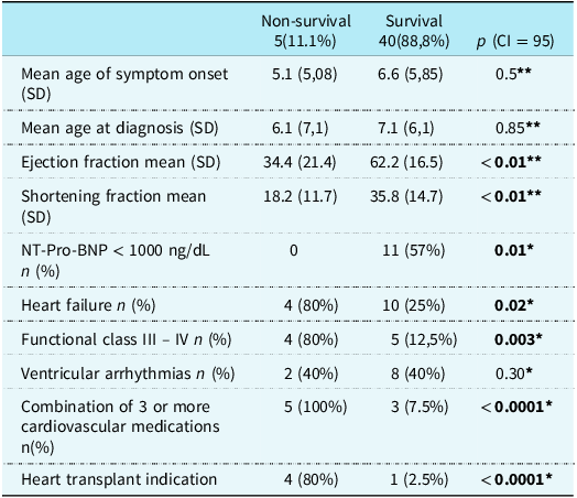
The overall mortality was 11.1%, predominantly among patients with dilated cardiomyopathy (p = 0.01). The presence of heart failure was significantly associated with an increased risk of death, with an odds ratio of 11.23 (p = 0.02; 95% confidence interval: 0.9–607). Similarly, functional class III/IV had a markedly higher risk of mortality (odds ratio= 24.6; p = 0.003; 95% confidence interval:1.9–1400). The small sample size limited the precision of risk estimates, as indicated by wide confidence intervals despite significant p-values.
Kaplan–Meier analysis revealed an overall 5-year survival probability of 88.9% (95% confidence interval: 80.2–98.6%) across all phenotypes. Patients with dilated cardiomyopathy had a significantly lower 5-year survival rate of 73.3% (95% confidence interval: 54.0–99.5%; p = 0.017). Log-rank showed significantly reduced survival in the following subgroups: ejection fraction below 30% (p < 0.0001, 95% CI 92.6–100%), functional class III/IV (p < 0.0001, CI 92–100%), heart failure (p = 0.0091, 95% CI 90.7–100%), use of three or more cardiovascular medications (p < 0.001, 95% CI: 15.3–91.7%), N-Terminal Pro-B-Type natriuretic peptide levels above 1000 pg/mL (p = 0.004, 95% CI: 63.3–100%), and medical indication for heart transplantation (p < 0.001, 95% CI: 92.7–100%) (Figure 2, table 2).
Kaplan-Meier survival curves for pediatric patients with cardiomyopathy, according to clinical and echocardiographic characteristics. Legend: Survival curves were compared using the Log-Rank test (p < 0.05 was considered statistically significant). ( a ) Overall survival, ( b ) Survival comparison between dilated cardiomyopathy and other phenotypes, ( c ) NT-pro-BNP levels > 1000 pg/mL versus ≤ 1000 pg/mL, ( d ) Left ventricular ejection fraction < 30% versus ≥ 30%, ( e ) Presence versus absence of heart failure, ( f ) Ross/NYHA functional class I–II versus III–IV, (g) Indication for heart transplantation versus no indication, ( h ) Use of three or more cardiovascular medications versus fewer than three.

Comparison between survival and non-survival groups

* Fisher´s exact test, **T-Test.
SD = standard deviation.
Discussion
CHARISMA describes important characteristics of paediatric cardiomyopathy in a metropolitan region of the State of Rio de Janeiro, Brazil. It is a pioneering study because there are no similar studies published in Brazil and Latin America so far, with only one retrospective study carried out in Colombia. Reference Huertas-Quiñones, Mestra, Peña-Trujillo, Gallo-Bernal, Villaveces and Alarcón-Forero19,Reference Zenteno, Peñafiel and Del Río20
The knowledge about the epidemiological characteristics of paediatric cardiomyopathy has been based on three large registries carried out in developed countries (Finland, the United States, and Australia). Reference Lipshultz, Sleeper and Towbin1–Reference Nugent, Daubeney and Chondros3 These studies defined an estimated incidence of 1:100.000 children/year. Although rare, cardiomyopathy represents high morbidity and mortality in paediatrics. Dilated cardiomyopathy is the most common phenotype in childhood, followed by hypertrophic, differing from the adult population, where hypertrophic cardiomyopathy is more frequent. Reference Lipshultz, Sleeper and Towbin1–Reference Nugent, Daubeney and Chondros3,Reference Huertas-Quiñones, Mestra, Peña-Trujillo, Gallo-Bernal, Villaveces and Alarcón-Forero19,Reference Arbelo, Protonotarios and Gimeno29
In the present study, there was a similar distribution between dilated and hypertrophic cardiomyopathy (33.3% each). Noncompaction cardiomyopathy presented 26.3% of the total, versus the 5%, 9.2%, 13.8%, and 14.7% found, respectively, in the Paediatric Cardiomyopathy Registry, Australian registry, EURObservational Research Programme Cardiomyopathy and Myocarditis Registry, and in a Chinese study. Reference Jefferies, Wilkinson and Sleeper21–Reference Kaski, Norrish and Gimeno Blanes24 Noncompaction cardiomyopathy represents the third main phenotype, Reference Lipshultz, Law and Asante-Korang4,Reference Rohde, Muslem and Kaya13,Reference De Angelis, Bobbo and Paldino25 and recently paediatric studies describe an increase in its incidence. Reference Petersen, Jensen and Aung14,Reference De Angelis, Bobbo and Paldino25
The distribution of phenotypes differed from previously described patterns, Reference Lipshultz, Sleeper and Towbin1–Reference Nugent, Daubeney and Chondros3,Reference Huertas-Quiñones, Mestra, Peña-Trujillo, Gallo-Bernal, Villaveces and Alarcón-Forero19,Reference De Angelis, Bobbo and Paldino25–Reference Arbelo, Protonotarios and Gimeno29 but corroborates with Kaski et al Reference Kaski, Norrish and Gimeno Blanes24 that hypertrophic cardiomyopathy may be more common in the paediatric population.
Cardiomyopathy occurs in all paediatric age groups. There was a peak in the first year of life, mainly due to hypertrophic, with a significant predominance of RASopathies (p < 0.001). This peak was associated with syndromic cardiomyopathy, while the second peak occurs during adolescence and is attributed to sarcomeric variants. Reference Lipshultz, Sleeper and Towbin1,Reference Lipshultz, Law and Asante-Korang4,Reference Kasai-Brunswick, Campos and Braga18,Reference Kaski, Norrish and Gimeno Blanes24,Reference Lee, Hsu and Kantor26,Reference Bagkaki, Parthenakis and Chlouverakis28–Reference Gal, Morales and Rojahn32.
This study confirms the heterogeneous clinical presentation of paediatric cardiomyopathy. Reference Kasai-Brunswick, Campos and Braga18,Reference Huertas-Quiñones, Mestra, Peña-Trujillo, Gallo-Bernal, Villaveces and Alarcón-Forero19,Reference Bagkaki, Parthenakis and Chlouverakis28 Although cardiovascular symptoms were the main reason for diagnosis, 39.1% were asymptomatic, mostly identified during evaluation for genetic syndromes. This highlights that non-cardiac manifestations may precede cardiovascular signs, reinforcing the importance of a systemic approach. Reference Arbelo, Protonotarios and Gimeno29,Reference Rapezzi, Arbustini and Caforio33–Reference Tsatsopoulou, Protonotarios and Xylouri35
MOGE(S) emphasises a multisystemic approach, and it improves diagnostic yield and associates with excellent survival when cardiomyopathies’ causes are identified and treated. Reference Arbustini, Narula and Dec17,Reference Hazebroek, Moors and Dennert36 CHARISMA supports this by showing that aetiology was clarified in most cases with multisystem involvement (77.7%, p = 0.017).
Evaluation with MRI occurred in approximately half of the cases and presented a higher proportion than the results in European study (46.6% versus 30.5%); however, the presence of myocardial fibrosis detected by delayed gadolinium enhancement was lower (33.3% versus 59.1%). Reference Kaski, Norrish and Gimeno Blanes24 Although less frequent in children, myocardial fibrosis is linked to increased risk of ventricular arrhythmias. Reference Shafqat, Shaik and Koritala37,Reference Spaapen, Bohte, Slieker and Grotenhuis38
The use of advanced techniques (MRI, 3D echocardiography, and strain analysis) for diagnosis and follow-up of cardiomyopathies is limited in low- and middle-income countries and challenging in children who require sedation. Reference Spaapen, Bohte, Slieker and Grotenhuis38–Reference Kozak, Afiune and Grosse-Wortmann41 Simple measures such as ejection fraction and fractional shortening remain key for follow-up and prognosis. CHARISMA confirms their association with mortality, aligning with prior studies. Reference Alvarez, Wilkinson and Lipshultz42–Reference Towbin, Lowe and Colan45
High N-Terminal Pro-B-Type natriuretic peptide levels correlated with ventricular dysfunctioned clinical status. Most paediatric studies involve mixed heart failure aetiologies, with limited data on specific cardiomyopathies phenotypes. Reference Agrawal, Janjua, Alsayed Ali Zeyada and Taher Elsheikh46–Reference Mangat, Carter, Riley, Foo and Burch48 Cantarutti et al. demonstrated that high values correlate with increased ventricular mass in hypertrophic cardiomyopathy and were shown to be an independent risk factor for implantable cardioverter-defibrillator implantation. Reference Cantarutti, Adorisio and Di Marzio49 In CHARISMA, high values (> 1000pg/dL) were significantly correlated with more advanced stages of HF, indication for heart transplantation, and death.
The majority of participants (66.7%) underwent genetic testing; this proportion is similar to that observed in the EORP (68%), Reference Kaski, Norrish and Gimeno Blanes24 and much higher when compared to the Colombian epidemiological study (25%). Reference Huertas-Quiñones, Mestra, Peña-Trujillo, Gallo-Bernal, Villaveces and Alarcón-Forero19
This high number, however, does not correspond to the Brazilian reality, since it is not available in SUS and represents a high cost in the private network. The fact that most of us have undergone genetic investigation is due to the opportunity of collaboration of RENOMICA, Reference Kasai-Brunswick, Campos and Braga18 an independent research project that studies the cost-effectiveness of performing genetic testing for cardiovascular diseases in SUS.
The CHARISMA cohort showed a positivity rate of 60%, identical to that reported by Kaski et al, Reference Kaski, Norrish and Gimeno Blanes24 and higher than the rates observed in studies by Ware et al (48%), Reference Ware, Wilkinson and Tariq6 Sun et al (40%), Reference Lee, Hsu and Kantor26 and Keisling et al (39%). Reference Keisling, Bedoukian and Burstein50
The presence of a family history did not correlate significantly with genetic test positivity. This is interesting to highlight because its absence does not exclude the possibility of a genetic cardiomyopathy. Reference Ware, Wilkinson and Tariq6,Reference Rapezzi, Arbustini and Caforio33
Dilated cardiomyopathy included most children with advanced heart failure (55.5%, p < 0.0001) and had the highest mortality (26.6%, p = 0.01). In contrast, hypertrophic and arrhythmogenic cardiomyopathy patients were more frequently associated with ventricular arrhythmias (p = 0.004) and showed higher genetic test positivity (64.2% for hypertrophic and 100% for arrhythmogenic cardiomyopathy) aligning with findings from other studies. Reference Huertas-Quiñones, Mestra, Peña-Trujillo, Gallo-Bernal, Villaveces and Alarcón-Forero19,Reference Sun, Guo and Zhang23,Reference Kaski, Norrish and Gimeno Blanes24,Reference Bagkaki, Parthenakis and Chlouverakis28,Reference Daubeney, Nugent and Chondros44,Reference Towbin, Lowe and Colan45,Reference Nugent, Daubeney and Chondros51
In CHARISMA, 66,7% of noncompaction cardiomyopathy were asymptomatic, which justifies the significant non-use of cardiovascular drugs in most of the group (p < 0.001). This is a debated phenotype, while European Society of Cardiology does not classify it as a cardiomyopathy. Clinical findings often include heart failure and arrhythmias, with higher genetic test positivity in children compared to adults. These observations underscore the importance of recognising myocardial hypertrabeculation even in asymptomatic children, given its potential progression to high-risk phenotypes. Reference Rath and Weintraub9,Reference Rohde, Muslem and Kaya13,Reference Shi, Betancur and Nugent22,Reference van Waning, Moesker, Heijsman, Boersma and Majoor-Krakauer52
Mortality was 11.1%, with a survival rate of 88.9% for all phenotypes within five years of follow-up. These data are similar to current studies published by Huertas-Quiñones and Bagkaki that demonstrated a mortality of 12.36% and 12.5%, respectively. Reference Huertas-Quiñones, Mestra, Peña-Trujillo, Gallo-Bernal, Villaveces and Alarcón-Forero19,Reference Bagkaki, Parthenakis and Chlouverakis28
Dilated cardiomyopathy had the worst prognosis, with mortality of 26.6% and probability of survival of 73.4% (p = 0.001). In a recent study, it showed a transplant-free survival of 69.8% over a period of 5 years, Reference Ciuca, Ragni and Hasan53 while previous studies showed survival ranging from 54 to 77%. Reference Daubeney, Nugent and Chondros44,Reference Towbin, Lowe and Colan45,Reference Alexander, Daubeney and Nugent54,Reference Tsirka, Trinkaus and Chen55 The probability of survival vary depending on the aetiology, with a worse prognosis for neuromuscular diseases (57%) and better rates for myocarditis (90%). Reference Towbin, Lowe and Colan45 In general, in the first year, transplant-free survival can vary from 75 to 85% and up to 62% after 10 years of diagnosis. Reference Ciuca, Ragni and Hasan53,Reference Alexander, Daubeney and Nugent54,Reference Fenton, Horne, Simmonds, Neligan, Andrews and Burch56
The main factors that were significantly correlated with the risk of death were ejection fraction less than 30%, N-Terminal Pro-B-Type natriuretic peptide levels above 1000 pg/mL (Figure 1c), heart failure, functional class III/IV, use of three or more cardiovascular medications at the time of diagnosis, and the medical indication for heart transplantation during the follow-up. These findings corroborate previous publications that mortality is mainly associated with cases of advanced heart failure. Reference Lipshultz, Law and Asante-Korang4,Reference De Angelis, Bobbo and Paldino25,Reference Lee, Hsu and Kantor26
Genetic testing was unavailable for 3 of the 5 patients who died, preventing correlation between genotype and mortality.
The CHARISMA registry is the first prospective study of paediatric cardiomyopathies in Brazil and Latin America, offering novel regional data. It shows a high genetic test positivity, especially in systemic cases, and identifies key mortality predictors for risk stratification. Collaborative networks between research and care are vital to improve outcomes. Better regional understanding can guide public health policies and cardiovascular care.
Study limitation
This study faces limitations common to rare disease research. Despite spanning five years in a large metropolitan area, the small sample size (expected given the low incidence) may limit representativeness and statistical power. Missing data from unequal access to diagnostic tests reflect real-world challenges in public healthcare systems of middle-income countries, highlighting barriers to health policy.
Acknowledgements
None.
Financial support
This research received no specific grant from any funding agency, commercial, or not-for-profit sectors.
Competing interests
The author(s) declare none.
Ethical standard
The authors affirm that all procedures related to data collection comply with the ethical standards of the relevant national guidelines for research involving human subjects (Resolution CNS 466/12, Brazil), and with the Helsinki Declaration of 1975, as revised in 2008, and were approved by the Research Ethics Committee of Federal Fluminense University, Faculty of Medicine, under protocol number CAEE: 93874218.2.0000.5243.