Introduction
The middle-ear ossicular chain is responsible for carrying sound into the inner ear. Middle-ear diseases can damage the ossicular chain, leading to transmission-type hearing loss.Reference Goebel and Jacob1 The most frequent among these diseases is chronic otitis media. Surgical treatment of this condition aims to eliminate the pathological process as well as to correct the hearing loss caused by the disease. For this purpose, ossiculoplasty techniques are used.
Geyer, Helms and Babighian are the first reported otologists to have used glass ionomer cement in otological surgery.Reference Rondini-Gilli, Grayeli, Borges Crosara, El Garem, Mosnier and Bouccara2–Reference Geyer, Helms, Yanagihara and Suzuki4 In the past 25 years, over 2000 reports of glass ionomer cement use have appeared. Many of these were for dental applications; only a small number concerned glass ionomer cement use in middle-ear surgery.Reference Rondini-Gilli, Grayeli, Borges Crosara, El Garem, Mosnier and Bouccara2
The most frequently seen ossicular chain rupture due to chronic otitis is caused by necrosis of the long arm of the incus in the incudostapedial joint.Reference Muller, Geyer and Helms5, Reference Mikaelian6 Glass ionomer cement is often used for the repair of this joint. The ossiculoplasty techniques that have been described generally involve bypassing the defective bone using autogenous and homogenous bone grafts, or alloplastic implants.Reference Rondini-Gilli, Grayeli, Borges Crosara, El Garem, Mosnier and Bouccara2, Reference Stea, Cervellati, Cavedagna, Savarino, Cenni and Pizzoferrato3 Unlike these techniques, glass ionomer cement allows the creation of mechanisms that are similar to natural anatomy and that ensure sound transmission.Reference Rondini-Gilli, Grayeli, Borges Crosara, El Garem, Mosnier and Bouccara2
Despite the availability of numerous different prostheses and specific surgical techniques, the success rates of ossiculoplasty methods are difficult to predict because of patient- and disease-related factors such as mucosal disease status, eustachian tube dysfunction and variation in anatomy. Although patient- and disease-related factors cannot be fixed surgically, the surgeon can determine the most suitable surgical technique and type of prosthesis to use, and establish the appropriate placement and stability of the prosthesis.
The existent clinical studies mainly focus on post-operative clinical observation. The air–bone gap, which indicates post-operative success, is affected by middle-ear ventilation and abnormal soft tissue infections, so it is not indicative of reconstructed ossicular chain function. A clinical study by Brask, which investigated ossicular chain reconstruction using glass ionomer cement, reported that 81.3 per cent of the 22 cases evaluated for incus long arm defects had a post-operative average air–bone gap of under 20 dB.Reference Brask7 Babu and Seidman reported using hydroxyapatite bone cement in 18 out of 264 tympanoplasty and mastoidectomy surgical procedures in chronic suppurative otitis media patients, and achieved considerable post-operative improvement of the air–bone gap.Reference Babu and Seidman8 Elsheikh et al. compared the use of hydroxyapatite bone cement in 82 ears of 62 patients against a control group consisting of 20 patients with partial Plastipore™ ossicular replacement prostheses.Reference Elsheikh, Elsherief and Elsherief9 Post-operatively, the hydroxyapatite bone cement group showed significantly better air–bone gap averages than the control group. In a study by Feghali et al., the use of glass ionomer cement in nine patients resulted in significant post-operative improvement in pure tone audiometry averages when compared with pre-operative values.Reference Feghali, Barrs, Beatty, Chen, Green and Krueger10
Successful use of glass ionomer cement in ossiculoplasty, and the assurance of reconstruction stability and continuous hearing gain, depend on: knowledge of the biomechanical characteristics of the prostheses and materials used, and the mixing proportions and preparation conditions for glass ionomer cement.
The biomechanical properties of glass ionomer cement when used in the incudostapedial joint have not yet been evaluated in any study. We used compression tests to evaluate the biomechanical properties and resistance force of glass ionomer cement samples (of two different lengths) used to repair incudostapedial joint rupture, and determined which defect length would be optimal for repair with glass ionomer cement.
Materials and methods
The study material comprised 30 incudes and 30 stapedes taken from cadavers that were donated for education and training to the Department of Anatomy, Cerrahpasa Medical Faculty, Istanbul University. The ethical committee of the Cerrahpasa Medical Faculty, Istanbul University, did not require approval for this cadaver study. The study was conducted in the laboratory of the Department of Metallurgy and Material Engineering, Yildiz Technical University. The laboratory temperature was maintained at 18 ± 2°C.
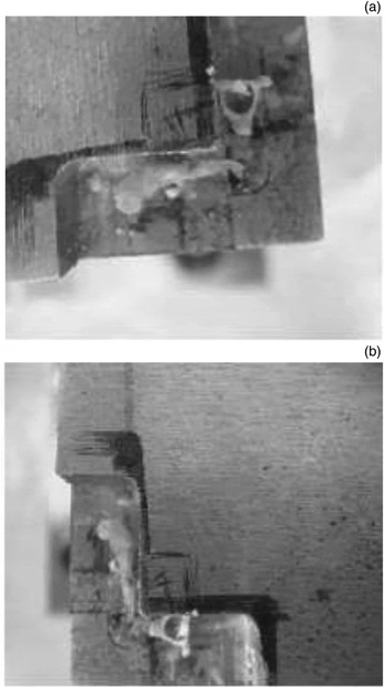
The incus and stapes were placed in a master block made from cold work tool steel (type 2080), and angled at 91° to approximate their natural position in the ear. This angle was constant in all experiments (the experimental geometry conforming to natural anatomy was obtained in the master block). The block had a right and a left chamber for the incus and stapes, as seen in Figure 1.
Placement of ossicles ((a) shows right side and (b) shows left side) in a master block, to obtain a 91° angle.

When the incus and stapes were placed into the block, a gap was left between the ossicles. Two experimental groups were created based on the size of the gap (1.0 mm in group 1 and 2.0 mm in group 2) by using appropriate mountings. Glass ionomer cement was then applied to these spaces. The glass ionomer cement used was Ketac™ Cem Radiopaque Permanent Glass Ionomer Luting Cement (33 g powder and 12 ml liquid). The powder and liquid were hand-mixed for 60 seconds on a glass slide with a needle tip.
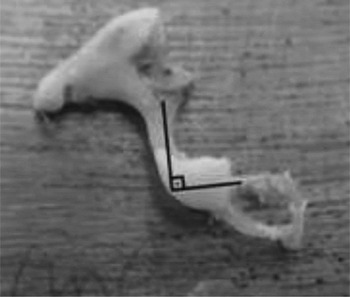
The incus and stapes were placed into the mechanism. The prepared glass ionomer cement mixture was poured drop by drop (by the same person (EAS)) with a straight hook into the space between the stapes head and incus. After the drop settled and several minutes had elapsed, another drop was applied, until the 1.0 and 2.0 mm spaces were filled. After the glass ionomer cement had solidified, the bonded incus and stapes were taken out of the block (Figure 2). The diameter of glass ionomer cement was measured with digital calipers (Mitutoyo Digimatic Caliper, model 500-151U-CD-15C; Mitutoyo, Andover, UK) at a sensitivity of 0.01 mm. The cross-sectional area was calculated using the equation: A = π.DReference Rondini-Gilli, Grayeli, Borges Crosara, El Garem, Mosnier and Bouccara2/4 (where A is area and D is diameter).
Incus and stapes bonded with bone cement at a 91° angle.

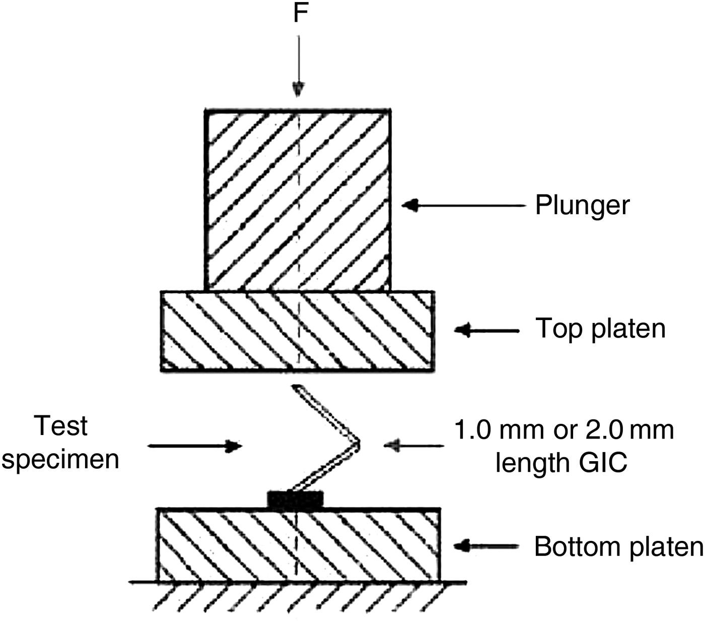
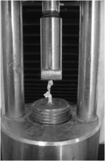
Compression forces affect the incus–stapes system in the ear; therefore, a compression test was applied to the experimentally bonded incus–stapes systems, as shown schematically in Figure 3. A compression test is a static test that determines the fracture behaviour of materials under compression loading. The tests were carried out using an Instron 3340 (Instron, High Wycombe, UK), a universal tensile–compression machine that applies vertical loading to test the mechanical properties of materials.Reference McCaghren, Retief, Bradley and Denys11 In our study, the compression-type loading mode was used. Before the test, the bonded ossicle system was placed in the testing device in a position consistent with normal ear anatomy, and fixed to the lower grip plate of the test device with polyester resin to ensure stability (Figure 4). The upper grip plate was free to contact the incus, thereby achieving uniaxial compression conditions.
Schematic illustration of the compression test performed on the bonded system. F = force; GIC = glass ionomer cement

Ossicles positioned in the testing device (Instron 3340) prior to the compression test.

The test speed was adjusted to 0.5 mm/minute and was constant in all experiments. Tests were performed on 15 incudes and stapedes pairs in each of the 2 groups; hence, there were 30 pairs in total. Compression force was applied until the bond fractured. The Instron 3340 Programmable Logic Control system automatically detected the force at fracture. The ultimate force at fracture (compressive force) was determined; this was then divided by the cross-sectional area of the glass ionomer cement sample to establish the strength of the bond (compressive strength). The compressive force and compressive strength values for each group were statistically compared.
After the bonds fractured, the ossicles were taken out of the test device. Before exposure to any external influences, the fractured surfaces were observed in a scanning electron microscope (model Jeol JSM 5410 LV; Jeol, Tokyo, Japan) and photographed. This fractographic analysis was performed to obtain information about fracture type and formation, and about the character of the material.
Results
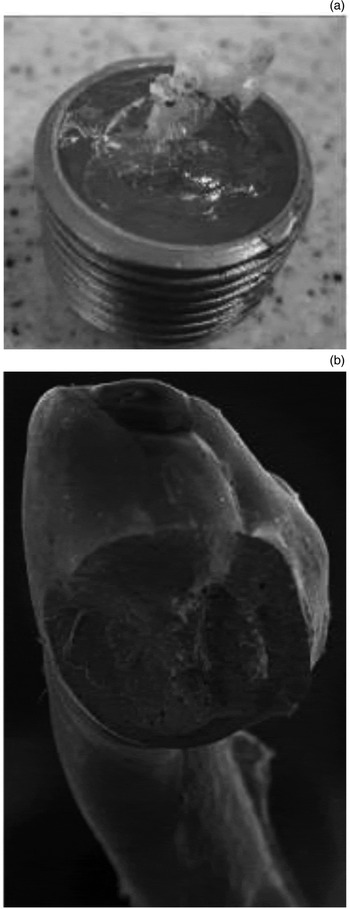
A total of 30 experiments were conducted, 15 in each group. Some of the compression tests in both groups produced skewed values because the fractures occurred between the bone and the cement (Figure 5). As the study aimed to investigate the properties of glass ionomer cement, these skewed values (six samples from each group) were excluded from the analysis. The fractured surface photographs of the included specimens were studied.
Images (a and b) of fractures that occurred between bones and cement.

Each specimen had a different cross-sectional area, which meant that comparison of the force values would lead to inaccuracies; consequently, the results were compared in terms of the more suitable strength values. Strength is the force that affects the unit area, so it is independent of the cross-sectional area of a sample. Brittle materials such as bones fail suddenly after their ultimate strength is exceeded. The maximum compressive strength is defined as the maximum stress that a material can withstand in compression before failure. It is determined by dividing the maximum force in compression by the original cross-sectional area of the test specimen.
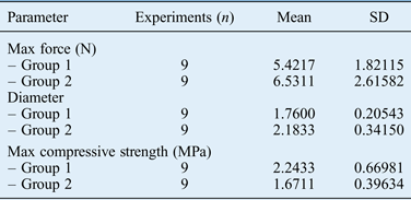
The average maximum force was 5.4217 N in group 1 (which had a 1.0 mm gap between the incus and stapes) and 6.5311 N in group 2 (which had a 2.0 mm gap). The average diameters of the samples were approximately 1.76 and 2.18 mm, respectively. The ultimate compressive strength was calculated to be 2.24 MPa for group 1 and 1.67 MPa for group 2 (Table I).
Maximum force, diameter and maximum compressive strength data

SD = standard deviation; max = maximum
No significant difference was found for maximum force (p = 0.312) between the two groups. The diameter was significantly higher in group 2 than in group 1 (p = 0.006). The maximum compressive strength value was significantly higher in group 1 than in group 2 (p = 0.042).
The ultimate compressive strength was 25.5 per cent lower in group 2 than in group 1. Glass ionomer cement exhibited brittle fracture behaviour at the bonded sections, which were at a 91° angle. When compression is applied to a material at this angle, the material is also affected by flexure. Thus, group 1 samples withstood 25.5 per cent more flexural and compressive stress than did group 2 samples.
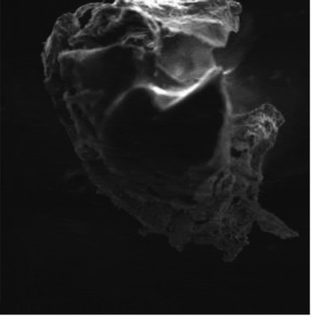
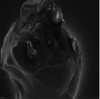
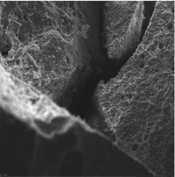
Scanning electron microscope observation of samples in both groups showed that the brittle fracture consisted of the formation and propagation of cracks through the cross-section of glass ionomer cement, in a direction perpendicular to the applied force. Scanning electron microscope micrographs of group 1 samples at magnitudes of 100 and 200 revealed that the glass ionomer cement fractures had brittle fracture characteristics (Figure 6); however, multipart fractures were also observed in group 2 (Figures 7 and 8). Multipart fractures are indicative of lower strength; therefore, strength was lower in group 2 than in group 1.
Image of a brittle fracture that occurred in group 1. (×100 magnification)

Image of a multipart fracture that occurred in group 2. (×100 magnification)

Image of a multipart fracture that occurred in group 2. (×200 magnification)

Discussion
The ease of use and other advantages of glass ionomer cement as a material have drawn the attention of otolaryngologists, and it is now being used for various indications. Partial and total ossicular replacement prostheses are commonly used to bypass or replace the defective incus. However, in cases where the incus defect is narrow, the use of bone cement is increasingly preferred. Use of this material allows the incus and stapes to grow back together; furthermore, bone cement is compatible with normal anatomy, easily applicable and accepted as biocompatible.Reference Rondini-Gilli, Grayeli, Borges Crosara, El Garem, Mosnier and Bouccara2, Reference Stea, Cervellati, Cavedagna, Savarino, Cenni and Pizzoferrato3 Glass ionomer cement has high sound energy conductivity, lacks the weak link issue common in prosthesis use and can ensure strong durability of the ossicular chain. The high-frequency gain obtained in patients using glass ionomer cement is connected to the vibration feature of the material.Reference Rondini-Gilli, Grayeli, Borges Crosara, El Garem, Mosnier and Bouccara2
Since the initial use of glass ionomer cement in otology, several clinics have published studies on the subject; however, no relevant experimental studies of the material's strength and reliability have been performed to date. A study by Bayazit et al. reported success rates of incus to stapes and malleus to stapes bridging of 78.6 and 87.5 per cent, respectively.Reference Bayazit, Ozer, Kanlikama, Durmaz and Yilmaz12 Although defect lengths were measured, the results for incus to stapes and malleus to stapes joints were reported as not statistically significantly different. In our study, the fragility of glass ionomer cement increased with increases in length, so that a 2.0 mm bone cement sample was 25.5 per cent more fragile than a 1.0 mm sample. Therefore, the length of the sample is one criterion that can affect the success of bone cement use.
Many chemical and physical factors influence the outcome of ossiculoplasties performed using glass ionomer cement. Among these are heat, powder particle size, and the amounts of powder and liquid used.Reference Stea, Cervellati, Cavedagna, Savarino, Cenni and Pizzoferrato3 In otological surgery, a change in heat is particularly important. We standardised the effect of heat in our study by performing our tests in an environment similar to operating theatre conditions, in an air-conditioned room with the temperature maintained at 18°C.
Mistakes made during the preparation of the glass ionomer cement may result in early hearing loss. Hearing reconstruction can be considered unsuccessful at an early stage if: the stiffness and rigidity of the bone cement decreases as a result of improper mixing, the surgery is ended before the bone cement solidifies, or the cement bridge is broken by a tampon.Reference Stea, Cervellati, Cavedagna, Savarino, Cenni and Pizzoferrato3
Bayazit et al. reported another potential cause of failure; specifically, after the tympanomeatal flap is closed and a tampon is inserted into the external ear canal, the force applied may increase the fragility of the bone cement.Reference Bayazit, Ozer, Kanlikama, Durmaz and Yilmaz12 For this reason, Bayazit et al. recommended that ossicular chain repair be conducted after the membrane or sponge is put in place. The same study also indicated that improper manipulations which occur after the temporalis muscle fascia or cartilage is put in place, especially in malleus to stapes rebridging with bone cement, can result in ossicular chain transmitted pressure, which in turn can lead to sensorineural hearing loss, tinnitus and vertigo.Reference Bayazit, Ozer, Kanlikama, Durmaz and Yilmaz12 The experimental mechanism we created in our study exposed the glass ionomer cement used in the incus to stapes repair to an average force of 7.25 N in group 1 (which had a 1.0 mm gap between the incus and stapes) and 6.53 N in group 2 (which had a 2.0 mm gap). The normal ear physiology incorporates energy-modifying systems like the stapedius tendon, ossicular ligaments, the oval and the round window, and the inner-ear fluid environment. The ossicular chain in our in vitro study rested on a firm surface, with none of these energy-modifying systems; thus, no energy loss occurred and the strength of the glass ionomer cement could be determined objectively.
The glass ionomer cement powder and liquid used in our study were prepared and brought to the right consistency by the same person. Interfacial adhesion between glass ionomer cement and bone directly depends on the surface roughness of bones, and on other interfacial factors such as blood and the ambient temperature of fibrous tissue.Reference Smith, Ratner, Hoffman, Schoen and Lemons13 In an in vitro study, the other interfacial factors have no effect. High surface roughness of the bones leads to an increase in the cross-sectional area, and thus to better adhesion. We had no direct knowledge about the surface roughness of the incudes and stapedes, which were taken from healthy individuals, so this factor was ignored in this study. We also disregarded the effect of the ligaments, muscles and the fibrous tissue in the middle ear. These are limitations that prevent generalisation to clinical situations.
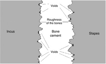
The aim of bone cementing is to establish a chemical bond between glass ionomer cement and bone, but provision of mechanical interlocking is also important in order to strengthen the effects of adhesion. This type of interlocking occurs when the bonding agent penetrates into surface irregularities or porosities in the substrate surface. The effect of adhesion is increased when the bonding agent fills these surface irregularities. The adhesive must therefore have high fluidity (low viscosity) at this point, in order to penetrate into the fine recesses. Otherwise, the porosities on the surface of ossicles will not be filled with glass ionomer cement, as seen in Figure 9, and voids between glass ionomer cement and the bone surface will form.Reference Smith, Ratner, Hoffman, Schoen and Lemons13–Reference Callister and Callister15 Six samples from each of the two groups were excluded from the study because of the occurrence of fractures between the bone and glass ionomer cement. These samples with skewed values were thought to have many voids between the surfaces of glass ionomer cement and bone due to imperfect interlocking.
• This study assessed glass ionomer cement used for incudostapedial rebridging
• Compression tests were conducted to evaluate biomechanical properties and resistance force of the cement (two different sample lengths)
• The defect length optimal for repair with glass ionomer cement was determined
• The fragility of bone cement used as a filler was 25.5 per cent higher for a 2 mm gap than for a 1 mm gap
• The use of bone cement in ossiculoplasty may be safer for repair of smaller incus to stapes defects
Schematic illustration of interlocking between bones and bone cement.

A study by Skinner et al. reported an angle of the incudostapedial joint of approximately 93.03° ± 8.27°.Reference Skinner, Honrado, Prasad, Nelson and Selesnick16 In our study, this was set at a constant angle of 91°. Because the bonding occurred at an angle of 91°, the stress imposed on the glass ionomer cement was not directly compressive. The resulting flexural and compressive stress affected the bonded system, causing completely brittle fracture to occur. Brittle fractures can be single-crack or multi-crack in metals, and are multipart in ceramic-based materials, such as glass ionomer cement, which show little to no strain. In our study, we observed multipart fractures in the tested glass ionomer cement samples.
Conclusion
The fragility of bone cement used as a filler was 25.5 per cent higher for a 2 mm gap than for a 1 mm gap. Transmission electron microscopy confirmed the experimental test results. Increases in the gap length lead to faster crack propagation and multipart fractures, causing glass ionomer cement bonding to fail. We speculate that the use of bone cement may be safer for the repair of smaller incudostapedial defects.