Introduction
The placenta in mammals acts as an interface between the mother and the foetus and regulates the in-utero environment for optimal foetal development. The placenta is a highly vascularised tissue, including both embryonic and maternal blood vessels. During pregnancy, the vascular networks within the placenta are responsible for exchanging respiratory gases, nutrients, and waste between the maternal and foetal systems (Ref. Reference Carter1).
Throughout gestation, the vasculature in the placenta is constantly evolving to adapt the growing demands of the foetus and can be directly influenced by some exogenous factors like maternal nutrition, environmental heat or cold stress, smoking, and medication use (Refs Reference Saben2–Reference Khalil4). Impaired placental vasculature leads to decreased placental blood flow, nutrient uptake, and foetal oxygen. Those changes can produce an adverse uterine environment resulting in pregnancy complications for both the mother and the foetus, such as miscarriage, foetal growth retardation, preeclampsia, gestational hypertension, stillbirth, or preterm delivery, and has been associated with a predisposition to chronic diseases in adulthood (Refs Reference Pereira5–Reference Longtine and Nelson11). Animal studies have demonstrated that angiogenesis and vasodilatation of the placenta is the key mechanism to increase placental blood flow during late gestation, an important change for normal foetal growth (Ref. Reference Reynolds12).
The angiogenic factors affecting placental development include the vascular endothelial growth factor (VEGF), placenta growth factor, fibroblast growth factor, the angiopoietin protein family, and their respective receptors (Refs Reference Klagsbrun and D'Amore13–Reference Levine17). In addition, several signalling pathways regulate placental angiogenesis, such as the MAPK, PI3K/Akt, and eNOS-NO pathways (Refs Reference Cargnello and Roux18–Reference Cooke20). With the bifunctional activities of arginine demethylase and lysyl hydroxylase activities, JMJD6 contributes to diverse disease pathogenesis. Multiple studies have demonstrated the function of JMJD6 in regulating cell proliferation and a wide range of tumorigenesis (Refs Reference Sharma, Kelly and Jones21–Reference Bagan, Sarrion and Jimenez23). Moreover, emerging roles of JMJD6 in infection, inflammation, and immune responses have been reported (Refs Reference Chen24–Reference Walsh, Lee and Choi27). Recently, the role of JMJD6 in placental angiogenesis and preeclampsia has been frequently reported (Refs Reference Boeckel28–Reference Palmer33). This review focuses on the effects of angiogenesis, as the importance of vasculogenesis and trophoblast mediated arterial remodelling have been reported previously (Refs Reference Demir, Seval and Huppertz34–Reference Maltepe and Fisher38).
Molecular functions of JMJD6
JMJD6 belongs to the large family of JmjC domain-containing metalloenzymes. The main enzymes of JmjC family are Fe(II)- and 2-oxoglutarate (2OG)-dependent oxygenases (Ref. Reference Pedersen and Helin39). In 2000, JMJD6 was originally identified as a phosphatidylserine receptor on the cell surface of macrophages, fibroblasts, and epithelial cells, having a possible role in the phagocytosis of apoptotic cells (Refs Reference Fadok40, Reference Fadok41). In 2004, Cikala et al. identified the distorted double stranded β-helix fold (DSBH) fold along with its possible nuclear localisation signals (NLS) (Ref. Reference Cikala42). According to the similarities between the predicted JMJD6 structure and other 2OG-dependent oxygenase enzymes, such as hypoxia-inducible factor (HIF)-alpha asparaginyl hydroxylase FIH, JMJD6 was suggested to be a 2-oxoglutarate (2OG)-dependent dioxygenase (Ref. Reference Cikala42). According to this prediction, it was named JMJD6. Meanwhile, JMJD6 knockout mice revealed neonatal lethality and serious morphological defects, including brain deformity, eye heart, lung, and kidney defects, and impaired cell differentiation, among others which cannot be explained by impaired apoptotic cell clearance (Refs Reference Bose43, Reference Kunisaki44). In 2007, Chang et al. first reported JMJD6's arginine demethylase activity on histone proteins (Ref. Reference Chang45). Later, Webby et al. showed its lysine hydroxylase activity on U2 auxiliary factor 65 (U2AF65), a splicing associated factor (Ref. Reference Webby46). These findings revealed the bifunctional enzymatic activity of JMJD6. Growing evidence indicates that JMJD6 acts at the transcriptional, splicing, posttranscriptional, and metabolic levels, although the detailed molecular mechanisms are not clear (Refs Reference Chang45, Reference Liu47, Reference Lee48).
JMJD6 structure
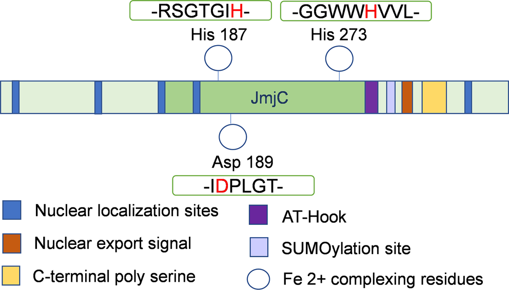
The structural analysis of the JMJD6 protein demonstrated that JMJD6 contains a JmjC domain, highly conserved from eukaryotes to bacteria, as well as other sequence motifs, such as three NLS, a DNA binding domain (AT-hook), a putative SUMOylation site, and a polyserine domain (polyS) (Fig. 1) (Refs Reference Cui49–Reference Bottger52). The core protein structural fold of all 2OG-dependent oxygenases comprises the typical cupin or DSBH surrounded by characteristic secondary structure elements (Ref. Reference Aik53). Crystallographic studies verified the DSBH structure, which forms a barrel-like structure with two β-sheets, with the Fe(II) binding site of the catalytic centre located at the opening of the barrel, and iron-binding and 2OG binding residues (Ref. Reference Mantri54). Electrophoretic mobility shift assays in full-length JMJD6 proteins revealed efficient JMJD6 binding to single stranded RNA (ssRNA) instead of double-stranded RNA (Ref. Reference Hong55). In addition, JMJD6 can exist in monomeric and larger oligomeric forms (Ref. Reference Vangimalla56).
Predicted structure of the protein domains and motifs of JMJD6. Diagram presentation of the full-length JMJD6 protein with central JmjC domain (green).

The metal is most frequently bound by the side chain of three residues (His187, Asp189, and His273), which form an HXD/E(X)nH motif and are indispensable for JMJD6's enzymatic activity (Refs Reference Bottger52, Reference Kwok57–Reference McDonough59). Five functional NLS can target JMJD6 to the nuclei either alone or together (Refs Reference Cikala42, Reference Cui49). The AT-hook was proposed to be a DNA binding motif; however, JMJD6 binds effectively to ssRNA instead of DNA (Refs Reference Hong55, Reference Huth60). Moreover, a three-dimensional structural model of the JMJD6 protein suggests that the AT-hook, the NLS, and the SUMOylation site may be able to interact with other proteins (Ref. Reference Hahn50). The polyS region is located at the C-terminus and may regulate its oligomeric structure. JMJD6 forms a ring-shaped overall oligomeric structure with the complete polyS domain, otherwise, JMJD6 forms a filamentous structure (Ref. Reference Wolf61). The polyS also has an impact on JMJD6's sub-nuclear localisation. With it, JMJD6 is mostly found in the nucleoplasm, while without it is mostly found in the nucleolus and nuclear speckles (Ref. Reference Vangimalla56).
JMJD6 localisation and enzymatic activity
The JMJD6 protein was located in the nucleus both in transfected cells and in cells expressing endogenous JMJD6 mRNA, by using mouse breast carcinoma and most of human tumour cell lines. However, JMJD6 can be present both in the nucleus and the cytoplasm, where it exists in a soluble form associated with intracellular vesicles. Moreover, it has also been reported to be a secreted protein in the extracellular matrix (Ref. Reference Miotti62). JMJD6 facilitates cell proliferation and migration in vitro, and promotes tumour growth in vivo (Refs Reference Zhou63, Reference Zhang64). JMJD6 overexpression is compatible with advanced clinicopathological stage, increased aggressiveness, and poor survival in patients like lung, colon, and hepatic carcinoma (Refs Reference Kunisaki44, Reference Zhang64–Reference Wan66).
Nowadays, JMJD6 is a well-recognised bifunctional enzyme contributing to the regulation of histone demethylation, transcriptional polymerase II promoter pause release, and mRNA splicing. It catalyses two types of reactions, N-methyl argininyl demethylation and lysyl hydroxylation on histone and non-histone protein substrates both in the nucleus and cytoplasm (Refs Reference Bose43, Reference Chang45). Both activities are post-translational events crucial for a large range of cellular processes (Ref. Reference Kwok57). In addition, JMJD6 can also work as a histone tyrosine kinase (Ref. Reference Liu67). In human embryonic kidney (HEK) 293 T cells, JMJD6 was found to interact with 40 such proteins, including proteins related to mRNA processing and splicing, helicases, DNA binding proteins, etc (Ref. Reference Webby46).
The role of JMJD6 in splicing regulation may be through its interaction with serine and arginine-rich (SR) proteins and SR-like splicing factors, particularly U2AF65, which binds to VEGF receptor 1 (Flt1) mRNA (Refs Reference Boeckel28, Reference Webby46). JMJD6 and U2AF65 directly bind to pre-mRNA and coregulate alternative splicing events (Ref. Reference Yi68).
JMJD6's role in angiogenesis
JMJD6 was proposed to regulate Flt1 splicing and control angiogenic sprouting (Ref. Reference Boeckel28). JMJD6 downregulation increased the expression of Flt1's alternative splice variant, leading to higher levels of a secreted protein in cell-culture supernatants that acts as a soluble VEGF- and PlGF-trapping molecule, thus inhibiting angiogenesis (Refs Reference Boeckel28–Reference Maynard30, Reference Olsson69). In contrast, neutralising soluble VEGF receptor (sFlt1) antibodies or saturating VEGF or PlGF ameliorate the angiogenesis inhibition in JMJD6 silenced cells. Accordingly, JMJD6 silencing by siRNA in vitro or in transgenic mouse models resulted in impaired angiogenic sprouting (Ref. Reference Boeckel28).
Oxygen sensor
Throughout gestation, proper development of new blood vessels is necessary to maintain the oxygen supply. Consistent low oxygen is a crucial factor in the pathogenesis of placental-associated disorders such as preeclampsia.
JmjC domain-containing proteins catalyse Fe(II) and 2-oxogluarate–dependent hydroxylation or dioxygenase reactions and, therefore, are mediators of hypoxic gene expression (Ref. Reference Tsukada70). Hewitson et al. showed that the factor inhibiting hypoxia-inducible factor 1, a JmjC domain-containing protein, regulates the recruitment of hypoxia-inducible factor (HIF1α)'s transcriptional coactivators (Refs Reference Hewitson71, Reference Lando72). In 2015, Alahari et al. described that the spatial and temporal pattern of JMJD6 expression in the human placenta is associated with changes to physiological oxygen levels. JMJD6 levels negatively correlated with changes in oxygen tension during early placental development, i.e., high at 7–9 weeks for low pO2 and subsequently declining with increasing pO2. Moreover, JMJD6 was determined to be a downstream target of HIF1, which consists of an inducible HIF1α subunit and a constitutively active β-subunit (Ref. Reference Alahari, Post and Caniggia73).
In normoxic environment, JMJD6 could normally hydroxylate a component of the splicing machinery U2AF65, and leading to produce the full-length membrane-bound Flt-1 transcript (Ref. Reference Boeckel28). However, under hypoxic conditions, the ability of JMJD6 to hydroxylate U2AF65 is decreased, causing the production of shorter alternatively spliced sFlt-1 transcripts from the Flt-1 transcript (Refs Reference Boeckel28, Reference Palmer33).
Moreover, hypoxia increases the transcription of certain JmjC domain-containing enzymes, especially of those involved in histone demethylation (Refs Reference Xia31, Reference Pollard74–Reference Krieg78). In 2018, Alahari et al. reported that JMJD6 regulates the expression and stability of the von Hippel-Lindau (VHL) protein, important to the proteasomal degradation of HIF1α. However, the persistent low oxygen in preeclampsia compromises JMJD6's enzymatic activity, impairing JMJD6-dependent histone demethylation and VHL lysyl hydroxylation, resulting in high HIF1α levels (Refs Reference Alahari32, Reference Alahari, Post and Caniggia73). In 2019, Farrell et al. demonstrated that JMJD6 positively regulates AMOT through oxygen-dependent lysyl hydroxylation. The low oxygen in preeclampsia inactivates JMJD6, which cannot regulate AMOT expression, resulting in AMOT downregulation and impaired trophoblast migration (Ref. Reference Farrell79).
JMJD6's role in preeclampsia
Preeclampsia is a severe pregnancy complication, occurring in 5–8% of all pregnancies, contributing significantly to maternal and perinatal morbidity and mortality, as well as preterm birth (Ref. 80). Despite numerous research efforts, its mechanism remains poorly understood. The dominant pathophysiology is poor placental implantation, leading to ischaemia and hypoxia in the placenta. Under low oxygen levels, the placenta releases excessive anti-angiogenic proteins into the maternal circulation, causing extensive endothelial dysfunction (Refs Reference Maynard30, Reference Nevo81).
A mostly recognised factor involved in preeclampsia is the soluble VEGF receptor, sFlt1 (Ref. Reference Maynard30). JMJD6 and U2AF65 have been implicated in sFlt-1 splicing, which increases in preeclampsia leading to angiogenic imbalance and endothelial dysfunction. However, using immortalised placental trophoblasts and primary endothelial cells, Eddy et al. found that U2AF65 has almost no effect on sFlt-1 splicing. The conflicting results pinpoint the possibility of different regulation patterns in various tissues and under different environmental conditions (Ref. Reference Eddy82).
In the human placenta, Alahari et al. discovered that JMJD6 regulates VHL gene expression. In preeclampsia, inhibited JMJD6 demethylase activity due to hypoxia and decreased Fe2+ bioavailability may cause VHL downregulation. Particularly, Fe2+ supplementation could rescue JMJD6 histone demethylase activity in the preeclamptic placenta of both human and mouse models (Ref. Reference Alahari32).
Recently, a novel mechanism of fibronectin homeostasis/function in preeclampsia was been proposed. Namely, in the preeclamptic environment like reduced oxygen and iron availability, an impaired JMJD6 lysyl hydroxylase function would lead to alterations in glycoprotein fibronectin expression and deposition, affecting cell migration. The ability of Hinokitiol to rescue fibronectin assembly and JMJD6 function reveals the possible direction of preeclamptic treatment (Ref. Reference Alahari83).
Summary
JMJD6 has been implicated in embryonic development, cellular proliferation and migration, tumorigenesis, infection, and immune responses. Although JMJD6 has been reported to interact with several factors involved in preeclampsia, the precise mechanism underlying these effects are not yet clear. More studies are necessary to elucidate the physiological functions of JMJD6 and the mechanisms by which JMJD6 contributes to regulation of angiogenesis and preeclampsia in clinic. Therefore, the impact of JMJD6 on impaired angiogenesis in placenta makes this enzyme an attractive target for therapeutic interventions.
Financial support
This work was supported by National Natural Science Foundation of China (Grant No. 81901487), and a grant of the Repronatal Foundation, Basel, Switzerland.
Conflict of interest
All authors declare no conflict of interest.