Introduction
Haemangiomas are rather common head and neck tumours in childhood but rarely develop in adults. About 65 per cent of adult haemangiomas are found in the head and neck area, but seldom develop in the sinonasal cavities.Reference Watson and McCarthy1 We analysed the clinical manifestations and treatment results for 22 cases of endoscopic excision of sinonasal haemangioma.
Materials and methods
The present study included 22 patients with sinonasal haemangioma who had undergone surgical tumour resection between March 1996 and February 2007 at the Department of Otorhinolaryngology of The Catholic University of Korea. A retrospective chart review assessed these patients' clinical manifestations, operative findings and surgical results.
Results and analysis
Demographics
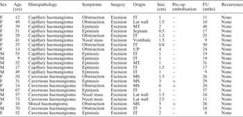
Patients' demographic data are shown in Table I. Of the 22 patients included, nine (41 per cent) were male and 13 (59 per cent) were female. Regarding age distribution, five patients (22.7 per cent) were in their second decade of life, two (9 per cent) were in their twenties, three (13.5 per cent) in their thirties, three (13.5 per cent) in their forties, five (22.7 per cent) in their fifties and two (9 per cent) in their seventies. One patient was younger than 10 years, and one was in their sixties. The average age was 38.3 years.
Haemangioma of nasal cavity and paranasal sinus: patients' demographic and clinical data

Yrs = years; pre-op = pre-operative; FU = follow up; mths = months; F = female; M = male; IT = inferior turbinate; lat wall = lateral nasal wall; MT = middle turbinate; UP = uncinate process; MS = maxillary sinus
Presenting symptoms and mass size
The most common presenting symptom was epistaxis (10 patients; 45.5 per cent). Nine patients (40.9 per cent) had nasal obstruction as the main complaint, whilst three (13.5 per cent) presented with a visible nasal cavity mass lesion as the chief complaint.
The mass size was defined as the length of the largest diameter of the surgically removed mass. The mean size was 2.1 cm long; the largest mass measured 6 cm long (Table I).
Diagnostic investigation
Pre-operatively, all patients underwent computed tomography (CT), and six underwent biopsy. Three cases were confirmed as haemangioma on pre-operative biopsy, but two were misdiagnosed as pyogenic granuloma and one as inflammatory nasal polyp.
Site of origin
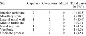
The haemangiomas' sites of origin are shown in Table II. The inferior turbinate was the most common site of origin (10 cases; 45.5 per cent), while four cases (18.2 per cent) arose from the maxillary sinus, three (13.6 per cent) from the lateral nasal wall and two (9.1 per cent) from the middle turbinate. The remaining three cases were found variously to originate from the nasal septum, uncinate process and the nasal vestibule. The three cases originating from the lateral nasal wall are of special interest; two originated from the superior meatus and the other from the mucosa overlying the lacrimal bone (between the inferior turbinate and the anterior edge of the middle turbinate).
Haemangiomas: site of origin and histological type

Data represent case numbers unless otherwise specified.
Treatment and histopathology
All 22 patients had their sinonasal mass surgically removed via an endoscopic approach. In cases in which en bloc resection was impossible, piecemeal resection was chosen as an alternative, and the mucosa and perichondrium or periosteum adjacent to the mass origin were resected as well. In the three cases in which the mass was judged to be large, the feeding vessels were embolised pre-operatively. In the two cases in which the mass originated from the maxillary sinus, the sinus tumour was completely removed with the help of trocar insertion through the canine fossa, after the intranasal portion of the tumour had been removed along with part of the inferior turbinate.
On histopathological examination, 12 tumours (54.5 per cent) proved to be capillary haemangiomas, while nine (40.9 per cent) were cavernous haemangiomas and one (4.5 per cent) was a mixed type haemangioma (Table II). Interestingly, three of the four haemangiomas arising from the maxillary sinus were found to be the cavernous type.
Clinical course
The patients were followed post-operatively for a mean 21 months (range eight to 48 months). Within this follow-up period, no patients showed any evidence of recurrence or complications.
Discussion
Haemangiomas are benign, vascular tumours commonly found in newborns and children, with 85 per cent being discovered within the first year of life.Reference Shpitzer, Noyek, Witterick, Kassel, Ichise and Gullane2 About 50 per cent resolve completely before the age of five years, and up to 70 per cent disappear by the age of seven years; this probably accounts for why haemangiomas are rarely encountered in adults.Reference Sasaki, Pang and Wittliff3
Haemangiomas of childhood are known to have a proliferative stage and an involution stage, and each stage can be subdivided into an early and late period. The proliferative stage takes place in the first year of life, as the endothelial cells actively proliferate and the number of vessels within the tumour increases; following this, the involution stage takes place, for up to 12 years.Reference Simpson4–Reference Bowers, Graham and Tomlinson6 Complications due to rapid growth are more common during the proliferative stage. Waner et al. Reference Waner, Suen and Dinehart7 reported that the prognosis is dependent on the management of the early proliferative stage, and that laser photocoagulation during this period is highly effective. The early involution stage takes place during the first three to four years after the proliferative stage, during which endothelial cells are flattened and a fibrofatty matrix become abundant.Reference Mulliken, Mulliken and Young8 The late stage of involution follows, in which the deposition of fatty tissue, collagen matrix and vessels differing from normal capillaries is predominant.Reference Mulliken, Mulliken and Young8 Gender predominance is known to be negligible.Reference Myer and Gluckman9
About 65 per cent of haemangiomas occur in the head and neck region, but the nasal cavity and paranasal sinuses are uncommon sites.Reference Shpitzer, Noyek, Witterick, Kassel, Ichise and Gullane2 OsbornReference Osborn10 reported the nasal septum (Little's area in particular) and the nasal vestibule as the most common sites of sinonasal haemangioma development, while Iwata et al. Reference Iwata, Hattori, Nakagawa and Tsujimura11 reported the nasal septum and the inferior turbinates as the most common sites. In the present study, the inferior turbinate was found to be the most common site of haemangioma origin.
The precise mechanism of haemangioma development still remains an enigma, but several theories exist. Several growth factors such as hormones are known to affect haemangioma growth,Reference Sasaki, Pang and Wittliff3 while congenital malformation, vascular hamartomas, haemodynamic instability and local trauma have also been suspected as underlying causes.Reference Pitanguy, Machado, Radwanski and Amorim12–Reference Ahn, Chang, Kim, Hong and Cha14
The main presenting symptoms reported in the literature are nasal obstruction, epistaxis and occasionally a visible nasal mass, all of which were also evident in the present study.
Histologically, haemangiomas can be divided into capillary, cavernous and mixed types, but clinical symptoms do not differ among the types.Reference Bataskis and Rice15, Reference Yao-Sui and Karl16 In children, capillary haemangiomas predominate, but adult haemangiomas are more likely to be the cavernous type. The reason for this difference probably lies in the fact that capillary haemangiomas are more prone to natural resolution, while cavernous haemangiomas are less likely to show complete resolution and thus more likely to persist into adulthood.Reference Mulliken and Glowacki17
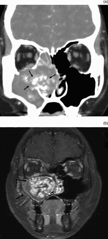
The differential diagnosis of sinonasal haemangiomas includes angiofibroma, organising haematoma, neuroma, inverted papilloma and malignant sinonasal tumours.Reference Jammal, Barakat and Hadi18 Diagnostic investigation includes detailed history-taking and physical examination, and imaging studies such as CT, magnetic resonance imaging (MRI) and angiography. Diagnostic confirmation is achieved through histopathological examination.Reference Shpitzer, Noyek, Witterick, Kassel, Ichise and Gullane2 Pre-operative biopsy is recommended, but was undertaken in only six of the 22 cases in the present study. This is probably due to the fact that office-based biopsies for highly vascular tumours may trigger uncontrolled bleeding. This is why many surgeons, including the authors, prefer intra-operative biopsy (such as frozen section biopsy) for cases in which pre-operative imaging studies reveal a tumour extent amendable to complete excision with routine surgical techniques. Computed tomography informs the surgeon of the anatomical location and local extent of the tumour, including bone destruction and calcification; however, when bone destruction is present discrimination from malignancy can be difficult (Figure 1a).Reference Hyung, Jung, Jae and Eui19 On MRI, a haemangioma can be suspected when the mass shows gadolinium enhancement on T1-weighted images and flow void on T2-weighted images (Figure 1b).
Radiological findings in a patient with sinonasal haemangioma. (a) Coronal, contrast-enhanced, computed tomography scan showing a large, enhanced mass with bone destruction (arrows) within the right maxillary sinus and nasal cavity. (b) Coronal, contrast-enhanced, T1-weighted, magnetic resonance imaging scan showing a heterogeneously enhanced mass (arrows).

The traditional method of surgical excision of a sinonasal haemangioma is via an open approach, utilising lateral rhinotomy or mid-face degloving approaches. However, recent advances in endoscopic techniques have enabled endoscopic excision of haemangiomas, offering better surgical visualisation and avoiding the surgical morbidity commonly encountered in open approaches (e.g. surgical scar, epiphora and facial anaesthesia).Reference London, Schlosser and Gross20 In cases involving the skull base, many authors prefer the craniofacial approach; however, Rodney et al. Reference Schlosser, Woodworth, Gillespie and Day21 recently reported that endoscopic en bloc resection is possible with preservation of the ethmoid roof and cribriform plate.Reference Kulkarni, Bonner and Abdo22 In the present study, an endoscopic approach was chosen as the primary surgical approach, but preparation for surgical conversion to an external approach with internal maxillary artery ligation was undertaken in all cases.
• This paper reports the clinical manifestations and results of endoscopic excision for 22 cases of sinonasal haemangioma
• The most common presenting symptom was epistaxis, and the most prevalent site was the inferior turbinate
• Traditional surgical excision of sinonasal haemangioma is via an open approach utilising lateral rhinotomy or mid-face degloving approaches
• Endoscopic excision of sinonasal haemangioma is a safe and effective substitute for conventional open approach surgery
Haemangiomas in adults require aggressive treatment because spontaneous regression is uncommon, and thorough pre-operative investigation, including imaging studies, is essential as profuse intra-operative bleeding may be encountered.Reference Kulkarni, Bonner and Abdo22 During surgical excision of sinonasal haemangiomas, a wide resection is recommended including a portion of the adjacent mucosa and perichondrium; pre-operative embolisation with Gelfoam or a coil is advised for huge tumours.Reference Azzolini, Bertani and Riberti23 Other therapeutic options include sclerotherapy, cryotherapy and excision with yttrium aluminium garnet laser,Reference Howerd24, Reference Hara, Hiyosi and Ogata25 while systemic steroids can be prescribed in cases of cutaneous haemangioma and laryngeal haemangioma.Reference Rossiter, Hendrix, Tom and Potsic26
Treatment standards have not been established for sinonasal haemangiomas, owing to the rarity of these lesions. In the present study, the most common presenting symptom was nasal bleeding, and the inferior turbinate was the most common site of tumour origin. In the 22 cases of endoscopic excision of sinonasal haemangioma in the present study, complete excision was possible and no evidence of recurrence was observed during the follow-up period. Hence, we believe that endoscopic excision of sinonasal haemangioma is a safe and effective substitute for conventional open approach surgery.
Acknowledgements
No author received financial support of any kind in regard to the present article.