Introduction
Over recent years, many studies have been carried out in an attempt to determine which conditions are needed during the in vitro maturation, fertilisation and culture processes to maximise embryo production (Shirazi et al., Reference Shirazi, Shams-Esfandabadi, Ahmadi and Heidari2010). In oocyte maturation, two events can be differentiated: cytoplasmic and nuclear maturation. Cytoplasmic maturation refers to the process that prepares the oocyte for activation, pronuclear formation, and preimplantation development, while nuclear maturation refers to the ability of the oocyte to progress through meiosis (Ptak et al., Reference Ptak, Loi, Dattena, Tischner and Cappai1999). The mechanism of meiotic arrest and subsequent resumption has received increasing attention, although the mechanism by which the stimulation operates remains largely unknown (Grøndahl et al., Reference Grøndahl, Ottesen, Lessl, Faarup, Murray, Gronvald, Hegele- Hartung and Ahnfelt-Ronne1998). The lack of competence of prepubertal animal oocytes has been reported in previous studies (O'Brien et al., Reference O'Brien, Dwarte, Ryan, Maxwell and Evans1996; Ledda et al., Reference Ledda, Bogliolo, Calvia, Leoni and Naitana1997), and all these reports suggested that developmental arrest of embryos derived from prepubertal oocyte was due to a range of aspects related with cytoplasm maturation. It has been shown that oocytes from prepubertal cattle have a lesser developmental competence, possibly due to deficiencies in cytoplasmic maturation, which impair further embryo development (Mermillod et al., Reference Mermillod, Le Bourhis, Lonergan, Khatir and Heyman1998; Salamone et al., Reference Salamone, Damiani, Fissore, Robl and Duby2001).
The in vitro culture systems available can be classified according to their formulation as follows: undefined, in which serum or/and co-culture are used; semi-defined, in which co-culture is omitted and serum is replaced by albumin; or fully defined, a protein-free system in which albumin is replaced by macromolecules such as polyvinyl alcohol and polyvinyl pyrrolidone (Marquant-Le Guinne and Humblot, Reference Marquant-Le Guienne and Humblot1998; Farin et al., Reference Farin, Crosier and Farin2001; Vanroose et al., Reference Vanroose, Van Soom and de Kruif2001). Oxygen tension also seems to be an important factor that affects oocyte maturation and embryo development. When oxygen effects have been investigated there are conflicting results. In the bovine model, IVM at 5% O2 dramatically decreased the viability of oocytes (Pinyopummintr and Bavister, Reference Pinyopummintr and Bavister1995; Hashimoto et al., Reference Hashimoto, Minami, Takakura, Yamada, Imai and Kashima2000), whereas oxygen levels appear to have no effect on pig oocytes (Park et al., Reference Park, Hong, Yong, Hwang, Lim and Lee2005). Furthermore, blastocyst development rates have been shown to both improve (Park et al., Reference Park, Hong, Yong, Hwang, Lim and Lee2005) or remain unchanged when porcine oocytes are matured in 20% oxygen (Kikuchi et al., Reference Kikuchi, Onishi, Kashiwazaki, Iwamoto, Noguchi, Kaneko, Akita and Nagai2002). Medium composition may play a role in these discrepancies, as Hashimoto et al. (Reference Hashimoto, Minami, Takakura, Yamada, Imai and Kashima2000) also reported that 5% O2 during IVM supports bovine embryo development if the glucose concentration is increased.
Mammalian cumulus cells play a very important role during oocyte growth and maturation. They are known to supply nutrients (Laurincík et al., Reference Laurincík, Kroslak, Hyttel, Pivko and Sirotkin1992) and/or messenger molecules for the development of the oocyte (Lawrence et al., Reference Lawrence, Beers and Guila1978; Buccione et al., Reference Buccione, Schroeder and Eppig1990) and to mediate the effects of hormones on the cumulus–oocyte complex (Zuelke & Brackett, Reference Zuelke and Brackett1990). In vertebrates, gonadotrophins trigger the production of paracrine hormones in the cumulus cells enclosing the oocytes (Byskov et al., Reference Byskov, Andersen and Leonardsen2002). Many of the factors secreted by gonadotrophin-stimulated cumulus cells of cumulus–oocyte complexes can affect the meiotic maturation of oocytes (Buccione et al., Reference Buccione, Schroeder and Eppig1990; Mattioli, Reference Mattioli1994; Xia et al., Reference Xia, Byskov and Andersen1994, Reference Xia, Kikuchi, Noguchi and Izaike2000). Although still not proven, growth factors may also stimulate cumulus granulosa cells to produce steroids, follicular fluid-derived meiosis-activating sterols (FF-MAS) and other factors that may play a role in promoting oocyte maturation (Jamnongjit & Hammes, Reference Jamnongjit and Hammes2005). The findings of Grøndahl et al. (Reference Grøndahl, Ottesen, Lessl, Faarup, Murray, Gronvald, Hegele- Hartung and Ahnfelt-Ronne1998) strengthened the evidence that FF-MAS could be an endogenous signalling molecule involved in the resumption of meiosis. FF-MAS are present in human pre-ovulatory follicular fluid and are an intermediary occurring naturally in the biosynthetic pathway between lanosterol and cholesterol (Byskov et al., Reference Byskov, Andersen, Nordholm, Thogersen, Xia, Wassmann, Andersen, Guddal and Roed1995). FF-MAS and T-MAS are produced by conversion of lanosterol (Grøndahl, Reference Grøndahl2008). Yamashita et al. (Reference Yamashita, Shimada, Okazaki, Maeda and Terada2003) showed that FF-MAS is synthesised in cumulus cells and does not accumulate, but is metabolised to cholesterol, which is then converted into progesterone, and accelerates ongoing germinal vesicle breakdown in porcine oocytes. However, little is known about the mechanisms that underlie the activity of FF-MAS or its analogues on either meiotic maturation or competence to complete preimplantation development (Marín-Bivens et al., Reference Marín-Bivens, Lindenthal, O'Brien, Wigglesworth, Blume, Grøndahl and Eppig2004). In the present study, the effect of oxygen tension and lanosterol addition on the in vitro maturation in semi-defined culture system of prepubertal ewe oocytes was investigated.
Material and methods
Chemicals
Unless otherwise stated, all chemicals were purchased from Sigma-Aldrich (Alcobendas, Madrid, Spain).
Oocyte collection and in vitro maturation
Ovaries from slaughtered prepubertal ewe (30–40 days old, body weight 6–10 kg) were transported from a local abattoir to the laboratory within 1.5 h in Dulbecco's phosphate buffered saline (DPBS) at a temperature around 30°C. After washing in fresh medium, ovaries were sliced using a micro-blade and the follicle content was released in TCM-199 medium supplemented with 25 mM HEPES, 50 IU/ml penicillin, 50 μg/ml streptomycin, and 0.1% (w/v) polyvinyl alcohol (PVA). Cumulus–oocyte complexes (COCs) that presented four to 10 layers of granulosa cells, uniform cytoplasm and homogenous distribution of lipid droplets were selected for these experiments. After three washes in the same fresh medium, the selected COCs were in vitro matured in TCM-199 supplemented with 4 mg/ml bovine serum albumin (BSA), 100 μM cysteamine, 0.3 mM sodium pyruvate, 0.1 UI/ml r-FSH (Gonal-F® 75 UI), 0.1 UI/ml r-LH (Lhadi® 75 UI,) and 1 μg/ml estradiol-17β, penicillin (3000 IU/ml) and streptomycin (3000 μg/ml), in accordance with Accardo et al. (Reference Accardo, Dattena, Pilichi, Mara, Chessa and Cappai2004). Thirty COCs in 500 μl of IVM medium were cultured. Maturation of COCs was performed for 24 h at 38.5 °C.
Experiment 1: Effect of oxygen tension on chromatin configuration
The gas mixtures used were 5% or 20% O2 (5% CO2 and balance of N). Hereafter, these gas mixtures are simply referred to as 5% and 20% O2. Following maturation, oocytes were partially stripped of surrounding cumulus cells using 300 UI/ml of hyaluronidase in HEPES-TCM199. Oocytes were fixed with 2% (v/v) glutaraldehyde and 2% (v/v) formaldehyde in DPBS. Oocytes were incubated in DPBS containing Hoechst 33342 (10 μg/ml) for 15 min and, after washing in fresh DPBS, and observed under an epifluorescence microscope with an excitation filter of 365 nm and a barrier filter of 410 nm. Each oocyte was individually classified according to chromatin configuration as germinal vesicle (GV), metaphase I (MI), metaphase II (MII) or degenerated/unidentifiable.
Experiment 2: Effect of lanosterol on in vitro maturation
Based on results obtained in the first experiment, the COCs were cultured in 5% CO2 in air at 38.5 °C in the maturation medium supplemented with lanosterol (10 and 50 μM). Dilutions of lanosterol were prepared from a stock solution containing 5 mg lanosterol dissolved in 1 ml ethanol. In order to obtain a 0 (control group), 10 and 50 μM lanosterol use concentration, 0 μl, 0.84 μl or 4.2 μl of lanosterol stock solution was added, respectively, together with 4.2 μl, 7.56 μl, or 0 μl ethanol, respectively, to give a final volume of 500 μl maturation medium. The final ethanol concentration in all culture media, including the control group, was 1.68%. The COCs were cultured for 24 h covered with mineral oil. After in vitro maturation, oocytes were denuded, fixed and stained with Hoechst 33342 as previously. Each oocyte was individually classified according to chromatin configuration as GV, MI, MII or degenerated/unidentifiable.
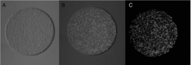
Oocytes after in vitro maturation were also denuded and fixed as previously. Later, oocytes were then incubated in 10 μg/ml Nile red solution (Molecular Probes, Inc.) dissolved in physiological saline (0.9% NaCl) with 1 mg/ml polyvinylpyrrolidone (Genicot et al., Reference Genicot, Leroy, Soom and Donnay2005; Marco-Jiménez et al., Reference Marco-Jiménez, Llobat and Vicente2010). The Nile red stock solution (1 mg/ml) was prepared by dilution in DMSO and stored at room temperature in the dark. Oocytes were stained overnight in the dark and at room temperature unless otherwise indicated. The oocytes had three final washes in DPBS containing 1 mg/ml polyvinylpyrrolidone and were mounted on glass slides. The amount of emitted fluorescent light of the whole oocyte was evaluated with an inverted fluorescence microscope (excitation: 400–500 nm and emission: 515LP) equipped with a digital camera (DMX1200F, Nikon). Individual photographs were taken of stained oocytes and arbitrary units of fluorescence measurement were quantified with Gene Tools software (Syngene, IZASA). Results were expressed in arbitrary units of fluorescence, a high fluorescence being associated with high lipid content (Fig. 1).
Representative in vitro mature prepubertal ovine oocytes stained by Nile red with a granulated cytoplasm under visible light (A); mixed visible light and after ultra-violet (UV) light excitation (B); and after UV light excitation (C) (×200 magnification).

Statistical analysis
The effects of oxygen tension on the percentage of oocytes that reached the metaphase II (MII) stage after IVM were analysed by chi-squared test. The effects of lanosterol on the percentage of oocytes that reached MI and MII stage after IVM were analysed by chi-squared test, while the lipid contents (cytoplasm maturation) were compared by analysis of variance (ANOVA) using the general linear model. All procedures were performed with the Statgraphics®Plus 5.1 (Statistical Graphics Corp.). Significance level was set at p < 0.05.
Results
Experiment 1: Effect of oxygen tension on chromatin configuration
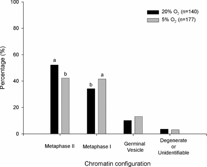
A total of 317 oocytes were evaluated. The presence of 20% O2 tension significantly increased the percentage of oocytes that reached metaphase II stage (52.2 vs. 42.2%, for 20% and 5% O2, respectively; Fig. 2) and decreased the percentage of oocytes that reached metaphase I stage. (34.2 vs. 41.6%, for 20% and 5% O2, respectively, Fig. 2). No differences were observed between groups for GV and degenerated or unidentifiable (10.1 vs. 13.1% and 3.5 vs. 3.1%, for 20% and 5% of O2, respectively; Fig. 2).
Effect of oxygen tension on chromatin configuration during in vitro maturation in semi-defined culture system of prepubertal ewe oocytes. Bars with different superscripts are significantly different (p < 0.05).
Experiment 2: Effect of lanosterol on in vitro maturation
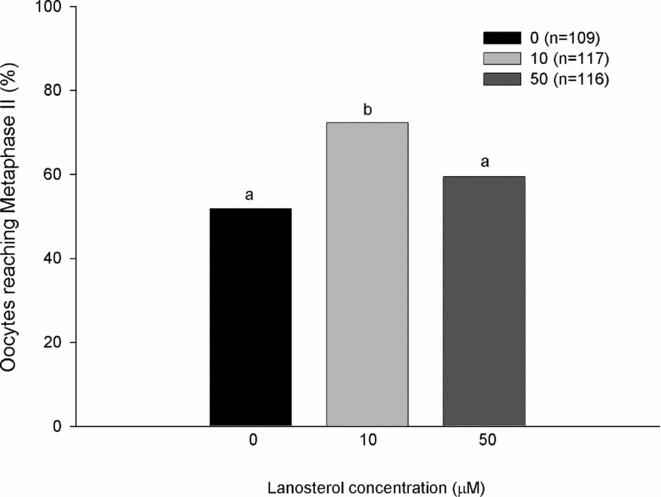
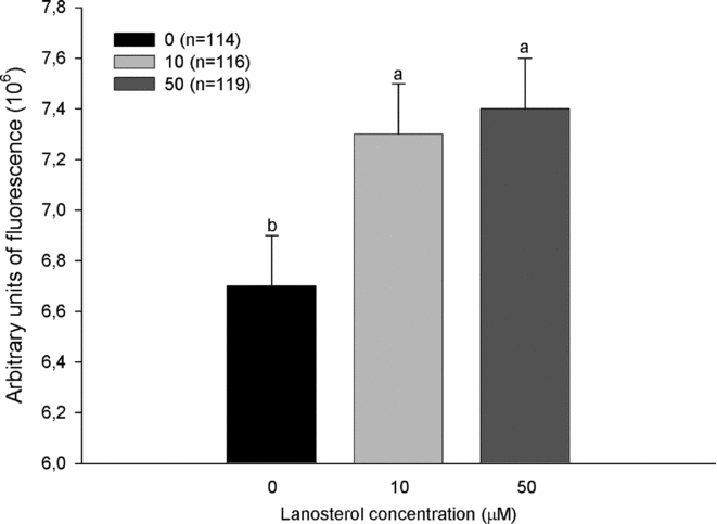
A total of 342 oocytes were evaluated using Hoechst 33342. As shown in Fig. 3, the presence of 10 μM of lanosterol promoted the oocyte meiosis resumption (MII) significantly compared with 50 μM and control groups (72.3 and 59.4% vs 51.8% for 10 and 50 μM of lanosterol vs control, respectively; Fig. 3). Significant reduction of percentage of MI stage was observed for 10 μM of lanosterol compared with the 0 and 50 μM groups (Fig. 3). No differences were observed between groups for the GV and degenerated or unidentifiable (9.5, 9.1 and 7.8% and 3.0, 2.5 and 0.7%, for control, 10 and 50 μM of lanosterol, respectively, Fig. 3). A total of 349 oocytes were evaluated using Nile red stain. Oocytes culture in the presence of lanosterol (10 and 50 μM) promoted an increase in emitted fluorescence light compared with the control group (7.3 ± 0.2 × 10⁶ and 7.4 ± 0.2 × 10⁶ vs. 6.7 ± 0.2 × 10⁶ arbitrary units of fluorescence, for 10 and 50 μM vs control group, respectively, Fig. 4).
Effect of lanosterol on nuclear maturation in in vitro mature prepubertal ovine oocytes. Bars with different superscripts are significantly different (p < 0.05).
Amount of emitted fluorescence of prepubertal ovine oocytes in vitro matured in different lanosterol concentrations stained with Nile red. Data represent the least square means ± standard error means. Bars with different superscripts are significantly different (p < 0.05).
Discussion
The most commonly used system is a culture medium, which is a complex nutrient mixture, supplemented with hormones, serum or albumin and growth factors (Younis et al., Reference Younis, Brackett and Fayer-Hosken1989; Bavister et al., Reference Bavister, Rose-Hellekant and Pinyopummintr1992; Thompson, Reference Thompson2000; Vanroose et al., Reference Vanroose, Van Soom and de Kruif2001). The choice of medium and supplements may affect the meiotic regulation and can have an impact on the regulation of mammalian oocyte growth and embryonic cell function (Downs & Mastropolo, Reference Downs and Mastropolo1997; Kito & Bavister, Reference Kito and Bavister1997; Mather Reference Mather1998). When using co-culture or semi-defined medium, repeatability can be a problem but sanitary measures must also be taken to prevent introduction of pathogens into the culture system (Vanroose et al., Reference Vanroose, Van Soom and de Kruif2001). Due to suspicions of detrimental effects of serum on embryonic and fetal development, the use of semi-defined culture systems obtained by replacing serum with albumin has been investigated (Bavister, Reference Bavister1995; Young et al., Reference Young, Sinclair and Wilmut1998). Moreover, the commonly used commercial gonadotrophins vary in purity, and contaminants in these products may influence experimental results (Zuelke & Brackett, Reference Zuelke and Brackett1990; Choi et al., Reference Choi, Carnevale, Seidel and Squire2001). In the last few years, some authors have reported results of recombinant gonadotrophins in different species (Törnell et al., Reference Törnell, Bergh, Selleskog and Hillensjö1995; Byskov et al., 1997; Cortvrindt et al., Reference Cortvrindt, Hu, Liu and Smitz1998; Anderiesz et al., Reference Anderiesz, Ferraretti, Magli, Fiorentino, Fortini, Gianaroli, Jones and Trounson2000; Modina et al., Reference Modina, Luciano, Scesi, Perazzoli, Lauria and Gandolfi2000; Ali & Sirard, Reference Ali and Sirard2002a, Reference Ali and Sirardb).
In the present study it was noted that oocytes cultured in semi-defined system in presence of low oxygen tension affect the progression of the meiotic cycle. Similar results were obtained in other experiments (Pinyopummintr & Bavister, Reference Pinyopummintr and Bavister1995; Hashimoto et al., Reference Hashimoto, Minami, Takakura, Yamada, Imai and Kashima2000). However, several studies have shown that efficiency of oocyte or embryo culture could be improved by decreasing oxygen tension (Eppig & Wigglesworth, Reference Eppig and Wigglesworth1995; Berthelot & Terqui, Reference Berthelot and Terqui1996; Izquierdo et al., Reference Izquierdo, Villamediana and Paramio1999; deMatos et al., Reference deMatos, Gasparrini, Pasqualini and Thompson2002). The achievement of meiosis observed in the present study was similar to previous results when culture medium was serum free (Khatir et al., Reference Khatir, Lonergan and Mermillod1998). Succu et al. (Reference Succu, Leoni, Berlinguer, Madeddu, Bebbere, Mossa, Bogliolo, Ledda and Naitana2007) reported higher values in prepubertal ewes when oestrus ewe serum was including in the IVM media. The addition of oestrus serum also supported high maturation rates in IVM of ewe oocytes (Rao et al., Reference Rao, Naidu, Amarnath, Vagdevi, Rao, Brahmaiah and Rao2002; Succu et al., Reference Succu, Leoni, Berlinguer, Madeddu, Bebbere, Mossa, Bogliolo, Ledda and Naitana2007). Accardo et al., Reference Accardo, Dattena, Pilichi, Mara, Chessa and Cappai(2004) reported no differences between hypophyseal and recombinant gonadotrophins on oocyte maturation in ewes. However, developmental competence of in vitro matured oocytes from prepubertal animals has been reported to be lower than oocytes derived from adult animals (Pinyopummintr & Bavister, Reference Pinyopummintr and Bavister1995; O'Brien et al., Reference O'Brien, Dwarte, Ryan, Maxwell and Evans1996; Ledda et al., Reference Ledda, Bogliolo, Calvia, Leoni and Naitana1997; Gandolfi et al., Reference Gandolfi, Vassena and Lauria2000; Leoni et al., Reference Leoni, Bebbere, Succu, Berlinguer, Mossa, Galioto, Bogliolo, Ledda and Naitana2007). No papers studying differences between recombinant and non-recombinant hormones on IVM of prepubertal ewes were found in the published literature. In addition, the research into supplementing recombinant hormones such as leptin, insulin-like growth factor, and vascular endothelial growth factor in IVM system has served to improve development of in vitro maturation in several species (Moreira et al., Reference Moreira, Paula-Lopes, Hansen, Badinga and Thatcher2002; Boelhauve et al., Reference Boelhauve, Sinowatz, Wolf, Paula-Lopes, Cao, Zhou, Luo, Zhao and Shi2005; Cao et al., Reference Cao, Zhou, Luo, Zhao and Shi2009; Kim et al., Reference Kim, Jeong, Lee, Jeong, Hossein, Hyun and Hwang2010). The advantage of semi-defined medium is that it eliminates the potential harmful effects of serum, hormones, etc and allows better control of culture conditions, facilitating studies designed to evaluate in vitro oocyte culture requirements. The type of gonadotrophins used in the current study (human recombinant) could be the reason for the observed discrepancy between the present results. However, human recombinant gonadotrophins (r-FSH and r-LH) have been demonstrated to similarly stimulate IVM in adult ewes (Accardo et al., Reference Accardo, Dattena, Pilichi, Mara, Chessa and Cappai2004). In addition, the researches supplementing recombinant hormones such as leptin, insulin-like growth factor, and vascular endothelial growth factor in IVM system have been used to improve development of in vitro maturation in several species (Moreira et al., Reference Moreira, Paula-Lopes, Hansen, Badinga and Thatcher2002; Boelhauve et al., Reference Boelhauve, Sinowatz, Wolf, Paula-Lopes, Cao, Zhou, Luo, Zhao and Shi2005; Cao et al., Reference Cao, Zhou, Luo, Zhao and Shi2009; Kim et al., 2010).
Lanosterol, first intermediary with a sterol ring, is structurally very homologous to meiosis-activating sterols (T-MAS and FF-MAS) which are intermediaries in the cholesterol biosynthetic pathway from lanosterol (Rozman et al., Reference Rozman, Cotman and Frangez2002; Bokal et al., Reference Bokal, Tacer, Vrbnjak, Leposa, Virant-Klun, Verdenik and Rozman2006). The vast majority of sterols assessed have proven ineffective in promoting meiosis (Byskov et al., Reference Byskov, Andersen and Leonardsen2002). However, FF-MAS has shown an effect on meiosis-promoting and the activity seems to be non-specific across species (Byskov et al., Reference Byskov, Andersen, Nordholm, Thogersen, Xia, Wassmann, Andersen, Guddal and Roed1995; Ruan et al., Reference Ruan, Gerst, Emmons, Shey and Schroepfer1997; Grøndahl et al., Reference Grøndahl, Ottesen, Lessl, Faarup, Murray, Gronvald, Hegele- Hartung and Ahnfelt-Ronne1998, Reference Grøndahl, Lessl, Faerge, Hegele-Hartung, Wassermann and Ottesen2000; Leemuis et al., Reference Leemuis, Van der Louw and Groen1998). Nevertheless, the mechanism whereby meiosis-activating sterols mediate in the resumption of meiosis in the oocyte is largely unknown (Grøndahl et al., Reference Grøndahl, Ottesen, Lessl, Faarup, Murray, Gronvald, Hegele- Hartung and Ahnfelt-Ronne1998). In the present study, the addition of 10 μM of lanosterol allowed a greater percentage of oocytes to reach metaphase II state. Previously, lanosterol has tested inactive on meiosis-promoting activity (Byskov et al., Reference Byskov, Andersen, Nordholm, Thogersen, Xia, Wassmann, Andersen, Guddal and Roed1995; Grøndahl et al., Reference Grøndahl, Ottesen, Lessl, Faarup, Murray, Gronvald, Hegele- Hartung and Ahnfelt-Ronne1998). However, recently, Marco-Jiménez et al. (Reference Marco-Jiménez, Llobat and Vicente2010) showed that exogenous lanosterol could be used by the cumulus cells to incorporate with the cholesterol biosynthetic pathway and increased the percentage of porcine oocytes that acquired meiotic competence. Grøndahl et al. (Reference Grøndahl, Ottesen, Lessl, Faarup, Murray, Gronvald, Hegele- Hartung and Ahnfelt-Ronne1998) evaluated different concentrations of lanosterol but without results, possibly due to the low concentrations assayed (0.07–7 μM). In this study, the addition of 10 and 50 μM of lanosterol increased the resumption of oocytes to reach MII state, although 10 μM appears to be a more suitable measure. Previous reports showed that lanosterol in naked oocytes was not effective (Byskov et al., Reference Byskov, Andersen, Nordholm, Thogersen, Xia, Wassmann, Andersen, Guddal and Roed1995; Grøndahl et al., Reference Grøndahl, Ottesen, Lessl, Faarup, Murray, Gronvald, Hegele- Hartung and Ahnfelt-Ronne1998). These results are in agreement with our hypothesis, because if lanosterol is incorporated in the cholesterol biosynthetic by the cumulus cells, in naked oocytes no effect should be expected (Marco-Jiménez et al., Reference Marco-Jiménez, Llobat and Vicente2010). Although oocytes exhibiting nuclear maturation can be fertilised, they may be developmentally incompetent because of a deficiency in the cytoplasmic factors needed for full development (Sun & Nagai, Reference Sun and Nagai2003). Niimura et al. (Reference Niimura, Takano, Onishi and Hosoe2002) considered that the transformation of lipids in the cytoplasm is closely related to the resumption of meiotic maturation, regardless of in vitro maturation. The role of lipids in oocyte maturation and embryo development is unclear, although they may have a potential role as reserve fuels, providing ATP for the protein synthesis that is necessary for continuation of cytoplasmic maturation and meiosis (Sturmey & Leese, Reference Sturmey and Leese2003). Lipids also adjust the physiological function (Fujihira et al., Reference Fujihira, Kishida and Fukui2004). Nile red technique is highly sensitive and repeatable and contrasts with previously described techniques to detect differences in lipid content (Ferguson and Leese, Reference Ferguson and Leese1999; McEvoy et al., Reference McEvoy, Coull, Broadbent, Hutchinson and Speake2000; Kim et al., Reference Kim, Kinoshita, Ohnishi and Fukui2001; Genicot et al., Reference Genicot, Leroy, Soom and Donnay2005; Marco-Jiménez et al., Reference Marco-Jiménez, Llobat and Vicente2010). To our best knowledge, no previous works using Nile red stain with ovine oocytes have been reported. The addition of lanosterol (10 or 50 μM) to the semi-defined culture medium suggested lipid accumulation after in vitro maturation, and it may be used as an energy source (McEvoy et al., Reference McEvoy, Coull, Broadbent, Hutchinson and Speake2000; Sturmey & Leese, Reference Sturmey and Leese2003). The availability of lipid reserves and lipase activity would enable the oocyte to use lipids as oxidative substrates after it is separated from cumulus cells during maturation (Cetica et al., Reference Cetica, Pintos, Dalvit and Beconi2002). However, further study is needed to determine the functional role of cytoplasmic lipid in immature ovine oocytes during maturation.
In conclusion, this study has demonstrated that the low oxygen concentration in in vitro maturation in semi-defined culture system has a detrimental effect on prepubertal ewe oocytes reaching metaphase II state. In addition, oocytes exposed to lanosterol increased the percentage of oocytes reaching metaphase II and the lipid accumulation. Nevertheless, it is clear that determination of the accurate dose of lanosterol will be crucial in order to draw proper conclusions concerning this compound. Additional experiments will be needed to understand the involvement of lanosterol in the cholesterogenic pathway.
Acknowledgements
This work was supported by funds from INIA (project RZ2006–00004) and Universidad Politécnica de Valencia (20081166). English text version revised by N. Macowan English language service.