Introduction
Anhydrous copper vanadates are abundant minerals in active fumaroles of scoria cones related to the Great Fissure Tolbachik eruption (Pekov et al., Reference Pekov, Zubkova, Zelenski, Yapaskurt, Polekhovsky, Fadeeva and Pushcharovsky2013; Vergasova and Filatov, Reference Vergasova and Filatov2016). Some of them contain chloride ions: averievite Cu5O2(VO4)2·nMX (Vergasova et al., Reference Vergasova, Starova, Filatov and Anan'ev1998), leningradite PbCu3(VO4)2Cl2 (Siidra et al., Reference Siidra, Krivovichev, Armbruster, Filatov and Pekov2007); yaroshevskite Cu9O2(VO4)4Cl2 (Pekov et al., Reference Pekov, Zubkova, Zelenski, Yapaskurt, Polekhovsky, Fadeeva and Pushcharovsky2013) and dokuchaevite Cu8O2(VO4)3Cl3 (Siidra et al., Reference Siidra, Nazarchuk, Zaitsev, Polekhovsky, Wenzel and Spratt2019). Copper arsenate minerals are also common in high-temperature fumarolic assemblages (Pekov et al., Reference Pekov, Koshlyakova, Zubkova, Lykova, Britvin, Yapaskurt, Agakhanov, Shchipalkina, Turchkova and Sidorov2018).
Herein we report on the composition, structure and properties of aleutite (Cyrillic = алеутит). The mineral is named for the Aleuts ethnic group who are the original inhabitants living on the Commander Islands, Aleutsky District, Kamchatka Krai. Both the mineral and the mineral name were approved by the Commission on New Minerals, Nomenclature and Classification of the International Mineralogical Association (IMA2018-014, Siidra et al., Reference Siidra, Nazarchuk, Agakhanov and Polekhovsky2018b). Type material is deposited at the Mineralogical Museum, St. Petersburg State University, St. Petersburg, Russia (catalogue no. 1/19689).
Occurrence and association
Aleutite occurs as a product of fumarolic activity. It was found in the summer of 2015 in Yadovitaya (‘Poisonous’) fumarole at the Second scoria cone of the Northern Breakthrough of the Great Fissure Tolbachik Eruption in Kamchatka, Russia. The Second scoria cone is located ~18 km SSW of the active shield volcano Ploskiy Tolbachik (Fedotov and Markhinin, Reference Fedotov and Markhinin1983). The temperature of gases at the sampling location was ~300°C. Aleutite could be deposited directly from the gas phase as a volcanic sublimate.
All the samples recovered were packed immediately and isolated to avoid any contact with atmosphere. Aleutite is very rare and closely associates with anhydrite. Other associated minerals are euchlorine, kamchatkite, langbeinite, lyonsite, pseudolyonsite, tenorite and hematite.
General appearance and physical properties
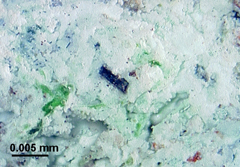
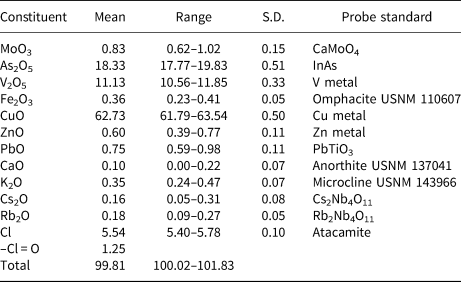
Aleutite occurs as individual crystals in masses of polycrystalline anhydrite (Fig. 1). Aleutite is dark red, with a reddish black streak and close to adamantine lustre. It is brittle with no cleavage observed. Parting was not observed and its fracture is uneven. The density could not be measured due to lack of sufficient material. The calculated density using the empirical formula derived from the analytical data given in Table 1 is 4.887 g cm–3.
Aleutite dark-red prismatic crystal associated closely with anhydrite (white) and euchlorine (green).

Composition (EPMA, wt.%) of the studied aleutite.

S.D. – standard deviation
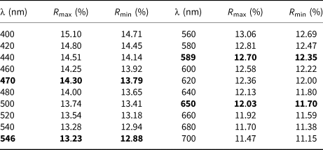
Despite the transparency of aleutite, its optical properties had to be investigated in reflected light because of the high values of refractive indices, typical of arsenates and vanadates. Reflectance measurements were made using a SiC standard in air in the range 400–700 nm (Table 2). In reflected light, aleutite is grey with a yellowish tint. It is non-pleochroic, with abundant, brown–red internal reflections and a weak bireflectance. The measured anisotropy is ΔR 589 = 0.35%.
Reflectance values for aleutite (SiC standard in air).

The reference wavelengths required by the Commission on Ore Mineralogy are given in bold.
Composition
Three crystals (60 μm ×15 μm ×10 μm) of aleutite checked previously by single-crystal X-ray diffraction were mounted in epoxy resin and polished with oil suspension. Six spot analyses on the grains were obtained using a JEOL Superprobe 733 scanning electron microscope equipped with an Oxford Instruments INCA Energy Dispersive Spectrometer. The electron beam accelerating voltage was 20 kV, and electron beam current 2 nA, measured with a Faraday cup. X-ray acquisition live-time was 30 s. System calibration was performed on Ni. A defocused beam (5 μm) was used for analyses. X-ray matrix correction was carried out automatically by the Oxford Instruments INCA version of the Pouchou and Pichoir (XPP) routine. No other elements with Z > 9 other than those reported in Table 1 were detected. The empirical formula calculated on the basis of (As + V+Mo + Fe3+) = 2 atoms per formula unit is Сu5.40Zn0.05Ca0.01As1.09V0.84Mo0.04Fe0.03K0.05Pb0.02Rb0.01Cs0.01O9.97Cl1.07 or (Сu4.94Zn0.05Ca0.01)Σ5.00O2.11[(As0.54V0.42Mo0.02Fe0.02)Σ1.00OΣ3.93]2(Cu0.46K0.05Pb0.02Rb0.01Cs0.01)Σ0.55Cl1.07. Taking into account structural data, the simplified formula is [Cu5O2](AsO4)(VO4)·(Cu0.5□0.5)Cl. Aleutite is soluble in hot H2O.
X-ray crystallography
Experiment
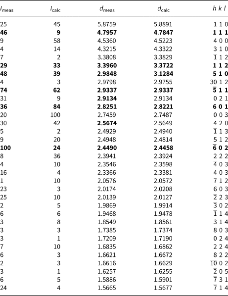
Powder X-ray diffraction data were collected using a Rigaku R-AXIS Rapid II single-crystal diffractometer equipped with a cylindrical image plate detector using Debye–Sherrer geometry (with d = 127.4 mm) (CoKα radiation) at the Department of Crystallography, St. Petersburg State University, Russia after crushing the crystal fragments used for the single-crystal analysis. Data (in Å) are given in Table 3. Unit-cell parameters refined from the powder data are as follows: monoclinic, space group C2/m, a = 18.0788(9) Å, b = 6.2270(5) Å, c = 8.2445(3) Å, β = 90.56(4)°, V = 928.09(7) Å3 and Z = 4.
Powder X-ray diffraction data (d in Å) for aleutite.

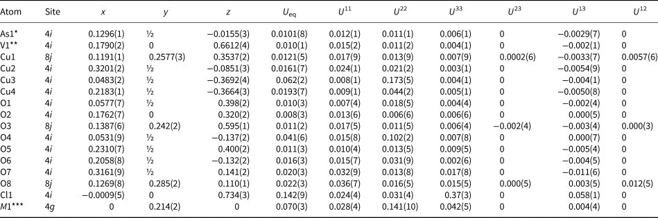
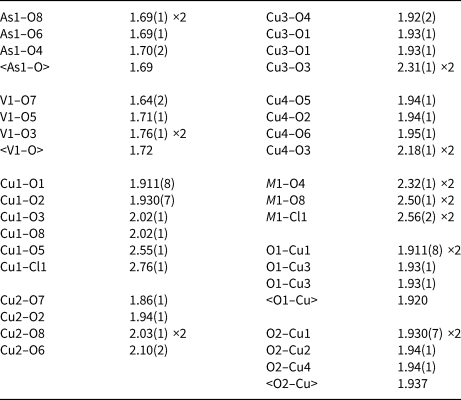
A dark-red prismatic crystal fragment of aleutite was mounted on a thin glass fibre for X-ray diffraction analysis using a Bruker APEX II DUO X-ray diffractometer with a micro-focus X-ray tube operated with MoKα radiation at 50 kV and 40 mA. The data were integrated and corrected for absorption using a multi scan type model implemented in the Bruker programs APEX and SADABS (Bruker-AXS, 2014). More than a hemisphere of X-ray diffraction data was collected. The structure was solved by direct methods and successfully refined with the use of the SHELX software package (Sheldrick, Reference Sheldrick2015). The refinement in the C2/m space group converged to R 1 = 0.066 (Table 4). The structure was refined as a 2-component twin (twin ratio 0.955:0.045). The final atomic coordinates and anisotropic displacement parameters are given in Table 5, and selected interatomic distances in Table 6. The crystallographic information files have been deposited with the Principal Editor of Mineralogical Magazine and are available as Supplementary material (see below).
Crystallographic data and refinement parameters for aleutite

Coordinates, isotropic and anisotropic displacement parameters (Å2) of atoms in aleutite.

*As0.82(3)V0.18(3); **V0.81(3)As0.19(3); ***fixed in agreement with microprobe data Cu0.46Pb0.02(K,Rb,Cs)0.04.
Selected interatomic distances (Å) in aleutite.

Cation coordination
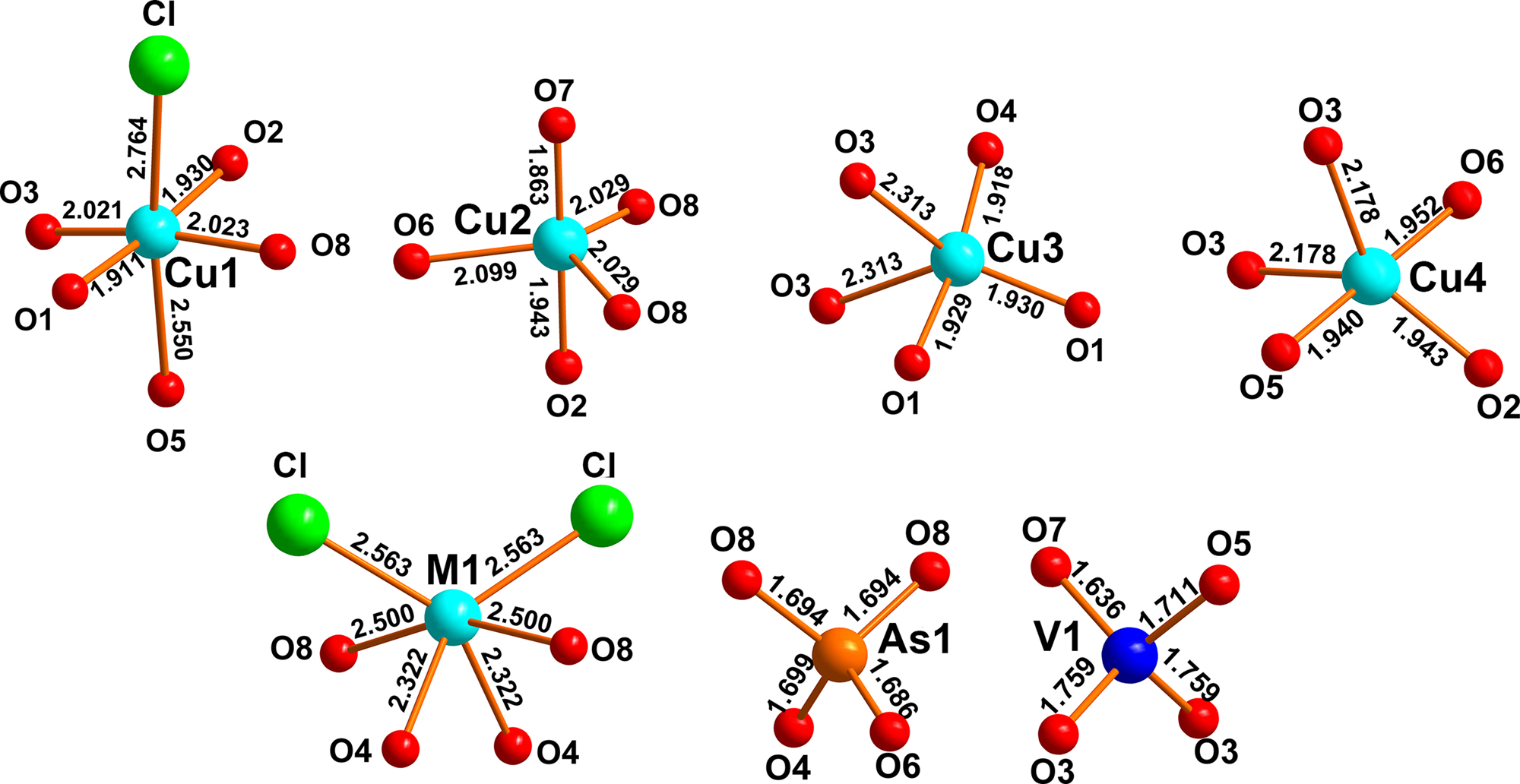
The crystal structure of aleutite contains four symmetrically independent Cu sites occupied by Cu2+ cations with different coordination environments (Fig. 2). The Cu1 site is coordinated by four O atoms to form an almost planar CuO4 square that is complemented by an apical Cl– anion and one O2– anion. As a result, an elongated CuO4OCl octahedron is formed. The Cu2, Cu3 and Cu4 sites are coordinated by five oxygen atoms each and as a result Cu2O5 regular trigonal bipyramid and strongly distorted Cu3O5 and Cu4O5 tetragonal pyramids are formed.
Coordination of atoms in aleutite.

There are two tetrahedral sites coordinated by four O anions each (Fig. 2, Table 6). The As1 site is occupied predominantly by As5+ cations, whereas the V1 site is occupied mostly by V5+ cations (Table 5). The <As1–O> distance of 1.693 Å correlates well with the <As5+–O> value of 1.685 Å reported in Majzlan et al. (Reference Majzlan, Drahota, Filippi, Bowell, Alpers, Jamieson, Nordstrom and Majzlan2014). The <V1–O> distance in the structure of aleutite demonstrates value of 1.716 Å, which is in a good agreement with the <V5+–O> distance of 1.721 Å reported for vanadates by Gopal and Calvo (Reference Gopal and Calvo1973). Note, AsO4 tetrahedra in the structure of aleutite are very regular and undistorted, whereas VO4 show a significant degree of distortion. The Baur (Reference Baur1974) distortion index in aleutite for AsO4 is 0.0015 and 0.025 for VO4.
There is one mixed and partially occupied M1 site in the structure of aleutite. The occupancy of this site refined using the atomic scattering factors of Cu is 0.45(1). The M1-site coordination is strongly distorted (Fig. 2) with four M1–O bonds and two M1–Cl bonds demonstrating similar bond-length values. Chemical analysis reveals a number of additional monovalent (K, Rb and Cs) and divalent (Pb) cations in aleutite, probably concentrated in the M1 site. The similar substitutions were reported previously in averievite (Vergasova et al., Reference Vergasova, Starova, Filatov and Anan'ev1998; Krivovichev et al., Reference Krivovichev, Filatov and Vergasova2015). On the final stages of the crystal structure refinement occupancy of the M1 site was fixed in agreement with microprobe data. The M1–M1 short distance of 2.67 Å along [010] is in agreement with the partial occupancy of the M1 site. We note large U 22 values for the M1, Cu3 and O4 atoms indicating positional disorder along [010] and an even larger U 33 value for Cl1 suggesting significant disorder of this atom along [001]. There is a possibility of significant ionic mobility with increasing temperature in the voids of the metal oxide framework, which might act as an ‘electron reservoir’.
Oxygen atoms O3, O4, O5, O6, O7 and O8 are strongly bonded in tetrahedral AsO43– and VO43– groups. We may term them as Ot. ‘Additional’ oxygen atoms O1 and O2 are bonded exclusively to Cu2+ cations and can be designated as Oa. Cu2+–Oa bonds are in the range of 1.911(8)–1.94(1) Å, whereas Cu2+–Ot range from 1.86(2) to 2.55(1) Å (Table 6). Because of the variability of the coordination polyhedra around Cu2+ cations and the strength of the Cu–Oa bonds compared to the weaker Cu–Ot and especially Cu–Cl bonds, one can describe the Cu-oxide substructure of aleutite in terms of oxocentred OCu2+4 tetrahedra.
Structure description
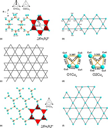
O1Cu4 and O2Cu4 tetrahedra have shared Cu–Cu edges and corners thus forming  ${}_\infty ^1 [\rm Cu_5O_2]^{6 +} $ bands (Fig. 3a). These bands grow parallel to the b axis. In order to describe the linkage of oxocentred tetrahedra with each other, connectivity diagrams (Siidra et al., Reference Siidra, Krivovichev and Filatov2008) depicted in Fig. 3a can be used. Each diagram represents a view from above onto a regular tetrahedron resting on one of its triangular faces. The following graphical indicators are used: (1) a corner shared with an adjacent tetrahedron is designated by a circle; (2) a shared edge is designated by a bold line. Identical or chiral diagrams are called equivalent. Thus the topology of
 ${}_\infty ^1 [\rm Cu_5O_2]^{6 +} $ bands is novel and has not been described before in minerals and synthetic materials.
 ${}_\infty ^1 [\rm Cu_9Pb_2O_4]^{14 +} $ bands described previously in the structures of complex synthetic oxyhalide selenites (Kovrugin et al., Reference Kovrugin, Colmont, Siidra, Mentré, Al-Shuray, Gurzhiy and Krivovichev2015; Siidra et al., Reference Siidra, Kozin, Depmeier, Kayukov and Kovrugin2018a) contain only corner sharing between the neighbouring OCu4 tetrahedra. However,
 ${}_\infty ^1 [\rm Cu_5O_2]^{6 +} $ bands in aleutite can be cut from the
 ${}_\infty ^2 [\rm Cu_6Pb_3O_4]^{10 +} $ layers described in [Cu2+6Pb3O4](Cu+Pb1.27Br3.54)(SeO3)4Br2 (Siidra et al., Reference Siidra, Kozin, Depmeier, Kayukov and Kovrugin2018a).
(a) Ball-and-stick and polyhedral representation of [Cu5O2]6+ oxocentred bands elongated along the b axis and connectivity diagrams for O1Cu4 and O2Cu4 oxocentred tetrahedra. (b) One-dimensional kagome-like fragment in aleutite, formed by Cu1–Cu1–Cu2 and Cu1–Cu1–Cu3 triangles. Cu2–Cu2–Cu3 triangles are complemented by additional Cu4 atoms above or below triangular faces, whereas Cu1–Cu1–Cu3 triangles are complemented by additional Cu3 atoms. (c) An ideal kagome network. (d) Cu–Cu distances in two symmetrically independent OCu4 tetrahedra. (e) [Cu5O2]6+ layers in the structure of averievite and connectivity diagrams for O1Cu4 tetrahedra. (f) Kagome sheets in averievite.

The structural architecture of the oxocentred  ${}_\infty ^1 [\rm Cu_5O_2]^{6 +} $ band in aleutite can be derived from a kagome (Mekata, Reference Mekata2003) network and represents a one-dimensional (Fig. 3b) slice from it (Fig. 3c). The kagome fragment in aleutite is formed by Cu1–Cu1–Cu2 and Cu1–Cu1–Cu3 triangles. Cu2–Cu2–Cu3 triangles are capped by additional Cu4 atoms above or below triangular faces, whereas Cu1–Cu1–Cu3 triangles are complemented by additional Cu3 atoms. It is of interest to consider the Cu···Cu distances of tetrahedral edges in Cu4 tetrahedra depicted in Fig. 3d. The Cu3···Cu3 edge in the O1Cu4 tetrahedron has a length of 2.762 Å, whereas two Cu3···Cu1 and one Cu1···Cu1 have almost identical edges of ~3 Å. The two other Cu3···Cu1 edges have significantly longer but identical values of 3.387 Å. The similar distribution of Cu···Cu distances is observed for O2Cu4 tetrahedron. Cu4···Cu2 and two Cu2···Cu1 edges are ~3 Å long. The Cu1···Cu1 edge is 3.210 Å and two Cu1···Cu4 are each 3.353 Å long.
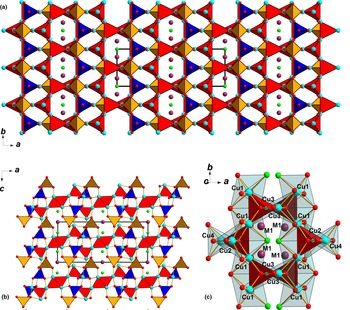
AsO4 and VO4 tetrahedra are attached to OCu4 tetrahedra in face-to-face fashion (Krivovichev et al., Reference Krivovichev, Mentré, Siidra, Colmont and Filatov2013) thus providing the assemblage of the bands into a porous  $\{ _\infty ^3 [\rm Cu_5O_2]({\rm As}{\rm O}_4)({\rm V}{\rm O}_4)\} ^0$ electroneutral framework shown in Fig. 4a,b. A two-dimensional system of channels with dimensions ~5.81 × 6.81 Å is occupied by metal–chloride species. Thus aleutite can be classified as a new mesoporous material.
General projection of the crystal structure of aleutite (a) along the c axis and (b) along the b axis. The tetrahedral framework is formed by OCu4 (red), VO4 (blue) and AsO4 (orange) tetrahedra. Channels (~5.81 Å × 6.81 Å) in the framework are filled by the different metals occupying the mixed M1 site (violet balls) and chloride ions (green balls). (c) A segment of the crystal structure of aleutite showing the complex arrangement of Cu-centred polyhedra (light blue) and six-membered rings formed by the corner and edge-sharing OCu4 tetrahedra (red).

Discussion
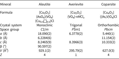
Aleutite is a new structure type. Chemically aleutite is an As,V-analogue of averievite (Table 7). Aleutite is also chemically related to coparsite (Starova et al., Reference Starova, Krivovichev and Filatov1998). No synthetic compounds directly related to aleutite are known to date.
Crystal chemical data of aleutite, averievite (Krivovichev et al., Reference Krivovichev, Filatov and Vergasova2015) and coparsite (Starova et al., Reference Starova, Krivovichev and Filatov1998).

The structure of aleutite is completely different from averievite (Starova et al., Reference Starova, Krivovichev, Fundamenskii and Filatov1997; Vergasova et al., Reference Vergasova, Starova, Filatov and Anan'ev1998; Krivovichev et al., Reference Krivovichev, Filatov and Vergasova2015). Six-membered rings in aleutite (Fig. 4c) are formed by both corner-sharing and edge-sharing OCu4 tetrahedra. OCu4 oxocentred tetrahedra via common corners only form  ${}_\infty ^2 [\rm Cu_5O_2]^{6 +} $ layers in averievite (Fig. 3e). Kagome-net derived layers (Fig. 3f) in synthetic averievite have outstanding magnetic properties described recently (Botana et al., Reference Botana, Zheng, Lapidus, Mitchell and Norman2018).
It is common for copper vanadate minerals of fumarolic origin to have a significant amount of arsenic substitution. Aleutite is unique amongst copper vanadates, as it has As5+ and V5+ cations ordered over two tetrahedral sites. Vasilseverginite Cu9O4(AsO4)2(SO4)2 (Pekov et al., Reference Pekov, Britvin, Yapaskurt, Krivovichev, Vigasina and Sidorov2015) is the only other known example of arsenates of fumarolic origin with segregation of tetrahedrally coordinated As5+ and S6+ cations.
The framework in aleutite has large elliptical channels occupied by the complex mixed cationic M sites and Cl– anions. This type of structural architecture in aleutite is similar to that observed in leningradite PbCu3(VO4)2Cl2 (Siidra et al., Reference Siidra, Krivovichev, Armbruster, Filatov and Pekov2007). The composition of mixed and partially occupied M sites in aleutite is somewhat similar to averievite (Vergasova et al., Reference Vergasova, Starova, Filatov and Anan'ev1998). However, the amount of alkali metals is significantly less in aleutite. The strong distortion of the M sites coordination environments in aleutite in comparison with averievite can be probably explained by the presence of Pb2+ cations with a stereochemically active lone electron pair in the former. A similar phenomenon is observed in the alunite-supergroup minerals. Pb has strongly distorted coordination environments in the structure of plumbogummite because of the presence of a stereochemically active 6s 2 lone electron pair (Mills et al., Reference Mills, Kampf, Raudsepp and Christy2009), but K is symmetrically coordinated in the structure of jarosite (Mills et al., Reference Mills, Nestola, Kahlenberg, Christy, Hejny and Redhammer2013). One symmetrically independent but disordered M site is observed in the structure of aleutite in contrast to two M sites in averievite (Krivovichev et al., Reference Krivovichev, Filatov and Vergasova2015). The ‘host–guest’ character of the aleutite structure may be the result of its formation from volcanic gases. The metal oxide species tend to organise in such a structure that allows inclusion of various metal chloride species into a basic oxide matrix.
Aleutite is also an interesting and complex example of a natural salt-inclusion phase. Such synthetic materials consisting of a covalent metal oxide framework containing voids filled by the simple ionic salt species have received considerable attention in the field of materials science and inorganic chemistry (Huang et al., Reference Huang, Ulutagay, Michener and Hwu1999; Ulutagay et al., Reference Ulutagay, Schimek, Hwu and Taye1998; Huang et al., Reference Huang, Hwu and Mo2001).
Acknowledgements
We are grateful to Peter Leverett and two anonymous reviewers for valuable comments. This work was supported financially by the Russian Foundation for Basic Research, grant no. 19-05–00413. Technical support by the SPbSU X-ray Diffraction Resource Centre is gratefully acknowledged.
Supplementary material
To view supplementary material for this article, please visit https://doi.org/10.1180/mgm.2019.42