

Introduction
Thelebolus microsporus (Thelebolaceae) is a psychrophilic, coprophilous ascomycete forming minute discoid apothecia (Kobayashi and others Reference Kobayashi, Hibatsuka, Korf, Tubaki, Aoshima, Soneda and Sugiyama1967). This is a common fungus in the Antarctic ecosystem and has been isolated from soil within skuas’ nesting areas; from dung of skuas, penguins and petrels; from feathers, lichens and moss; and from aquatic mud, trachea of skua and biomats of lakes (Wicklow and Malloch Reference Wicklow and Malloch1971; Azmi and Seppelt Reference Azmi and Seppelt1998; Leotta and others Reference Leotta, Pare, Sigler, Montalti, Vigo, Petruccelli and Reinoso2002; de Hoog and others Reference De Hoog, Gottlich, Platas, Genilloud, Leotta and Brummelen2005). Microfungi living in different Antarctic environments are generally well adapted to high-stress conditions such as extreme low temperatures, high UV irradiance and low water and nutrient availability; for this reason they could be investigated in order to explore the colonisation, succession and limits of fungal life (Onofri and others Reference Onofri, Selbmann, Zucconi and Pagano2004).
Bio-pigments are a special group of natural pigments that have high economical and therapeutic potential (Mukherjee and Singh Reference Mukherjee and Singh2011). Fatty acids from fungi are also gaining importance in dietary supplements. The physiological and ecological mechanisms by which the cold-tolerant fungi produce these metabolites are not completely understood (Russell Reference Russell1990; Weinstein and others Reference Weinstein, Montiel and Johnstone2000). A combination of mechanisms (such as cold-active enzymes or unsaturated membrane lipids, besides intracellular sugars, polyols and antifreeze proteins), however, are recognised to be necessary for psychrophiles to function (Robinson Reference Robinson2001). Industrially, cold-active fungal enzymes find several applications in the food, medicine and detergent industries (Schiraldi and De Rosa Reference Schiraldi and De Rosa2002; Feller and Gerday Reference Feller and Gerday2003; Gawas-Sakhalkar and Singh Reference Gawas-Sakhalkar and Singh2011). The present study was therefore conducted to screen Thelebolus microsporus for enzyme, fatty acid and pigment analysis.
Materials and methods
Sampling site
Sampling was done at McLeod Island, Larsemann Hills, in the Prydz Bay area of east Antarctica. The Larsemann Hills (latitude 69°20′S to 69°30′S; longitude 75°55′E to 76°30′E) contain hundreds of freshwater lakes of varying size, depth and biology. It is the second largest (50km2) of only four major ice-free oases found along east Antarctica's 5000km coastline. It consists of two main peninsulas on the two extremities, namely, Broknes Peninsula and Stornes Peninsula. In between these, there are a number of islands of varying dimensions and some unnamed promontories. McLeod Island is one such island, situated about 2km north of Stornes Peninsula from where samples for the present study were collected by one of the authors (S.M. Singh) during the Indian Expedition to Larsemann Hills, Antarctica and Southern Ocean in February 2006. The samples of dead skua skulls and adhered soils were collected from McLeod Island, stored in sterile plastic bags and brought to the laboratory in an ice bag at a temperature of −20°C.
Isolation of fungi
A small quantity of sample (1 g soil) was ground with the help of a sterile pestle and serially diluted up to 10−7. A soil suspension (1ml) was used as the inoculum for isolation of fungi on two different culture media, malt extract agar (MEA) and potato dextrose agar (PDA), in triplicates by pour plate and spread plate methods. A small amount (0.1mg mL−1) of penicillin was added to suppress the bacterial growth. The inoculated agar plates were incubated at 4°C, 10°C, 15°C and 20°C for 20 days in a Bio Multi Incubator (LH-30–8CT, Japan). Emerging colonies were observed after fifteen days of incubation. About two to three slow-growing colonies of Thelebolus microsporus in each plate were observed, which were then transferred onto fresh MEA and PDA plates and incubated at 4°C, 10°C, 15°C and 20°C. Lactophenol cotton blue slide mounts were prepared and observed under Olympus CX41 and BX51 microscopes for morpho-taxonomical studies. Photomicrographs of ascomata, asci and ascospores were taken using an Camedia Digital Olympus C-4000 camera. The fungal organism was identified down to generic/species level based on morphological characters and authenticated by 18S rDNA sequences. Pure culture was deposited in the National Fungal Culture Collection of India (NFCCI-WDCM 932), with accession number NFCCI1777.
Taxonomic description
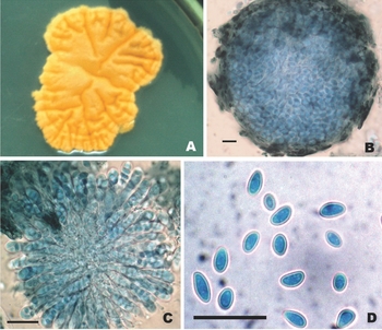
Colonies on MEA were slow growing (5cm diameter in 20 days at 15°C), smooth, flat with ridges and furrows, bright orange to orangish brown when old, developing a centre with numerous rounded ascomata with age, with a creamish to yellowish periphery and irregular margins (Fig. 1, Table 1). Colonies on PDA appeared light orange to brown coloured with flat and folded mycelium; ascomata were rarely developed. Ascomata were globose to oval, thin walled, pale brown, and measured 70–125μm × 70–112.5μm. Asci were club shaped, hyaline, eight ascospored, and measured 20–25μm × 7–7.5μm. Ascospores were arranged in ellipsoidal manner in an ascus, ovate to obovate or variable in size, double walled, hyaline, and measuring 5.5–9.0μm × 3.5–5μm.
Thelebolus microsporus (Berk. & Broome) Kimbr. [NFCCI 1777] A. Pigmented colony on MEA at 20° C, B. Cleistothecial ascoma, C. Asci, D. Ascospores. (Bar = 10 μm)

Phenotypic characteristics and enzyme activity of Antarctic strain Thelebolus microsporus (Berk. & Broome) Kimbr.

++ positive, + weak positive, − negative, n = 3.
DNA isolation, polymerase chain reaction amplification and sequencing
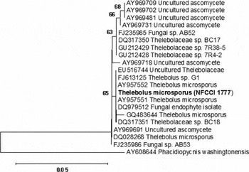
DNA was extracted from pure culture grown on PDA at 15°C and the mycelia homogenised in a FastPrep®-24 tissue homogeniser (MP Biomedicals GmbH, Germany) and then using the cetyl trimethylammonium bromide method of Graeser and others (Reference Graeser, Fari, Vilgalys, Kuijpers, de Hoog, Presber and Tietz1999). For the internal transcribed spacer (ITS-)PCR, the universal primers ITS4 (5′ TCC TCC GCT TAT TGA TAT GC 3′) and ITS5 (5′ GGA AGT AAA AGT CGT AAC AAG G 3′) amplifying a DNA fragment of about 700bp of the rDNA gene were used (White and others Reference White, Bruns, Lee, Taylor, Innis, Gelfand, Sninsky and White1990). The PCR mixture contained reaction buffer (10mM Tris-HCl pH8.0, 50mM KCl, 1.5mM MgCl2), 200μM each of deoxynucleoside triphosphates (Genei, Bangalore, India), 50pmol each of primers, 1U of Taq polymerase (Genei), and 25ng of template DNA. Samples were overlaid with sterile mineral oil and amplified through 30 cycles in a thermocycler (Mastercycler®, Eppendorf AG, Hamburg, Germany) as follows: initial denaturation for 5 min at 95°C, denaturation for 1 min at 95°C, annealing for 1 min at 56°C, and extension for 1 min at 72°C. This was followed by a final extension step for 10 min at 72°C. The resulting PCR product was checked on 1.2% agarose gel (Sigma-Aldrich, MO, USA). PCR products were cleaned with AxyPrep™ PCR Cleanup Kit (Axygen Scientific, CA, USA) and sequenced using primers ITS4 and ITS5 (White and others Reference White, Bruns, Lee, Taylor, Innis, Gelfand, Sninsky and White1990) on an automated DNA sequencer ABI 3130 (Applied Biosystems, USA). The evolutionary history was inferred using the neighbour-joining method (Saitou and Nei Reference Saitou and Nei1987). The evolutionary distances were computed using the maximum composite likelihood method (Tamura and others Reference Tamura, Nei and Kumar2004) and are in the units of the number of base substitutions per site (Fig. 2). All positions containing gaps and missing data were eliminated from the dataset (complete deletion option).
Phylogenetic tree of Thelebolus microsporus (NFCCI 1777), which explains the evolutionary history of the culture using the neighbour-joining method. The optimal tree with the sum of branch length = 0.14636173 is shown. The percentages of replicate trees in which the associated taxa clustered together in the bootstrap test (1000 replicates) are shown above the branches. The tree is drawn to scale, with branch lengths in the same units as those of the evolutionary distances used to infer the phylogenetic tree. There were a total of 442 positions in the final dataset. Phylogenetic analyses were conducted using MEGA4 (Tamura and others Reference Tamura, Dudley, Nei and Kumar2007).

Screening procedure
Pure culture was subjected to growing at different temperatures from its optimal temperatures for growth. Optimum was determined by measuring rates of growth, ranging from 4°C to 20°C in triplicate. Qualitative tests for enzymatic activities were performed on solid media using the standard technique (Hankin and Anagnostakis Reference Hankin and Anagnostakis1975). Quantitative amylase production was determined from broth culture. Thelebolus microsporus (NFCCI1777) was grown in a medium containing (per litre): starch, 20.0g; dextran, 8.0g; L-asparagine, 6.0g; KH2PO4, 3.0g; K2HPO4, 2.0g; MgSO4.7H2O, 0.5g; CaCO3, 3.0g; trace elements 1ml (trace elements in μg ml−1: CoCl2, 200; FeSO4.7H2O, 500; MgSO4.7H2O, 196; ZnCl2, 166). Pure culture was inoculated and incubated in a rotary shaker (180rpm) at different temperatures (10°C, 15°C and 20°C) for 10 days. Shake-flask cultures were carried out in 250ml Erlenmeyer flasks, containing 50ml medium.
Enzyme activity assays
α-amylase activity was determined with soluble starch as a substrate by the starch-iodine method (Mishra and Maheshwari Reference Mishra and Maheshwari1996) and by the dinitrosalicylic acid (DNSA) method (Miller Reference Miller1959) for measuring reducing sugars. One unit of enzyme activity was defined as the amount of enzyme that catalyzes the hydrolysis of 1mg starch per min at 15°C and pH5.0. Proteins were determined by the method described by Lowry and others (Reference Lowry, Rosebrough, Farr and Randall1951).
High-performance liquid chromatography analysis and gas chromatography-mass spectrometry profile of pigments
Pigments were extracted from a fungal mat growing on PDA. The fungal mat was homogenised in 100% methanol using a glass homogeniser. The homogenate was centrifuged at 200g for 5 min and the supernatant was filtered through a 0.13mm filter prior to loading (20μl) on the high-performance liquid chromatograph (Waters Spherisorb®). Identification of pigments was carried out using standard methods and a spectral profile of individual peaks was created using a PDA detector (Waters 2996). The pigments were separated using the method of Bhandari and Sharma (Reference Bhandari and Sharma2006).
For the gas chromatography-mass spectrometry (GC-MS) profile a concentration of 0.85mg/g of fresh weight of Thelebolus microsporus was harvested. In GC-MS conditions, an MS-ion source temperature of 220°C and a GC-oven temperature of 40°C to 220°C with ramping of 5°C/min were maintained. The injector temperature of 220°C and auxiliary temperature of 220°C were fixed. The carrier gas used was helium with a flow rate of 1ml/min. The column TR5 MS (in a Thermo Fisher Scientific ITQ 900) was used.
Fatty acid analysis
Fatty acid from the fungal mat grown on the solid medium was extracted using methanol:chloroform:water in a 2:1:0.5 ratio according to Christie (Reference Christie1982). Methyl esters were prepared according to Bhandari and Sharma (Reference Bhandari and Sharma2006) and 1μl of methyl esters was run on a gas chromatograph (Shimadzu GC-2014) equipped with a hydrogen flame ionisation detector. Fatty acids were separated according to Vaz and Sharma (Reference Vaz and Sharma2010) using heptadecanoic acid as the internal standard.
Result and discussion
Thelebolus microsporus produced a bright orange to yellow-orange pigment in culture. This may be an important feature as the isolate is cold-tolerant (psychrotrophic) in nature. Based on the morphological characteristics and sequencing of the ITS region, a subsequent BLAST (basic local alignment search tool) search showed 99% sequence homology to Thelebolus microsporus AY957552 (strain CBS 109799) and AY957551 (strain CBS 109909). A search in the GenBank for ITS sequences resulted in many accessions. A neighbour-joining tree showing the phylogenetic relationship of related species of Thelebolus microsporus NFCCI- 1777 was constructed (Fig. 2).
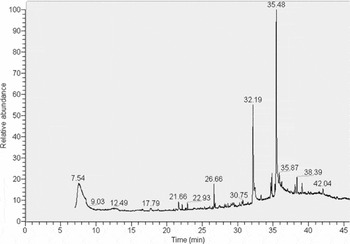
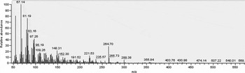
In order to characterise the pigment production, high-performance liquid chromatography (HPLC) analysis was carried out. The HPLC profile at 445nm showed five peaks, four of which could be identified as carotenoids based on their spectral profile. β-carotene was found to be predominant among carotenoids present in the extract. Other carotenoid peaks, though quantitatively insignificant, could be characterised as lutein. The exact chemical nature of the two other, quantitatively insignificant, carotenoid peaks could not be ascertained. The fifth peak showed spectral maxima in both blue and red regions and the chemical nature of this peak could not be ascertained either. A chromatogram for Thelebolus microsporus acetone extract exhibits a strong β-carotene peak with retention time at 35.48 min (Fig. 3a). The MS profile of β-carotene available in the NIST library is given in figure 3b. It is assumed, based on the earlier studies on lichens, mushrooms and bacteria that reveal that under the extreme cold and ultraviolet light stress existing in the Arctic and Antarctic area, the pigment and antioxidant production, improves the organisms’ stress tolerance (Kranner and Birtic Reference Kranner and Birtic2005; Dieser and others Reference Dieser, Greenwood and Foreman2010; S.M. Singh and others Reference Singh, Singh and Ravindra2011a; P. Singh and others Reference Singh, Singh, D'Souza, Roy and Singh2012).
Chromatogram for Thelebolus microsporus acetone extract. The peak at retention time 35.48 minutes is the peak for β-carotene.

MS profile for β-carotene. β-carotene in Figure 3a was identified by matching its MS profile that had been obtained with other profiles available in the NIST library.

The fatty acid profile of the culture extract showed various peaks indicating the presence of myristic acid, palmitic acid, heptadecanoic acid, stearic acid, linoleic acid and linolenic acid. Except linolenic acid, which was present in a substantial quantity, all other fatty acids were quantitatively insignificant. Linolenic acid is a polyunsaturated fatty acid (PUFA). PUFAs are known for modulating membrane fluidity, which is a physiological strategy for cold adaptation. High levels of unsaturated fatty acids are also reported in psychrophilic yeasts; that is, Candida, Leucosporidium and Torulopsis (Kerekes and Nagy Reference Kerekes and Nagy1980). Evidence suggests that membrane composition is critical for the ability of microorganisms to grow over specific temperature ranges (Robinson Reference Robinson2001). Previous studies report Microdochium nivale to possess polyunsaturated fatty acids (18:2 and 18:3) that enhance the ability of this fungus to survive at low temperatures (Istokovics and others Reference Istokovics, Morita, Izumi, Hoshino, Yumoto, Sawada, Ishizaki and Okuyama1998).
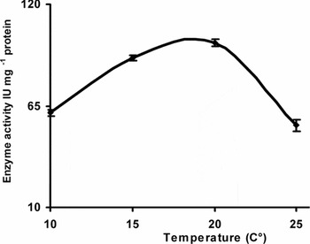
Cold-tolerant enzymes find applications in health, agriculture and industrial sectors (Feller and Gerday Reference Feller and Gerday2003; Gawas-Sakhalkar and Singh Reference Gawas-Sakhalkar and Singh2011; S.M. Singh and others Reference Singh, Yadav, Singh, Singh, Singh and Ravindra2011b; Gawas-Sakhalkar and others Reference Gawas-Sakhalkar, Singh, Naik and Ravindra2012; P. Singh and others Reference Singh, Singh, D'Souza, Roy and Singh2012) and therefore are a key area of investigation. However, studies on the cold-active enzymes in the polar regions are still fragmentary (Medigue and others Reference Medigue, Krin, Pascal, Barbe, Bernsel, Bertin, Cheung, Cruveiller, D'Amico, Duilio, Fang, Feller, Ho, Mangenot, Marino, Nilsson, Parrilli, Rocha, Rouy, Sekowska, Tutino, Vallenet, von Heijne and Danchin2005; Männistö and Häggblom Reference Männistö and Häggblom2006; Bej and Mojib Reference Bej, Mojib, Bej, Aislabie and Atlas2010). The results of the preliminary screening experiments for the production of various extracellular enzymes such as amylase, cellulase, chitinase, lipase and protease (qualitatively) are given in Table 1. Out of five enzymes studied, amylase exhibited significantly positive activity, and hence, was subjected to quantitative estimation. Quantification of α-amylase activity was carried out in triplicate and the values depicted are the mean values along with standard deviation. The isolate showed a maximum α-amylase production activity at 20°C (99.16IU/mg protein) and further increase in temperature led to decrease in activity (Fig. 4). Cold-adapted amylase production was also reported from an actinomycete, Nocardiopsis sp. (Zhang and Zeng Reference Zhang and Zeng2007). Cold-active amylases can be effectively applied as a detergent additive, in textile processing and in the food industry (Männistö and Häggblom Reference Männistö and Häggblom2006; Margesin and others Reference Margesin, Neuner and Storey2007; Zhang and Zeng Reference Zhang and Zeng2007; Bej and Mojib Reference Bej, Mojib, Bej, Aislabie and Atlas2010).
α-amylase production by Thelebolus microsporus, grown at different temperatures for 10 days.

To the best of the authors’ knowledge this is the first work on analysing Thelebolus microsporus for its pigments, fatty acids and enzymes.
Acknowledgements
We are grateful to the directors of the National Centre for Antarctic and Ocean Research (NCAOR), Goa, and the Agharkar Research Institute, Pune, for encouragement and facilities. We are grateful to Professor S.K. Dey, IIT-Kharagpur, for providing the chromatogram. Thanks are due to Professor C.H. Robinson and an anonymous reviewer for their valuable suggestions to improve the quality of the manuscript. This is NCAOR publication No. 33/2012.