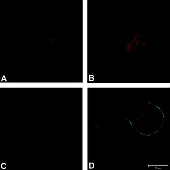
The wrong version of Figure 2, on page 121, was published. The publishers apologise for this error. The correct version appears below.
Figure 2
Detection of CD9 protein on the surface of growing oocytes. (A) An oocyte in the diameter range 10–13 μm. (B), (C) Oocytes between 13 and 17 μm in diameter. (D) Oocytes in the diameter range 17–22 μm. Pictures are single slices taken at the level of the centre of the oocytes. CD9 labelling is visualized in green, the nuclei of growing oocytes are shown in red. Scale bar represents 10 μm.