Introduction
Embryonic development is a process consisting of several different stages that lead to successive mitotic replications and transformations that are the origins of specialized, complex and functional cells that give life to individuals (Arrau et al., Reference Arrau, Roblero and Cury1981; Balinsky, Reference Balinsky1983; Kunz, Reference Kunz2004; Avery et al., Reference Avery, Killen and Hollinger2009; Akiyama et al., Reference Akiyama, Tero and Kobayashi2010). The success of this process will depend on the quality of the gametes and the numerous events that occur before and during embryonic development. Bobe & Labbé (Reference Bobe and Labbé2010) have described the molecular and cellular characteristics that affected the quality of male and female gametes, before, during and after fertilization in fish. ‘Before’ fertilization, there are mechanism that are responsible for initiation of sperm motility, and also the morphology and quality of the plasmatic membrane of spermatozoa; the physical and chemical parameters of ovary or coelomic fluid, and the appearance of the oocyte during its first stages of development (Kjørsvik et al., Reference Kjørsvik, Mangor-Jensen and Holmefjord1990; Bromage et al., Reference Bromage, Bruce, Basavaraja and Rana1994; Penney et al., Reference Penney, Lush, Wade, Brown, Parrish and Burton2006; Pavlov & Emel′yanova, Reference Pavlov and Emel′yanova2008); the volume of the egg and the lipid drop of species such as Seriola lalandi (Moran et al., Reference Moran, Smith, Gara and Poortenaar2007); and the composition of lipids and fatty acids in fish eggs and semen of species such as Oncorhynchus mykiss (Vassallo-Agius et al., Reference Vassallo-Agius, Watanabe, Yoshizaki, Satoh and Takeuchi2001), Dexten dexten (Giménez et al., Reference Giménez, Estévez, Lahnsteiner, Zecevic, Bell, Henderson, Piñera and Sanchez-Prado2006) and Ictalurus punctatus (Sink & Lochmann, Reference Sink and Lochmann2008). ‘During’ embryonic development, the characteristics and abnormalities of the blastomeres have been used as criteria to discriminate between good and bad quality embryos (Kjørsvik et al., Reference Kjørsvik, Mangor-Jensen and Holmefjord1990, Reference Kjørsvik, Hoehne-Reitan and Reitan2003; Bromage et al., Reference Bromage, Bruce, Basavaraja and Rana1994; Shields et al., Reference Shields, Brown and Bromage1997; Penney et al., Reference Penney, Lush, Wade, Brown, Parrish and Burton2006; Pavlov & Emel′yanova, Reference Pavlov and Emel′yanova2008; Avery et al., Reference Avery, Killen and Hollinger2009; Bobe & Labbé, Reference Bobe and Labbé2010; Vásquez et al., Reference Vásquez, Llanos-Rivera and Castro2010; Effer et al., Reference Effer, Sanchez, Ubilla, Figueroa and Valdebenito2012; Valdebenito et al., Reference Valdebenito, Sanchez, Effer and Ubilla2012), whereas in Hippoglossus hippoglossus different characteristics of the blastomeres (symmetry, cell size, adhesion between blastomeres, margins and inclusions) are correlated positively with the percentage of hatched larvae (Shields et al., Reference Shields, Brown and Bromage1997). In Scophthalmus maximus the symmetry of blastomeres is correlated positively with hatching rates, larval tolerance towards stress and larval survivability, metamorphosis and pigmentation (Kjørsvik et al., Reference Kjørsvik, Hoehne-Reitan and Reitan2003). In Limanda ferruginea, eggs with abnormal blastomeres have the highest mortalities when compared with eggs that have normal blastomeres (Avery & Brown, Reference Avery and Brown2005). In Zebrasoma scopas, Pavlov & Emel′yanova (Reference Pavlov and Emel′yanova2008) used symmetry, blastomere adhesion, cellular division, cleavage synchronization, and the formation of syncytia as criteria to evaluate fish egg quality, and in Gadus morhua, Avery et al. (Reference Avery, Killen and Hollinger2009) related egg mortality, hatching success, larval deformations and mortality to abnormal cleavage patterns.
In salmonids, there is a lack of studies on the symmetry of the first blastomeres. The first characterizations of blastomeres was made for brown trout (Salmo trutta fario) using drawings (Kopsch, Reference Kopsch1911 in Kunz, Reference Kunz2004) or entirely with microphotography using embryonic discs (Killeen et al., Reference Killeen, McLay and Johnston1999), in the same manner as for rainbow trout (Salmo gairdneri) by Ballard (Reference Ballard1973, Reference Ballard1985). More resent studies by Valdebenito et al. (Reference Valdebenito, Sanchez, Effer and Ubilla2012) and Effer et al. (Reference Effer, Sanchez, Ubilla, Figueroa and Valdebenito2012) have detailed for the first time morphometric aspects of blastomeres in rainbow trout and Atlantic salmon, respectively. In Atlantic salmon, Gorodilov (Reference Gorodilov1996) characterized embryonic development and observed the first cleavage after 19 h of incubation at 5 °C, but this study lacked information on the morphometrics of the first blastomeres.
Due to the importance of salmon farming in Chile and in the rest of the world, there is a need to study the first stages of embryonic development in this species, to use such preliminary information to create diagnostics tools to measure the quality of gametes and/or embryos. The present investigation describes the morphometric analysis of the first blastomeres in Atlantic salmon (Salmo salar) embryos.
Materials and methods
Procurement of the gametes and artificial fertilization
Oocytes were collected in May 2012 from a group of Atlantic salmon (S. salar) broodstock of 3.6 years of age from Comau fish farm (Aquagen - Chile), Los Lagos Region-Chile, and then put into freshwater approximately 1 month before spawning at the Chesque Alto fish farm. Once sexual maturity was obtained, the female and male gametes were extracted by abdominal incision and massage, respectively. Fertilization was carried out at 7.5 °C, with a pool of eggs from 10 females and semen from four males, using the standard fish farming methods for reproduction. Eggs were then grouped into a Zoug jar type incubator at 7.5 °C in the dark.
Determination of the first cleavages
Eggs were fixed in Stockard solution at 12, 14, 16, 20 and 24 h post fertilization. Then, eggs were transported to the Reproduction Laboratory at the Aquiculture School of the Universidad Católica de Temuco, Chile, for later analysis.
Procurement and processing of embryonic discs
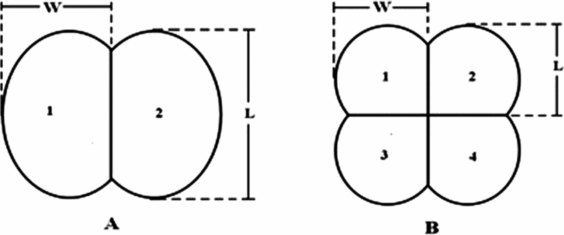
The chorion of the eggs was removed with tweezers under a stereoscopic Olympus SZ60 (Japan) magnifying lens with a cold light (Olympus LG PS2), equipped with a Micropublisher 3.3 RTV digital camera (Canada). Twenty blastodiscs from each sampled time period (12, 14, 16, 20 and 24 h) were observed and photographed at ×56 magnification, and were classified according to their morphology and frequency of appearance. The images were processed using Q Capture Pro 5.0 software (Canada), by which the diameter of the unfertilized blastodisc, and the length and width of each blastomere were measured (Fig. 1) following the criteria proposed by Effer et al. (Reference Effer, Sanchez, Ubilla, Figueroa and Valdebenito2012). For the morphometric description of the blastodisc with four blastomeres, we proceeded in the same way for each blastodisc and evaluated symmetry by comparison of the ratios of the length (L1/L2, L1/L3, L1/L4, L2/L3, L2/L4, L3/L4) and width (W1/W2, W1/W3, W1/W4, W2/W3, W2/W4, W3/W4; Fig. 1B), using the highest value as the denominator of both the ratio analyses.
Criteria used to determine length (L) and width (W) of blastomeres, and the designation of number of each blastomere in embryos with two (A) or four blastomeres (B). Reproduced from Effer et al. (Reference Effer, Sanchez, Ubilla, Figueroa and Valdebenito2012).

Twenty additional blastodisc with four blastomeres, plus symmetrical cleavages and of normal appearance were selected visually as controls (BC: control blastodiscs) following the criteria proposed by Shields et al. (Reference Shields, Brown and Bromage1997). Then morphometric symmetry was determined, as described in the previous paragraph, to compare the blastodiscs that had been characterized visually as abnormal or asymmetrical.
Statistical analysis
Values for ratios of length and width of the symmetrical or normal blastomeres, and for the asymmetrical or abnormal blastomeres, were analysed with statistical software GraphPad Prism® version 5.0 (GraphPad Software, San Diego, California, USA). A one-way analysis of variance (ANOVA) test was used for the non-parametric samples. Additionally, and in a parallel manner, a Dunnet's multiple comparisons test was done to evaluate any statistically significant differences between the ratios of length and width of the blastodiscs sampled (20 and 24) from the control group (BC). The level of significance was fixed at a P-value < 0.05 (n = 20 replicates).
Results
Determination of cleavages
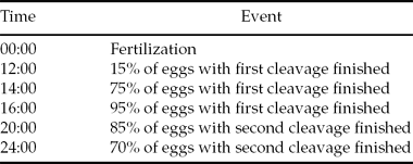
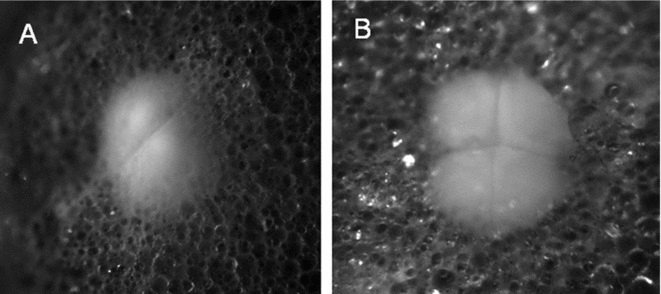
Cleavage (Fig. 2) was observed in 15, 75, 95, 85 and 70% of embryonic eggs after 12, 14, 16, 20 and 24 h of incubation, respectively (Table 1).
Eggs with cleavage (%), hours after fertilization in Atlantic salmon (Salmo salar) incubated at 7.5°C
Microphotography of embryonic discs of S. salar from first cleavage at 14 h (A) and second cleavage at 20 h (B) and incubated at 7.5 °C. Magnification ×56.
Morphological characterization
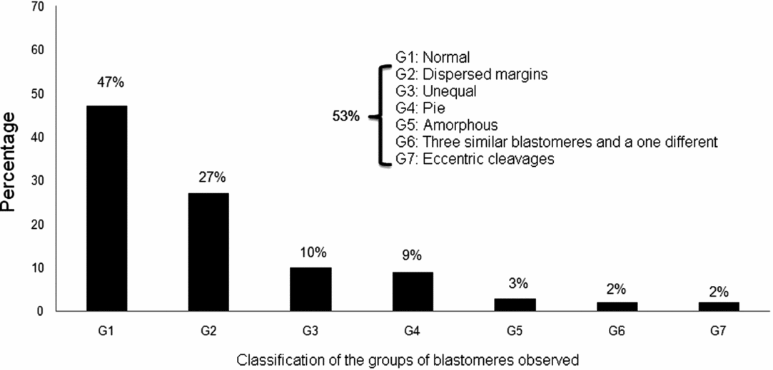
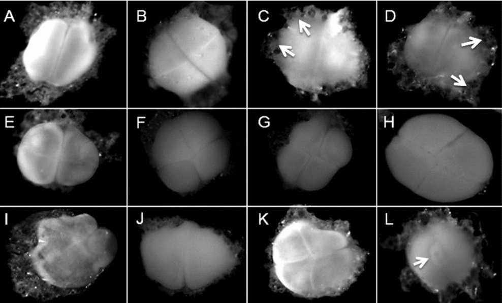
Figure 3 shows the seven groups of blastomeres as classified by morphology and frequency of appearance in embryos of S. salar, from which six groups were considered to be of abnormal morphologically. Around 47% of the embryos analyzed presented normal blastomeres (Category 1: G1) (Fig. 4A,B); 27% had dispersed margins (G2) (Fig. 4C,D); 10% had unequal blastomeres (G3) (Fig. 4E,F); 9% had ‘pie-shaped’ blastomeres (G4) (Fig. 4G,H); 3% had amorphous blastomeres (G5) (Fig. 4I,J); 2% had three similar and one different blastomere (G6) (Fig. 4K) and 2% had eccentric cleavage (G7) (Fig. 4L).
Morphological categories and percentage appearance of the observed groups of blastomeres in Atlantic salmon (S. salar). n = 100.
Microphotographs of normal and abnormal blastomeres of Atlantic salmon (S. salar). (A, B) Normal (G1). (C, D) Blastomeres with dispersed margins (G2) (arrows). (E, F) Unequal blastomeres (G3). (G, H) ‘Pie-shaped’ (G4). (I, J) Amorphous blastomeres (G5). (K) Three similar blastomeres and one different blastomere (G6). (L) Blastomeres with eccentric cleavage (G7) (arrow). Magnification ×56.
Morphometric characterization
The mean diameter of the blastodisc of S. salar found in this study was 1015.58 ± 109.79 μm; for those terminated at first cleavage the means of length and width from each blastomere was 826.85 ± 17.57 μm and 455.49 ± 16.65 μm, respectively. The mean values obtained from the second segmentation for length and width were 446.43 ± 9.79 μm and 430.25 ± 17.71 μm, respectively.
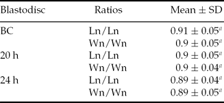
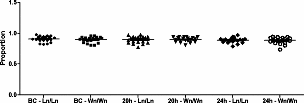
Mean values of either length (0.91 ± 0.05) or width (0.9 ± 0.05) of the control blastodiscs (CB) were compared with those of the other sampled groups (Table 2), but no statistically significant differences were found (P > 0.05) (Figure 5).
Mean values of the ratios of lengths and width of blastomeres of blastodiscs of S. salar
aNo significant difference compared with the control group (BC). SD, standard deviation.
Ln/Ln and Wn/Wn: ratios of lengths and widths of blastomeres of a same blastodisc, where ‘n’ is the random number of a blastomere.
Mean values of the ratios (proportions) of lengths and widths of blastomeres of control blastodiscs (BC), blastodiscs with 20 and 24 h of S. salar. Ln/Ln and Wn/Wn: ratios of lengths and widths of blastomeres of the same blastodisc, where ‘n’ is the random number of blastomeres (n = 20).
Discussion
This study describes a morphometric analysis of the first blastomeres of S. salar eggs incubated at 7.5 °C. Temperature is the main factor that regulates embryonic development in fish (Ballard, Reference Ballard1985; Billard, Reference Billard and Barnabé1990); our findings on the first and second cleavages after 16 or 20 h of incubation (120 and 150 Accuulated thermal units (AUT), respectively) resemble those obtained by Gorodilov (Reference Gorodilov1996) in the same species (95 and 13 AUT, for the first and second cleavage, respectively) and incubated at 5 °C. One reason for the differences obtained in both investigations could be the quality of the water used (not a factor considered in both studies) and/or the criteria used by the authors to define when the first or second cleavage ended or began (in this study the criteria used to determine when the first cleavage began was the time at which the highest percentage cleavage was determined, before the start the second cleavage).
Seven groups were determined based on blastomere morphology, from which six had visually abnormal cleavage patterns. These patterns could have originated in several ways such as: (1) elevated temperatures during the incubation phase (Pavlov & Moksness, Reference Pavlov and Moksness1994); (2) overmaturity or ageing of the eggs (Valdebenito et al., unpublished data); and/or (3) triploidy induction (Rani, Reference Rani2005; Effer & Valdebenito, unpublished data). The highest percentage of abnormal pattern was 27% for G2 (dispersed margins, Fig. 4C,D). This pattern is reported in Hippoglossus hippoglossus by Shields et al. (Reference Shields, Brown and Bromage1997). These authors determined that there was a correlation between lower numbers of blastomeres with margin dispersal and a higher hatching percentage. In contrast, the G3 (unequal blastomeres, Fig. 4E,F) and G4 patterns (pie-shaped, Fig. 4G,H) have been reported by Avery et al. (Reference Avery, Killen and Hollinger2009) in Gadius morhua, which produced viable larvae without significant differences in mortality compared with eggs with symmetrical blastomeres.
All in all, these findings show that not all abnormal cleavage patterns are severe, and therefore could result in viable larvae; i.e. during embryonic development, these anomalies can be corrected (Shields et al., Reference Shields, Brown and Bromage1997; Avery et al., Reference Avery, Killen and Hollinger2009). In salmonids it is very difficult to relate anomalies in the first blastomeres with per cent survival, hatching success and/or larval quality of individual fish eggs, due to the opacity of the chorion that does not allow the visualization of the blastodisc; therefore at present it is necessary to sacrifice the embryo to observe its blastodisc. For future studies, we suggest use of a search correlation between the percentage of asymmetries in blastomeres and indicators of embryonic quality such as larval survivability, per cent mortality, and survivability at 150 accumulated thermal units (ATU) etc., to establish a solid basis for the creation of an early evaluation tool for the quality of embryos in salmonids.
Authors such Kjørsvik et al. (Reference Kjørsvik, Mangor-Jensen and Holmefjord1990; Reference Kjørsvik, Hoehne-Reitan and Reitan2003); Shield et al. (Reference Shields, Brown and Bromage1997); Avery & Brown (Reference Avery and Brown2005); Penney et al. (Reference Penney, Lush, Wade, Brown, Parrish and Burton2006); Moran et al. (Reference Moran, Smith, Gara and Poortenaar2007); Pavlov & Emel′yanova (Reference Pavlov and Emel′yanova2008); Avery et al. (Reference Avery, Killen and Hollinger2009); Hamoutene et al. (Reference Hamoutene, Lush, Drover and Walsh2009); and Bobe & Labbé (Reference Bobe and Labbé2010), among others, have questioned and studied blastomere symmetry, but only from a morphological point of view. For this study in S. salar, a morphometrical criterion was used for the first time to evaluate these asymmetries, an approach similar to that considered in Oncorhynchus kisutch by Effer et al. (Reference Effer, Sanchez, Ubilla, Figueroa and Valdebenito2012). For this criterion, the ratio of all lengths and widths of blastomeres incubated (BC, 20 and 24 h groups), was close to 1 (Fig. 5).
When comparing the mean values of the ratios of length and width of the control blastodisc to other sampled groups, statistically significant differences were not found, despite the fact that 53% of total blastodisc in this study had abnormal cleavage patterns (Figure 3). These findings could indicate that: (1) some asymmetry is not only due to size (quantitative) but to qualitative differences such as position inside the blastodisc, as for G3: ‘pie-shaped’ (data not shown) in which there is asymmetry by position of blastomeres inside of the blastodisc (Fig. 4G,H); and (2) although morphometric asymmetry exists, only linear measurements (large and width) are considered. In future work, the ratios of volume between blastomeres of a same blastodisc should be measured, thereby being closer to the solid basis for an early evaluation tool for the quality of embryos in salmonids.
Acknowledgements
We are grateful to the Comau, Aquagen and Chesque Alto fish farms in Chile for their collaboration and proyect FONDECYT 1120705 for financial support.