Introduction
The diagnosis of Ménière's disease can be elusive, as it relies on subjective reporting of symptoms and no definitive diagnostic test is currently available. Non-specific symptoms can occur in the early stages and the disease progression can also fluctuate unpredictably. Furthermore, the condition may affect the cochlear and vestibular compartments of the inner ear differentially, potentially leading to variable clinical manifestations characterised by solely audiological or vestibular symptoms.Reference Pyykko, Nakashima, Yoshida, Zou and Naganawa1 It has been reported that the classic triad of hearing loss, tinnitus and vertigo is found in only 40 per cent of early Ménière's disease cases.Reference Belinchon, Perez-Garrigues and Tenias2
Endolymphatic hydrops has long been considered to be the structural correlate of Ménière's disease, in which the smaller central endolymphatic compartment expands into the surrounding perilymphatic chambers. Since Hallpike and Cairns first demonstrated the presence of endolymphatic hydrops in post-mortem studies of subjects with a history of the classical symptoms of Ménière's disease,Reference Hallpike and Cairns3 a number of subsequent histopathological studies have supported this notion.Reference Lindsay4–Reference Altmann and Kornfeld8
However, the precise relationship between endolymphatic hydrops and the clinical manifestation of Ménière's disease remains unclear. Idiopathic and secondary endolymphatic hydrops have been found in temporal bone studies on subjects with no history of the classical symptoms of Ménière's disease,Reference Rauch, Merchant and Thedinger9–Reference Fraysse, Alonso and House11 whilst cases of Ménière's disease without endolymphatic hydrops on autopsy have also been reported.Reference Kariya, Cureoglu, Fukushima, Kusunoki, Schachern and Nishizaki12 In addition, various animal models of endolymphatic hydrops to date have not been able to reproduce clinical features representative of Ménière's disease, although the latest approach with vasopressin in guinea pigs with surgically induced endolymphatic hydrops has produced more promising results.Reference Egami, Kakigi, Sakamoto, Takeda, Hyodo and Yamasoba13,Reference Kim and Kim14 It has even been suggested that endolymphatic hydrops be regarded as an epiphenomenon rather than the underlying aetiology of Ménière's disease.Reference Merchant, Adams and Nadol10 Nevertheless, a meta-analysis of temporal bone studies by Foster and Breeze puts forward a convincing theory that, whilst endolymphatic hydrops probably causes Ménière's disease, it alone is insufficient to cause clinical symptoms and there must be one or more additional cofactors that cause asymptomatic hydrops to become symptomatic Ménière's disease.Reference Foster and Breeze15
Over the last few decades, much effort has been made to develop diagnostic tests for Ménière's disease, especially for early stages of the disease, when the clinical presentation may not be complete with a definite presence of the triad of symptoms. One of the most widely studied tests is electrocochleography. In 1977, Gibson et al. described broadening of the summating potential/action potential waveform in Ménière's disease, using transtympanic electrocochleography.Reference Gibson, Moffat and Ramsden16 Subsequent studies concentrated on the summating potential/action potential ratio; as summarised in a recent review article by Hornibrook,Reference Hornibrook17 the sensitivity of electrocochleography with a click stimulus ranges from 40 per cent to 85 per cent, depending on the electrode placement (extratympanic or transtympanic) and the summating potential/action potential ratio criterion used (SP/AP ratio = 0.33–0.50), although the specificity, reported only in some studies, is much higher (94–97 per cent). An alternative approach of transtympanic electrocochleography using a tone burst stimulus offers significantly higher sensitivity,Reference Iseli and Gibson18 but clear, reliable tone burst responses can only be obtained at 100 dBnHL, which cannot be achieved consistently with newer model audiology evoked response systems.Reference Hornibrook17 It has been reported that clinical utilisation of electrocochleography for the diagnosis of Ménière's disease is not widespread among specialist otologists in the USA because of the perceived variability in results and a lack of correlation with symptoms.Reference Nguyen, Harris and Nguyen19
In the absence of a clearly established in vivo biological marker for endolymphatic hydrops, the diagnosis of Ménière's disease has long remained a clinical one. However, recent developments in magnetic resonance imaging (MRI) have allowed the demonstration of endolymphatic hydrops, and the clinical application of these imaging studies is being explored. There is now over 10 years of accumulated data on the diagnostic accuracy and utility of MRI in the setting of endolymphatic hydrops and Ménière's disease.Reference Nakashima, Naganawa, Sugiura, Teranishi, Sone and Hayashi20–Reference Attye, Eliezer, Boudiaf, Tropres, Chechin and Schmerber25 In view of this, our institution has started to perform MRI with a specific hydrops protocol in patients presenting with audiovestibular symptoms suggestive of potential endolymphatic hydrops. In this article, we report our experience thus far of hydrops imaging in patients with Ménière's disease and outline the significance of these findings.
Materials and methods
The study underwent local institutional review and was approved.
Magnetic resonance imaging is performed on a 3 Tesla Siemens Magnetom® Skyra scanner. Scanning is performed 4 hours after the intravenous administration of double-dose gadolinium-based contrast agent (0.2 mmol/kg). Hydrops images are obtained using an isotropic (0.7 mm voxel size) high-resolution three-dimensional fluid-attenuated inversion recovery (‘3D FLAIR’) sequence (repetition time = 6000 ms, echo time = 180 ms, inversion time = 2000 ms, number of excitations = 1, flip angle = 180°, echo train length = 27, pixel spacing = 0.7 mm, slice thickness = 0.7 mm, 256 × 240 matrix).
The sequence underwent an incremental development phase under the guidance of MRI physicists, prior to its application as a clinical tool. The MRI protocol was based on previous literature values, and these were also discussed with other international centres that routinely perform hydrops imaging. Non-gadolinium-enhanced sequences were initially optimised by scanning volunteers. Multiple differing voxel sizes and inversion times were trialled in the initial gadolinium-enhanced patient studies. Two head and neck radiologists judged the optimum parameters required to achieve adequate spatial resolution and perilabyrinthine enhancement contrast to noise, whilst achieving multi-planar reformatting with isotropic voxels.
Additional routine sequences are a T2-weighted sequence of the whole brain, and high-resolution isotropic (0.3 mm voxel size) T2-weighted sampling perfection with application optimised contrasts using different flip angle evolution (‘SPACE’) sequence of the temporal bones, with additional sequences depending on the clinical scenario.
Structured radiology reports incorporate a semi-quantitative analysis. Multi-planar reformats of the 3D FLAIR and T2-weighted sequences are directly compared for anatomical localisation. The degree of hydrops in each cochlea and vestibule is graded on a 3-point Likert scale (0 = no hydrops, 1 = mild hydrops, 2 = marked hydrops) using the Nakashima grading system.Reference Nakashima, Naganawa, Katayama, Teranishi, Nakata and Sugiura26 This method calculates the ratio of the area of the endolymphatic space to that of the entire vestibule in the axial plane. Vestibular endolymphatic hydrops is defined as absent if less than 33 per cent, grade 1 if 33–50 per cent and grade 2 if over 50 per cent. The degree of cochlear endolymphatic hydrops is categorised as grade 1 if there is displacement of Reissner's membrane, and grade 2 if it exceeds the area of the scala vestibuli. The saccule to utricle area ratio is also routinely recorded, as the saccule to utricle ratio inversion (‘SURI’) has been shown to be a useful qualitative marker of endolymphatic hydrops.Reference Attye, Eliezer, Boudiaf, Tropres, Chechin and Schmerber25
Hydrops imaging in our centre constitutes a limited clinical service involving a specific group of clinicians from otology, audiovestibular medicine and neuroradiology. The results are systematically recorded and closely audited. The prospective data collected include demographics, key symptoms and their duration, pure tone audiometry, current working diagnoses, and treatment responses where relevant. Currently, the indications for hydrops MRI in our centre are: (1) unilateral Ménière's disease, when invasive therapies are being considered; (2) Ménière's disease with bilateral aural symptoms; (3) episodic vertigo equivocal for Ménière's disease, where an alternative or overlapping diagnosis is being considered; (4) sudden-onset or fluctuating sensorineural hearing loss (SNHL); and (5) fluctuating aural fullness and tinnitus.
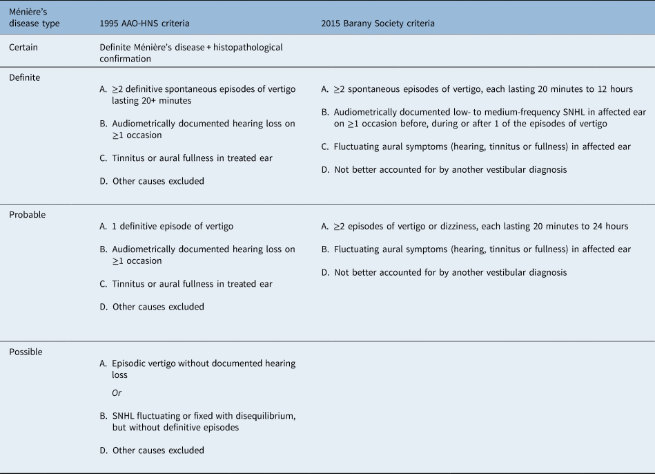
Ménière's disease cases are classified according to the 2015 Barany Society criteria into definite or probable Ménière's disease (Table 1).Reference Lopez-Escamez, Carey, Chung, Goebel, Magnusson and Mandalà27 For this study, the grading for every case was repeated by a neuroradiologist, who was blinded to the diagnosis and laterality of the symptoms. No hydrops imaging of control subjects was possible given the ethical implications of double-dose gadolinium administration without any diagnostic benefit.
Comparison between 1995 AAO-HNS criteria and 2015 Barany Society criteria for Ménière's diseaseReference Lopez-Escamez, Carey, Chung, Goebel, Magnusson and Mandalà27

AAO-HNS = American Academy of Otolaryngology–Head and Neck Surgery; SNHL = sensorineural hearing loss
Results
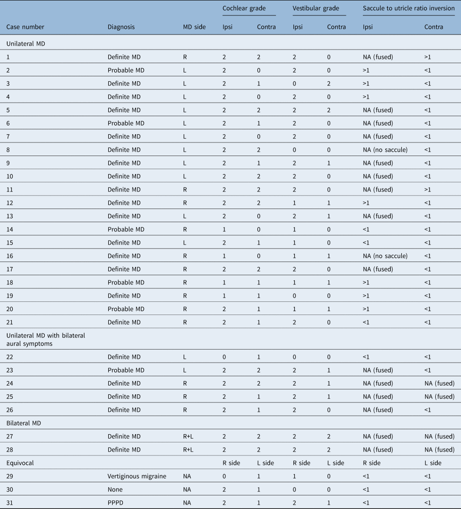
A total of 41 hydrops MRI investigations were performed between September 2017 and January 2019. Of these, 32 scans were performed for investigation of episodic vertigo. One case was non-diagnostic because of poor imaging quality and was excluded from the study, leaving 31 patients for analysis (19 females and 12 males). Patients’ mean age was 52 years (median = 51 years, range = 30–78 years). The details of imaging findings are shown in Table 2.
Details of hydrops MRI findings in 31 patients presenting with episodic vertigo

MRI = magnetic resonance imaging; MD = Ménière's disease; Ipsi = ipsilateral; Contra = contralateral; R = right; NA = not applicable; L = left; PPPD = persistent perceptual postural dizziness
Unilateral Ménière's disease
Twenty-one of the patients had unilateral Ménière's disease (16 with definite and 5 with probable Ménière's disease). The mean interval between disease onset and imaging was seven years (median = 5 years, range = 0.5–25 years).
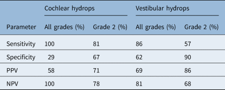
Grade 1 or 2 cochlear hydrops was demonstrated in the symptomatic ear in 100 per cent of the cases (21 out of 21) and in the asymptomatic contralateral ear in 71 per cent (15 out of 21). Of these, grade 2 cochlear hydrops was found in 81 per cent of symptomatic ears (17 out of 21), but in only 33 per cent of asymptomatic contralateral ears (7 out of 21). In 57 per cent of the cases (12 out of 21), there was asymmetry between the two sides; in every asymmetrical case, more marked hydropic changes were observed in the clinically symptomatic ear. The sensitivity and specificity of all grades of cochlear hydrops for Ménière's disease were 100 per cent and 29 per cent respectively. When only grade 2 changes were considered, the sensitivity and specificity changed to 81 per cent and 67 per cent respectively (Table 3).
Sensitivity, specificity, and positive and negative predictive values

PPV = positive predictive value; NPV = negative predictive value
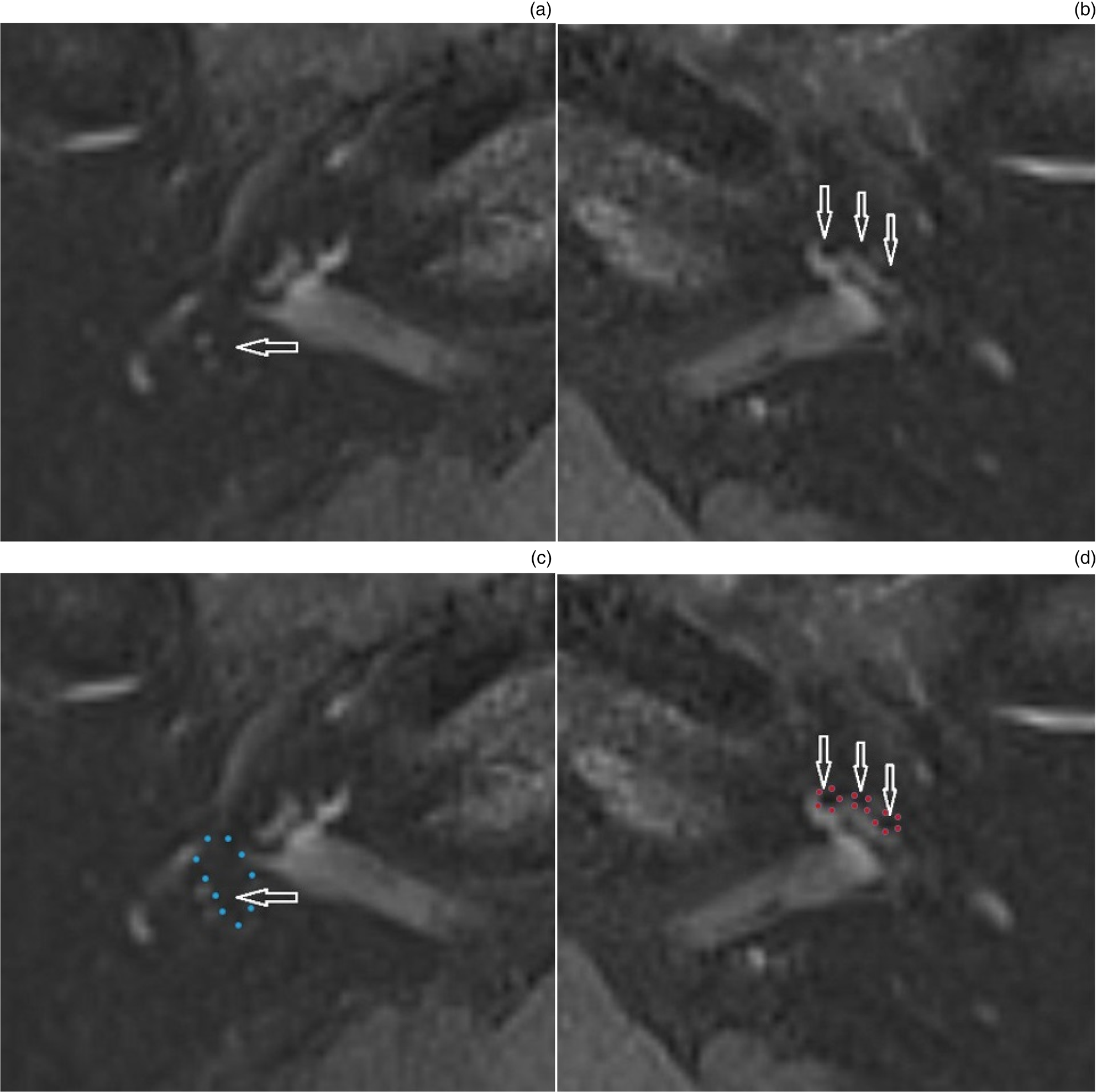
Grade 1 or 2 vestibular hydrops was detected in the symptomatic ear in 86 per cent of the cases (18 out of 21) and in the contralateral ear in 38 per cent (8 out of 21). Of these, grade 2 vestibular hydrops was found in 57 per cent of symptomatic ears (12 out of 21), but in only 10 per cent of asymptomatic contralateral ears (2 out of 21). Asymmetry in the vestibular grading was observed in 67 per cent of the cases (14 out of 21), with the MRI changes lateralising to the symptomatic ear in all cases (Figure 1) but one. The sensitivity and specificity of all grades of vestibular hydrops for Ménière's disease were 86 per cent and 62 per cent respectively. When only grade 2 changes were considered, the sensitivity and specificity changed to 57 per cent and 90 per cent respectively (Table 3).
Axial magnetic resonance imaging showing: (a) & (c) grade 2 endolymphatic hydrops in the right ear, and (b) & (d) a normal left ear. The right ear (a & c) demonstrates grade 2 cochlear hydrops (the cochlear duct is dilated and exceeds the area of the scala vestibule – vertical filled arrows) and grade 2 vestibular hydrops (more than 50 per cent cross-sectional area of the vestibule – horizontal filled arrows) on Nakashima criteria.Reference Nakashima, Naganawa, Katayama, Teranishi, Nakata and Sugiura26 The left ear (b & d) demonstrates perilymphatic space enhancement with no endolymphatic hydrops. Note the normal sized utricle (horizontal open arrows) and saccule (vertical open arrows), with endolymphatic structures less than 33 per cent of the vestibular cross-sectional area.

In two cases (10 per cent), there was no visible saccule on the symptomatic side, suggestive of saccular rupture. SURI could not be determined in 52 per cent of the cases (11 out of 21), because of either fusion of the vestibular endolymphatic structures (9 out of 21) or absence of the saccule (2 out of 21). SURI of greater than 1 was found in 33 per cent of the cases (7 out of 21) of the symptomatic ears and in 10 per cent (2 out of 21) of the contralateral ears (Figure 2). When the vestibular grading, fusion or rupture of vestibular structures and SURI were interpreted in conjunction, MRI detected and lateralised vestibular endolymphatic hydrops to the clinically symptomatic ear in 95 per cent of the cases (20 out of 21). When the cochlear hydrops grading was added, this figure rose to 100 per cent.
Axial magnetic resonance imaging showing: (a) & (c) a normal right ear, and (b) & (d) vestibular endolymphatic hydrops in the left ear, represented by saccule enlargement relative to the utricle (saccule to utricle ratio inversion of more than 1). The normal right (a & c) saccule (filled black arrows) is smaller than the utricle (solid white arrows). The left (b & d) saccule (open black arrows) is larger than the utricle (open white arrows).

Ménière's disease with bilateral symptoms
Seven patients (6 with definite and 1 with probable Ménière's disease) had previously been diagnosed with unilateral Ménière's disease between five and 30 years previously (mean = 17 years, median = 20 years) and had subsequently developed symptoms in the contralateral ear. Two of these patients had bilateral Ménière's disease clinically, whilst the other five appeared to have unilateral Ménière's disease but bilateral fluctuating aural symptoms.
Bilateral grade 2 cochlear and vestibular hydrops, together with fusion of the vestibular structures, were observed in both cases of bilateral Ménière's disease (Figure 3). Predominantly grade 2 cochlear and vestibular hydrops were demonstrated in four out of the five cases of unilateral Ménière's disease with bilateral aural symptoms, and fusion of the vestibular structures was seen in six out of the eight ears. Overall, hydropic changes were seen to be more marked on the side with Ménière's disease and a longer duration of aural symptoms. In the other patient, who had been in remission from acute attacks since saccus surgery 30 years previously, the only change was grade 1 cochlear hydrops in the contralateral ear, in which fluctuating SNHL, tinnitus and aural fullness had recently developed.
Axial magnetic resonance imaging scans (a) – (d) showing bilateral grade 2 endolymphatic hydrops. Both sides demonstrate grade 2 cochlear hydrops (the cochlear duct is dilated and exceeds the area of the scala vestibule – (b & d) vertical open arrows on the patient's left side) and grade 2 vestibular hydrops (more than 50 per cent cross-sectional area of the vestibule – (a & c) horizontal open arrows on the patient's right side) on Nakashima criteria.Reference Nakashima, Naganawa, Katayama, Teranishi, Nakata and Sugiura26

No clinical diagnosis of Ménière's disease
Three patients had presented with episodic vertigo lasting between 20 minutes and 12 hours, but clinically Ménière's disease was not felt to be the likely diagnosis. One patient, who was later diagnosed with vestibular migraine, had grade 1 cochlear hydrops in one ear and grade 1 vestibular hydrops in the other. Another patient with non-specific symptoms that improved spontaneously had grade 1 and 2 hydrops involving the cochleae only. The other patient with symmetrical, mild SNHL was found to have grade 2 cochlear and vestibular hydrops lateralising to one ear, but the clinical diagnosis remained as persistent perceptual postural dizziness. SURI of less than 1 was found bilaterally in all three cases.
Discussion
Although high-resolution T2-weighted sequences are widely used for the demonstration of labyrinthine anatomy and pathology, they are unable to distinguish the endolymphatic from perilymphatic compartments, and hence unable to depict endolymphatic hydrops. The potential role of gadolinium in delineating the endolymphatic chamber first became apparent in animal studies, when gadolinium was seen to accumulate in the perilymph but was excluded from the endolymph by the impermeable tight junctions.Reference Barath, Schuknecht, Naldi, Schrepfer, Bockisch and Hegemann24,Reference Attye, Eliezer, Boudiaf, Tropres, Chechin and Schmerber25
The intratympanic administration of gadolinium was initially explored, whereby high concentrations could enter the labyrinth through round window diffusion. However, the evolution of clinically applicable high-resolution sequences at 3 Tesla has allowed for gadolinium to be administered by the less invasive intravenous route, such that both ears may be imaged. Gadolinium undergoes slow accumulation in the perilymph by crossing the blood labyrinthine barrier, but does not penetrate the endolymph, which therefore demonstrates lower signal on the delayed gadolinium-enhanced images. A 4-hour delay is felt to be optimal to allow the maximum enhancement of the perilymph.Reference Naganawa, Yamazaki, Kawai, Sone and Nakashima28 Imaging requires sequences that are particularly sensitive to low concentrations of gadolinium-based contrast agent, and both 3D FLAIR and 3D inversion recovery turbo spin echo imaging with real reconstruction (3D real IR) have been recommended.
There are a number of reasons for the increasing interest in hydrops imaging. Firstly, radiological confirmation of endolymphatic hydrops may support or dissuade from the diagnosis of Ménière's disease in cases with incomplete phenotypes, and where there are other diagnostic considerations, such as vestibular migraine or autoimmune ear disease. Secondly, it may facilitate early identification, thus guiding future treatment options for patients who have traditionally been required to take a watchful waiting or empirical approach to management. In future long-term follow-up studies, it will be interesting to see if there are any correlations between the imaging findings, clinical diagnosis and responses to various treatments. Thirdly, identification of bilateral endolymphatic hydrops on imaging in a patient with unilateral fluctuating aural symptoms might predict future bilateral disease and potentially influence therapeutic approaches, especially if vestibular-destructive treatments are being considered. Fourthly, it offers the potential for an objective diagnosis of Ménière's disease that does not critically depend on the patient's description of symptom patterns.
Although the semi-quantitative scale we describe is the most widely applied,Reference Nakashima, Naganawa, Katayama, Teranishi, Nakata and Sugiura26 the publications to date are characterised by variations in MRI techniques, analytical methods or scoring, and alternative scales and endolymphatic:perilymphatic ratios are the subject of ongoing evaluation.Reference Barath, Schuknecht, Naldi, Schrepfer, Bockisch and Hegemann24,Reference Sepahdari, Ishiyama, Vorasubin, Peng, Linetsky and Ishiyama29 There is also potential to introduce additional items (e.g. asymmetrical perilymphatic enhancement) to the imaging evaluation in order to maximise diagnostic accuracy and reproducibility.
The finding of vestibular hydrops has been shown to be highly sensitive for Ménière's disease, and our finding of 86 per cent grade 1 or 2 vestibular hydrops in symptomatic ears is consistent with previously published values.Reference Sano, Teranishi, Yamazaki, Isoda, Naganawa and Sone30–Reference Yoshida, Sugimoto, Teranishi, Otake, Yamazaki and Naganawa32 More variable incidences of grade 1 or 2 cochlear hydrops in symptomatic ears have been reported,Reference Attye, Eliezer, Boudiaf, Tropres, Chechin and Schmerber25,Reference Sano, Teranishi, Yamazaki, Isoda, Naganawa and Sone30,Reference Tagaya, Yamazaki, Teranishi, Naganawa, Yoshida and Otake31 which likely reflects the technical difficulties in imaging the small cochlear duct, although our results attest to high detection rates. However, it should be borne in mind that high rates of mild (grade 1) hydrops are also demonstrated in asymptomatic ears as well as in some normal subjects; our rates of 71 per cent (cochlear) and 38 per cent (vestibular) in asymptomatic ears in unilateral Ménière's disease are within the range of previously published data.Reference Sano, Teranishi, Yamazaki, Isoda, Naganawa and Sone30–Reference Yoshida, Sugimoto, Teranishi, Otake, Yamazaki and Naganawa32 In fact, the limited studies of healthy volunteers have shown grade 1 or 2 vestibular hydrops in up to 90 per cent of ears.Reference Attye, Eliezer, Boudiaf, Tropres, Chechin and Schmerber25 This provides an explanation for the high incidence of bilateral grade 1 hydrops found in our study, which clearly exceeds the recognised incidence of bilateral Ménière's disease. It is therefore likely that the presence of grade 1 hydrops is of limited value in diagnosing Ménière's disease or differentiating it from other otological conditions. However, its absence has a high negative predictive value. This is in keeping with the findings of a meta-analysis of temporal bone studies by Foster and Breeze, in which 98.8 per cent of Ménière's disease cases that met the 1995 American Academy of Otolaryngology–Head and Neck Surgery criteria were found to have endolymphatic hydrops in at least one ear.Reference Foster and Breeze15 The absence of endolymphatic hydrops on MRI therefore will be useful in excluding Ménière's disease as the principal diagnosis.
Severe (grade 2) hydrops is considered a more specific but less sensitive finding for Ménière's disease. Our relatively low rate of grade 2 vestibular hydrops in symptomatic ears (57 per cent) is also similar to the 46 per cent and 60 per cent incidences previously reported, and we concur that grade 2 vestibular hydrops is rare in asymptomatic ears.Reference Attye, Eliezer, Boudiaf, Tropres, Chechin and Schmerber25,Reference Pakdaman, Ishiyama, Ishiyama, Peng, Kim and Pope33 Hence, the specificity of grade 2 vestibular hydrops indicates that it is a useful finding to diagnose and differentiate Ménière's disease from other entities. Again, more variable rates of grade 2 changes have been demonstrated in the context of cochlear hydrops.Reference Attye, Eliezer, Boudiaf, Tropres, Chechin and Schmerber25,Reference Sano, Teranishi, Yamazaki, Isoda, Naganawa and Sone30,Reference Tagaya, Yamazaki, Teranishi, Naganawa, Yoshida and Otake31
There are ongoing studies that seek to determine more accurate methods for diagnosing Ménière's disease on imaging studies. Morphological changes may ultimately prove more useful than overall changes in the size of the endolymphatic space. SURI is a qualitative comparison of the area of the saccule relative to that of the utricle, for which a combination of oblique sagittal and axial reformats are used. SURI of greater than 1 on MRI assessment has proved a reliable qualitative marker.Reference Attye, Eliezer, Boudiaf, Tropres, Chechin and Schmerber25 Asymmetry in the avidity of perilabyrinthine enhancement, and other grading scales or thresholds for the endolymphatic:perilymphatic space area or volume ratios, have also been explored.Reference Barath, Schuknecht, Naldi, Schrepfer, Bockisch and Hegemann24,Reference Sepahdari, Ishiyama, Vorasubin, Peng, Linetsky and Ishiyama29,Reference Yoshida, Sugimoto, Teranishi, Otake, Yamazaki and Naganawa32
Nonetheless, we have found to date that there is a very good correlation between the clinical symptoms and lateralisation of hydropic changes on MRI. Our current diagnostic approach is to emphasise the presence of grade 2 hydropic changes and asymmetric higher scores that correspond to the symptomatic ear. It should also be noted that endolymphatic hydrops may be secondary to disease processes other than Ménière's disease, such as viral infection, trauma, semicircular canal dehiscence, otospongiosis, autoimmune disease and vestibular schwannoma; such clinical entities may still need to be considered even when unilateral symptoms correspond to ipsilateral hydrops on MRI.
• Recent advances in magnetic resonance imaging (MRI) have enabled in vivo demonstration of endolymphatic hydrops
• There is a good correlation between clinical symptoms and lateralisation of hydropic changes on MRI
• Mild hydrops is often seen in clinically asymptomatic ears, the significance of which has yet to be elucidated
• Further refinements of imaging techniques and grading system will likely improve diagnostic accuracy and clinical utilisation of hydrops MRI
Gadolinium has an excellent safety record when used at low doses (0.1–0.3 mmol/kg) in patients with normal renal function. However, a causative relationship between gadolinium-based contrast agents and nephrogenic systemic fibrosis has been described in patients with renal insufficiency. In addition, there is increasing evidence that they deposit in the human brain after multiple administration, although the clinical significance of this remains uncertain.Reference Ramalho, Semelka, Ramalho, Nunes, AlObaidy and Castillo34 Some authors have used alternative gadolinium-based contrast agents, to enable adequate depiction of the endolymphatic structures with a single-dose technique and hence improve the safety profile.Reference Eliezer, Poillon, Gillibert, Horion, Cruypeninck and Gerardin35
There is also considerable interest in the evaluation of non-gadolinium-enhanced MRI techniques to image the enlargement of endolymphatic structures. These currently employ high-resolution 3D T2-weighted gradient echo sequences, such as constructive interference in steady state (‘CISS’) and fast imaging employing steady-state acquisition (‘FIESTA’), and have focused on the assessment of saccule size. Although the reliability and reproducibility of these techniques requires confirmation,Reference Simon, Guichard, Kania, Franc, Herman and Hautefort36,Reference Eliezer, Poillon, Horion, Lelion, Gerardin and Magne37 the potential for non-gadolinium-enhanced MRI sequences to demonstrate some endolymphatic structures would help justify not only the inclusion of a control group of healthy subjects but also the use of serial imaging studies in order to evaluate any fluctuation or progression of imaging changes with duration of disease, and to allow an assessment of imaging response to interventions.
We acknowledge that our study has a number of limitations. As hydrops imaging in our centre began 20 months ago as part of specific clinical protocols, the sample size is naturally small. The authors also feel that, until newer MRI techniques become available that require no intravenous gadolinium or only a standard dose, performing hydrops imaging on asymptomatic subjects as controls is ethically difficult to justify. This is true of most published studies, with very limited data being available from normal controls.Reference Attye, Eliezer, Boudiaf, Tropres, Chechin and Schmerber25 Finally, the clinical implications of any imaging findings will only become fully evident when long-term follow-up data emerge, potentially enabling a more complete characterisation of the subtypes of Ménière's disease.Reference Phillips, Murdin, Rea and Sutton38
Conclusion
Magnetic resonance imaging now allows in vivo demonstration of endolymphatic hydrops for the first time, and is perhaps one of the most significant recent advances in the field of Ménière's disease. In our experience so far, there has been a very good correlation between the clinical picture and the lateralisation of hydropic changes on MRI, despite high rates of mild hydrops in asymptomatic ears. Refinement of imaging techniques and classification systems is of great interest for future development. There is potential for MRI to contribute to diagnostic criteria and subtyping to define homogeneous cohorts for longitudinal studies of natural history and therapeutic response.
Competing interests
None declared