

Introduction
The Dilatata group of Paspalum is a polyploid complex native to the grasslands of temperate South America. The complex contains several informal taxonomic entities that will be generally referred to as biotypes in this paper. Paspalum dilatatum Poir. ssp. dilatatum, a trihybrid pentaploid apomict (Bashaw and Forbes, Reference Bashaw and Forbes1958; Bashaw and Holt, Reference Bashaw and Holt1958), is a widely recognized forage crop. This biotype has been assigned the IIJJX genomic formula (Burson, Reference Burson1983). The Dilatata group also includes several sexual allotetraploids and tetra- and hexaploid apomictic entities. The sexual allotetraploids (P. urvillei Steud., P. dasypleurum Kunze ex Desv., P. dilatatum ssp. flavescens Roseng. Arr. et Izag., and biotypes Virasoro and Vacaria of P. dilatatum) have been shown either directly or indirectly to share the IIJJ genomic formula (Burson et al., Reference Burson, Lee and Bennett1973; Burson, Reference Burson1979; Caponio and Quarín, Reference Caponio and Quarín1990; Quarín and Caponio, Reference Quarín and Caponio1995), and their interfertility has been either directly assessed by hybridization (Caponio and Quarín, Reference Caponio and Quarín1990; Quarín and Caponio, Reference Quarín and Caponio1995) or inferred from the occurrence of natural hybrids (Valls and Pozzobon, Reference Valls and Pozzobon1987). The apomictic hexaploids (P. dilatatum biotypes Uruguaiana and Chirú) have been assigned the IIJJXX2 and IIJJXX genomic formulae, respectively (Burson, Reference Burson1991b, Reference Burson1992), but no information is available for P. dilatatum Torres, an asynaptic apomictic hexaploid (Moraes-Fernandes et al., Reference Moraes Fernandes, Barreto and Salzano1968) and P. pauciciliatum, an apomictic tetraploid (Bashaw and Forbes, Reference Bashaw and Forbes1958). Recently, Machado et al. (Reference Machado, Valls, Peñaloza and dos Santos2005) have shown that there may be several additional pentaploid apomictic entities in the group which have not yet been described or named. Most of our knowledge of the group has been derived from chromosome numbers and pairing behaviour in hybrids between biotypes; however, only recently has it been possible to obtain further cytological information through the modification of cytological techniques that allow chromosome identification and karyotyping (Speranza et al., Reference Speranza, Vaio and Mazzella2003; Vaio et al., Reference Vaio, Speranza, Valls, Guerra and Mazzella2005). In spite of this, cytogenetics alone cannot answer many of the questions that must be addressed in this group.
Interest in breeding the common pentaploid biotype has been the main motivation for the study of this group. Pentaploid (IIJJX) resynthesis by hexaploid (IIJJXX) × tetraploid (IIJJ) hybridization has been suggested as a possible breeding strategy for the group (Burson, Reference Burson1983) and vigorous synthetic pentaploids have been successfully obtained by this kind of cross (Burson, Reference Burson1991a, Reference Burson1992; P. Speranza, unpublished data). In spite of the availability of extensive collections of the most widely used tetraploid (P. dilatatum ssp. flavescens), the assessment of the genetic variability they contain has not been successful particularly because qualitative morphological variability is notably lacking. Efficient utilization of these genetic resources, accurate descriptions of their breeding systems and genetic structure, and a meaningful interpretation of the variability represented by the natural apomicts are highly dependent on the development of the appropriate markers.
As most of the apomicts in the complex have been hypothesized to be inter-biotypic combinations, the markers required to address questions about their genetic structure and origin must be transferable among all of the putative parents involved and preferably co-dominant. Assessment of parentage would be best achieved with markers that are stable within biotypes, while the study of the genetic structure of the sexual components of the complex requires high levels of variability. Allopolyploidy adds an additional level of complexity to the genetic interpretation of molecular markers: the assessment of homology versus homeology among markers may not be straightforward, particularly in complex inter-biotypic combinations.
Attempts have been previously made to use molecular markers for the study of these species, but even when some degree of genetic differentiation between the biotypes and intra-biotypic variability were confirmed (isozymes: Hickenbick et al., Reference Hickenbick, Flores, Cavalli-Molina, Weber, Kersting, Costa, Souza-Chies and Albarus1992; Pereira et al., Reference Pereira, Sabbia, Fajardo and Speranza2000; T. Souza-Chies, unpublished data; amplified fragment length polymorphism (AFLP): P. Speranza, unpublished data; randomly amplified polymorphic DNA (RAPD): Casa et al., Reference Casa, Mitchel, Lopes and Valls2002), the levels of variability have not been high enough or their interpretability has been very limited. Microsatellite markers can potentially provide the best tools for the genetic study of this species complex. In addition, the generalized use of simple sequence repeat (SSR) enrichment and PCR-based protocols has greatly facilitated the development of microsatellite markers (Kandpal et al., Reference Kandpal, Kandpal and Weissman1994; Kijas et al., Reference Kijas, Fowler, Garbett and Thomas1994; Fischer and Bachmann, Reference Fischer and Bachmann1998; Jakše and Javornik, Reference Jakše and Javornik2001). Microsatellites not only are more informative due to their co-dominant nature, but they usually tend to be highly variable. Mutation rates of nearly 1 × 10− 3 have been directly observed in maize (Vigouroux et al., Reference Vigouroux, Jaqueth, Matsuoka, Smith, Beavis, Smith and Doebley2002). In well-studied selfing grass amphiploid systems like wheat, microsatellites are capable of revealing high genetic variability (Röder et al., Reference Röder, Plaschke, König, Börner, Sorrells, Tanksley and Ganal1995) where isozyme, RFLP and AFLP markers show a high degree of marker conservation (Kim and Ward, Reference Kim and Ward2000; Hazen et al., Reference Hazen, Leroy and Ward2002).
Finally, determining the directionality of hybridizations within the species complex may be crucial to understanding the mechanisms by which new genetic combinations are being generated. Chloroplast microsatellites, typically (T/A)n, have been successfully used to elucidate directional formation of allopolyploids in grasses (Ishii and McCouch, Reference Ishii and McCouch2000). Putatively universal primers for grass chloroplast microsatellites have been reported in the literature which can be assessed for variability (Provan et al., Reference Provan, Biss, McMeel and Mathews2004).
Sixteen variable nuclear and one variable chloroplast microsatellite loci for P. dilatatum ssp. flavescens were developed and characterized in this study. Their genetic behaviour and transferability among all the taxonomic entities with the IIJJ genomic formula of the Dilatata group and to P. dilatatum ssp. dilatatum (IIJJX) was assessed. Finally, their utility for addressing populational and phylogenetic studies at different levels is discussed.
Materials and methods
Microsatellite capture
A genomic DNA library consisting of Sau3AI fragments of P. dilatatum ssp. flavescens was enriched for putative microsatellite-containing sequences following the procedures of Ernst et al. (Reference Ernst, Branch, Clarck and Hokit2004) with minor modifications. Briefly, genomic DNA was extracted with Sigma Genelute™ kit (Sigma-Aldrich, St Louis, MO, USA) and digested with Sau3AI. Fragments smaller than approximately 400 bp were removed by fractioning using Chroma Spin® columns (Clontech Laboratories). Sau3AI linkers were ligated to the remaining fragments which were then amplified by PCR. The amplified fragment library was enriched for (GT)n-containing sequences by binding to a Vectrex® Avidin D matrix (Vector Laboratories, Burlingame, CA, USA) to which a biotinylated (CA)n oligonucleotide probe had been previously bound. The eluted fragments were reamplified by PCR using primers for the Sau3AI linkers, ligated into pCR® II-TOPO® plasmids (Invitrogen) and transformed into ONE shot®Escherichia coli competent cells. Colonies were screened by binding them to Magnacharge nylon transfer membranes (Osmonics). The membranes were probed with labelled (CA)n and positive colonies detected with Lumi-Phos 480 (Lifecodes, Stamford, CT, USA). All probe labelling, hybridization and detection was carried out with a Quick-Light™ system (Lifecodes). Positive colonies were grown overnight in a liquid medium and plasmids were purified with QIAprep Spin Miniprep Kit (Qiagen). Plasmid isolates were screened a second time by dot-blotting serial dilutions on nylon membranes and hybridizing to a (CA)n probe. Isolates showing consistent hybridization signal through the dilutions were sequenced and used for primer development.
Plasmid isolates were sequenced on a CEQ 8000 capillary sequencer (Beckman-Coulter, Fullerton, CA, USA) using CEQ™ 2000 Dye Terminator Cycle Sequencing in one-quarter reaction volumes with the addition of 80 mM Tris and 2 mM MgCl2 (pH 9) to complete the volume of a full reaction. The sequences were edited manually using Sequencher™ version 4.1.4 (Genecodes, Ann Arbor, MI, USA).
Primers were designed for sequences containing repeats longer than (GT)10 with Primer 3 (Rozen and Skaletsky, Reference Rozen, Skaletsky, Krawetz and Misener2000). Low-complexity regions were excluded for primer design when possible.
Plant material
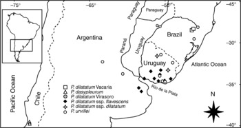
Seed samples were obtained from the Southern Regional Plant Introduction Station, Griffin, GA, USA and the Germplasm Bank at the Facultad de Agronomía, Montevideo, Uruguay. The geographic origin of the accessions used in this study are shown in Fig. 1. Potential variability of microsatellites was assessed by analysing a total of 28 accessions representing different species of the Dilatata group. To assess intraspecific variability, we analysed one individual from each of 10 accessions of P. dilatatum ssp. flavescens and P. urvillei. Accessions were chosen to represent as much of the native range of the species as possible (Fig. 1). To assess transferability, two accessions of P. dasypleurum, P. dilatatum ssp. dilatatum, and biotypes Virasoro and Vacaria of P. dilatatum were also included.
Geographical distribution of the accessions used in this study. The inset shows the magnified area.

DNA was also extracted from 43 F2 individuals derived from a P. dilatatum ssp. flavescens × P. dilatatum Virasoro hybrid to assess marker segregation patterns and linkage among loci. Deviation from expected segregation ratios and linkage between loci were assessed using Joinmap 3.0 (Van Ooijen and Voorrips, Reference Van Ooijen and Voorrips2001). When there were indications that more than one locus had been amplified by a primer pair (see below), the loci were considered as putative homeologs and the absence of linkage between them regarded as a test for their homeology.
F1 hybrids were obtained by manually emasculating a plant of P. dilatatum ssp. flavescens and pollinating it with pollen from an individual of P. dilatatum Virasoro. Emasculation and pollination were carried out about 1 h after sunrise. The plants to be emasculated were placed at approximately 20°C and 100% relative humidity to delay anther dehiscence after anthesis was initiated each morning. Mature full seeds were counted and germinated in Petri dishes on filter paper in an incubator with alternating temperatures (16 h at 30°C light, 8 h at 20°C dark). Germinators were placed at 4°C for 4 days prior to incubation to break dormancy and homogenize germination. The resulting progeny were grown and the hybrids were identified by the high number of nerves in glume II and lemma I which characterize the pollen donor. Selfed seed of one F1 hybrid was collected and treated as described above to establish the segregating F2 progeny used in this study.
Chloroplast markers
Sequences for six chloroplast non-coding regions were obtained for all the commonly recognized entities in the Dilatata group: P. dilatatum ssp. flavescens, P. dilatatum ssp. dilatatum, P. dilatatum biotypes Torres, Uruguaiana, Virasoro and Vacaria, P. dasypleurum, P. pauciciliatum and P. urvillei. Six regions were analysed: the trnT(UGU)-trnL(UAA) spacer, the trnL(UAA) intron, the PsbA-trnH spacer, the atpB-rbcL spacer, the trnG(UCC) intron and the trnL(UAA)-trnF(GAA) spacer. PCR and sequencing conditions and primers were those reported in Vaio et al. (Reference Vaio, Speranza, Valls, Guerra and Mazzella2005) except for the trnT(UGU)-trnL(UAA) spacer which was amplified and sequenced using primers A and B (Taberlet et al., Reference Taberlet, Gelly, Pavtou and Bouvet1991). Primers were designed flanking two poly-A repeats located in the trnT(UGU)-trnL(UAA) spacer. All primers reported by Provan et al. (Reference Provan, Biss, McMeel and Mathews2004) were also tested. A second poly A-tract not reported by Provan et al. (Reference Provan, Biss, McMeel and Mathews2004) was detected near the trnL(UAA)3′ exon but no length variability was observed among the available sequences, and no further analysis was performed on it. Primer design, labelling, amplification and detection procedures were performed as for the nuclear SSRs described below.
Microsatellite amplification and scoring
For all plant materials DNA was extracted from fresh leaves or silica gel-dried leaves using a Sigma Genelute™ kit (Sigma-Aldrich) according to the manufacturer's instructions.
Amplification, labelling and separation conditions were adjusted for all primer pairs following Boutin-Garnache et al. (Reference Boutin-Garnache, Raposo, Raymond and Deschepper2001). Forward primers were extended by adding an M13 tail (5′-CACGACGTTGTAAAACGAC-3′), and M13 primers were labelled with D4 (Beckman Coulter). All PCR amplifications were carried out in 10 μl reactions containing 0.2 units of NEB Taq polymerase (New England Biolabs, Beverly, MA, USA), 1.5 mM MgCl2, 0.15 μM reverse primer and labelled M13 primer, 0.01 μM extended forward primer, and 0.1 mM each dNTP in the manufacturer's buffer. Amplification was carried out in a Biometra® T3 Thermoblock with the same two-step programme for all primer pairs. The PCR profile consisted of an initial denaturing step of 5 min at 94°C, followed by 40 cycles of 15 s at 94°C and 3 min at 53°C, and a final extension step of 5 min at 72°C. Labelled microsatellite products were separated in a CEQ 8000 capillary sequencer (Beckman-Coulter) by loading 0.75 μl of the PCR product and 0.35 μl of CEQTM DNA Size Standard Kit-400 (Beckman Coulter) in 25 μl of formamide per well. Chromatograms were visualized on CEQ TM Genetic Analysis system software (Beckman Coulter) and scored manually.
Alleles in different size ranges from different biotypes or subspecies were sequenced to assess homology. For primer pairs amplifying a single locus, alleles were amplified and sequenced directly from the PCR products of homozygous individuals. For primer pairs amplifying more than one putative locus, PCR products were separated in 2% agarose, and the bands were cut, purified with Wizard® SV Gel and PCR Clean-up System (Promega, Madison, WI, USA) and sequenced directly. When multiple bands could not be separated by electrophoresis, gel slices containing several bands were cut and combined products cloned with a TOPO TA Cloning® kit.
Results
Capture and amplification success
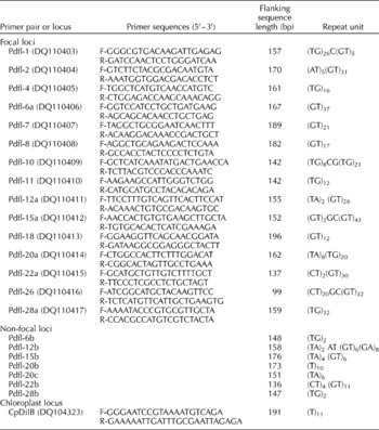
A total of 24 clones containing (GT)n repeats were captured and sequenced. Four clones were redundant. Primer pairs were designed for all clones, and all loci were amplified with the same two-step PCR profile detailed above. Fifteen of these primer pairs successfully amplified interpretable band patterns, and 12 of them were selected for further analysis based on a preliminary assessment of amplification success under the given conditions. Primer pairs for loci Pdfl-1, Pdfl-2 and Pdfl-26 are reported but not further characterized (Table 1). Primer sequences and corresponding Genbank accession numbers, flanking sequence lengths, and repeat units for all successfully amplified loci are shown in Table 1.
Primer sequences and structure for all the microsatellite loci reported in this study

When sequence information is available for more than one allele, the variable repeat motif is reported. GenBank accession numbers for the originally cloned sequence used to design each primer pair are given in parentheses.
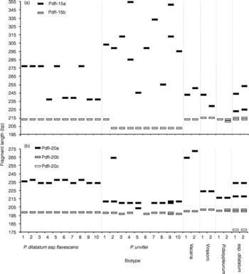
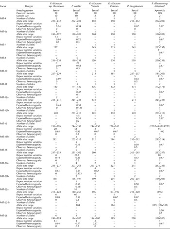
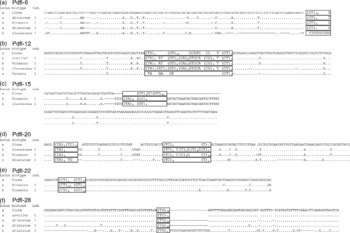
Primer pairs Pdfl-6, Pdfl-12, Pdfl-15, Pdfl-20 and Pdfl-22 amplified more than one band in all or some tetraploid individuals, most of which appeared homozygous for all the other loci. Results for representative primer pairs Pdfl-15 and Pdfl-20 are shown graphically in Fig. 2. In these cases, representative bands from both size ranges were cloned, sequenced and compared to the originally captured sequences (Fig. 2). Two other extra bands, a Pdfl-12 190 bp band in P. dilatatum ssp. flavescens and a 140 bp band in P. urvillei (data not shown) did not contain the forward primer when sequenced. These bands were in fact reproduced by using just the labelled M13 primer and the reverse primer for each locus. In all the other cases, the extra bands showed extensive sequence similarity to the captured alleles and differed mostly in the organization and length of the repeat units (Fig. 2). These fragments were interpreted as amplification products from a putative homeologous locus (see Discussion below). The loci for which the primers were originally designed will be referred to as the focal loci. When two different loci were detected, the focal locus was identified by adding ‘a’ to the primer pair name and ‘b’ or ‘c’ to the additional loci. Alleles shown in Table 2 were assigned to each locus based on the sequences shown in Fig. 2. For most non-focal loci, fragment sizes were smaller than the captured bands, and the microsatellite repeat was either absent or showed a lower number of repeats. The only exception was the Pdfl-12a 140 bp allele in Vacaria, which was smaller than the alleles at the non-focal locus Pdfl-12b (Table 2). For Pdfl-20 (Fig. 2b) and Pdfl-28, an extra band (177 and 178 bp, respectively) was present in the apomictic pentaploid P. dilatatum ssp. dilatatum while absent in all tetraploids. These bands were sequenced, and they showed again extensive similarity to the captured loci (Fig. 3d, f).
A schematic representation of the band patterns obtained with primer pairs Pdfl-15 (a) and Pdfl-20 (b). Size range and nucleotide sequence were used to assign individual fragments amplified by a given primer pair to different loci.

Summary of the fragments amplified for all the variable nuclear microsatellite loci reported in the tetraploid biotypes (IIJJ) of the Dilatata group of Paspalum

a Genotypes instead of allele size ranges are shown for the pentaploid apomict P. dilatatum ssp. dilatatum.
Alignment of nucleotide sequences of representative alleles for the non-focal loci compared to the sequences originally cloned from Paspalum dilatatum ssp. flavescens. Focal locus alleles are also included when their lengths differ significantly from those found in P. dilatatum ssp. flavescens. Alleles are identified by their source biotype and individual. All putatively homologous variable and non-variable repetitive sequences near or around the main microsatellite are highlighted for comparison.

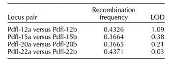
Homeologous loci are expected to be completely unlinked because they are on different chromosomes. Linkage analysis based on the segregating F2 population showed no evidence of linkage between loci amplified by the same primer pair with recombination frequencies ranging from 0.3664 to 0.4371 and LOD scores from 0.03 to 1.09 (significance threshold LOD equal to 3) (Table 3).
Estimated recombination frequency between pairs of loci amplified by the same primer combination, and LOD score for the test of independent segregation between loci (significant threshold LOD=3)

In addition, no significant linkage was observed when considering the full set of 16 loci (including non-focal loci), even at a low LOD threshold level of 2. Although the test for independence between loci as implemented in Joinmap is robust against segregation distortions, it is reassuring that all but one locus (Pdfl-15a) showed segregation ratios in agreement with the expectations for normal disomic inheritance (data not shown).
Most loci were amplified in all materials tested with a few exceptions (Table 2). Paspalum urvillei could not be scored for Pdfl-7 and Pdfl-10. Pdfl-7 was not amplified at all in most P. urvillei samples whereas for Pdfl-10, weak peaks were observed but could not be reliably scored probably due to interference during PCR from the M13-primed fragment mentioned above. No fragments were amplified in P. dilatatum Virasoro for Pdfl-18 and Pdfl-12a, and P. dasypleurum did not produce fragments for Pdfl-15. Locus Pdfl-22b amplified consistently only in P. dilatatum Virasoro and P. dilatatum ssp. dilatatum (Table 2).
Variability in nuclear loci
All biotypes showed polymorphisms for at least two loci. On the other hand, several alleles were mostly fixed within biotypes but variable among biotypes. Notably Pdfl-7 provided excellent biotype-specific markers for this sample (Table 2).
At least two different alleles were cloned and partially sequenced from the focal biotype for Pdfl-4, Pdfl-6a, Pdfl-11, Pdfl-12a, Pdfl-22a, Pdfl-15a and Pdfl-28a. Despite the effect of the long repetitive tract on the quality of the sequence, it could be clearly seen that variation in these focal loci is due to expansion/contraction of the (GT/CA)n repeat (data not shown).
Despite being the source biotype, P. dilatatum ssp. flavescens does not display the greatest variability nor allele sizes in this sample (Table 2). The average number of alleles per locus for P. dilatatum ssp. flavescens is 2.5 with an average size range of 9.9 bp. Paspalum urvillei showed a higher number of alleles per locus (3.9) and a higher size range (28.9 bp); furthermore, P. urvillei showed polymorphisms for all the scored loci, and 70% of the individuals were heterozygous for at least one locus whereas in P. dilatatum ssp. flavescens only one individual appeared heterozygous for locus Pdfl-6a. The two individuals sampled from Virasoro and Vacaria were completely homozygous, whereas heterozygosity was observed in both individuals of P. dasypleurum (Table 2). The two individuals of P. dilatatum ssp. dilatatum appeared heterozygous for all loci except individual 2 at locus Pdfl-22b.
In summary, 19 variable nuclear microsatellite loci were investigated, with one locus monomorphic in all the accessions (Pdfl-6b). Among the 18 variable loci, 11 were successfully amplified and interpreted in all the biotypes in this sample. Of the original 12 focal loci, four could not be scored in one of the biotypes: Pdfl-7 and Pdfl-10 in P. urvillei, Pdfl-18 in Virasoro, and Pdfl-15a in P. dasypleurum. Three of the non-focal variable loci (Pdfl-12b, Pdfl-15b and Pdfl-20b) were successfully amplified and scored for all biotypes, while Pdfl-22b was only amplified in Virasoro, the pentaploids and one individual of P. urvillei, and finally, two loci (Pdfl-20c and Pdfl-28b) were only amplified in the pentaploids.
Chloroplast loci
All regions were successfully amplified and sequenced except for the trnT(UGU)-trnL(UAA) spacer for which low-quality sequences were obtained due to the presence of poly-A tracts near both ends (GenBank accession numbers DQ104273–DQ104323). No further efforts were made to improve the quality of the sequences of the trnT(UGU)-trnL(UAA) spacer because they were considered appropriate for the purposes of this study. Overall, no sequence variability was found among the biotypes of the Dilatata group except for the length of the poly-A tract in the trnT(UGU)-trnL(UAA) spacer, and a G-T transversion in the trnL(UAA) intron in the Vacaria individual. No repetitive sequences were found except for poly-A/T tracts. Fragment sizes were not variable for any of the loci reported by Provan et al. (Reference Provan, Biss, McMeel and Mathews2004) or the poly-A repeat located near the trnL(UAA)5′ exon in the trnT(UGU)-trnL(UAA) spacer. Only the poly-A repeat located near trnT(UGU) (cpDilB) was variable as observed in the original sequences. Fragment lengths (after subtracting the M13 tail) were 198 bp for P. dilatatum ssp. flavescens, ssp. dilatatum, Virasoro and Vacaria, 199 bp for P. urvillei and 197 bp for P. dasypleurum. No intra-biotypic variability was observed except for individual 2 in P. urvillei, which contained the 198 bp allele.
Discussion
Capture efficiency
Among the loci reported here, Pdfl-2, Pdfl-12a, Pdfl-15b and Pdfl-20a were found to be compound (CA/GT)n (AT/TA)n repeats (Table 1). In a genome-wide survey in rice, Temnykh et al. (Reference Temnykh, Park, Ayres, Cartinhour, Hauck, Lopovich, Cho, Ishii and McCouch2000, Reference Temnykh, DeClerck, Lukashova, Lipovich, Cartinhour and McCouch2001) found that (CA)n repeats were frequently associated with (TA)n repeats.
In genome-wide surveys of grasses, (CA)n repeats have been reported to be relatively short compared to other dinucleotides (Temnykh et al., Reference Temnykh, Park, Ayres, Cartinhour, Hauck, Lopovich, Cho, Ishii and McCouch2000). During the enrichment phase of the capture protocol, this may have led to the retention of a limited number of longer repeats, which may explain the high level of redundancy (four out of 24) of the captured clones. A strong bias towards long repeats may be advantageous because the length of the perfect repeats is expected to be associated with higher degrees of variability (Weber, Reference Weber1990; Symonds and Lloyd, Reference Symonds and Lloyd2003). Here, loci with more than 30 perfect repeats (Pdfl-6, Pdfl-15a, Pdfl-22 and Pdfl-28) revealed the highest number of alleles per locus in both P. dilatatum ssp. flavescens and P. urvillei.
Amplification profile
Low temperatures during the PCR extension step have been suggested to reduce the generation of frameshift products (commonly known as ‘stutter’), particularly for fragments containing (CA)n repeats (Hite et al., Reference Hite, Eckert and Cheng1996). Under standard PCR conditions like those used here, though, extension temperature cannot be reduced below the desired annealing temperature. In preliminary amplifications a noticeable reduction in the number of stutter peaks was observed, particularly for long alleles, when extension was carried out at 53°C rather than at 72°C, while even lower temperatures resulted in the production of locus-nonspecific products. The use of a single, robust amplification profile does not require the adjustment of annealing and extension temperatures for each primer pair individually, and greatly increases logistic efficiency when simultaneously working with multiple primer pairs.
Non-focal loci
Most studies focus on primer pairs that amplify highly variable single loci. In this study, all bands were taken into account because stable, biotype-specific, co-dominant markers could be extremely useful for hybrid analyses within the complex. Sourdille et al. (Reference Sourdille, Tavaud, Charmet and Bernard2001) analysed a set of wheat microsatellite primer pairs including primers that amplified more than one locus taking into account their known chromosomal locations and transferability. In that study, 54% of the primer pairs amplified more than one locus, including cases in which the extra bands were monomorphic, independently segregating variable loci or co-segregating linked markers. These results are very similar to the ones obtained in this study, in which 50% of the primer pairs amplified more than one putative locus. In this study, however, extra bands were either monomorphic or independently segregating, but no putative tandem duplications were found. Definitive assessment of homeology would require a genetic map showing that the loci are located in syntenic homeologous chromosome segments.
The number of products amplified was always equal to or less than the number of loci expected for a tetraploid amphiploid, always consistent with the interpretation that primer pairs were amplifying products from either one or both the I or J genomes. Remarkably, primer pair Pdfl-20 amplified a number of bands exactly corresponding to the ploidy level in heterozygous individuals (Fig. 2b).
Nuclear loci
At least two variable loci were identified for each of the biotypes including the three sexual tetraploids represented by only two individuals. It is likely then that if more individuals were analysed, this set of loci could contain useful markers for population structure and breeding system assessment of all the biotypes in the group. Overall, the focal species showed less variation than P. urvillei. Only in 25% of the loci (Pdfl-4, Pdfl-6a, Pdfl-20a and Pdfl-28) did P. dilatatum ssp. flavescens show consistently longer repeats than P. urvillei. It is typically expected that due to selection for long repeats during library enrichment, longer repeats and higher variability are more likely to be found in the focal biotype (Ellegren et al., Reference Ellegren, Primmer and Sheldon1995), an artifact known as ascertainment bias. However, P. urvillei consistently showed more variability and a much higher level of heterozygosity for most loci. A higher number of alleles was implified in P. urvillei, even at loci for which fragment lengths were clearly lower than in P. dilatatum ssp. flavescens (Table 2) which contradicts the accepted consensus that repeat number and variability are associated regardless of the causes of this correlation (Schlöterer, Reference Schlöterer2000). A statistical comparison of variability within different biotypes is beyond the scope of this paper; however, the clear differences in variability and heterozygosity between P. dilatatum ssp. flavescens and P. urvillei are likely to be real despite the small sample size presented here. Amos et al. (Reference Amos, Sawcer, Feakes and Rubinsztein1996) claim that heterozygosity may lead to an increase in mutation rates at microsatellite loci. This may seem to be the case when comparing P. dilatatum ssp. flavescens and P. urvillei; however, the stochastic effects of the restricted distribution of P. dilatatum ssp. flavescens and its apparently extreme selfing rate may deserve further investigation as putative explanations for the observed ‘reverse ascertainment bias’. The relative evolutionan distance between the species analysed should also be taken into account to interpret meaningfully cross-amplification and ascertainment bias. The I, J and X genomes within the Dilatata polyploids can be considered to represent different species because they are implicitly assumed to have diverged independently as different diploid species between their coalescence time and the polyploidization event that brought them back together. When the putative homeologous sequences shown in Fig. 2 are compared, a strong ascertainment bias is evident in all of them. Similar flanking sequences combined with shorter and imperfect repeats like those found in the non-focal loci in this study were found by Chen et al. (Reference Chen, Cho and McCouch2002) when they amplified microsatellites developed for Oryza sativa in congeners containing different genomes.
Variation was also found between the two individuals of P. dilatatum ssp. dilatatum. These two individuals share 13 heterozygous allele combinations, making it very unlikely that the three differences found in Pdfl-15a and Pdfl-22b are due to a sexual recombination event or independent origins. However, enough mutations seem to have accumulated in this clonal biotype to observe variability within this set of microsatellite loci.
The transferability, sequence similarity, correspondence in allele sizes and number of alleles amplified in P. dilatatum ssp. dilatatum for each locus clearly indicate that, except for those bands that are exclusive to it (Pdfl-20c and Pdfl-28b), the rest of the loci are located on the I and J genomes. Alleles in loci Pdfl-20c and Pdfl-28c, on the other hand, would be located on the X genome and could be used to identify putative donors for this unknown genome. Alleles found in P. dilatatum ssp. dilatatum in eight loci are also present in Virasoro, suggesting that this tetraploid could have been involved in the origin of the pentaploid biotype. In spite of this, the pentaploids could not have arisen directly from a cross involving the Virasoro genotypes analysed here because they are heterozygous for loci for which Virasoro shows null alleles (Pdfl-12a and Pdfl-18). More intra-biotypic variability must be analysed, but the markers developed in this study seem to have great potential for assessing the relationships among the sexual and apomictic components of the Dilatata group.
Clustering and uneven genomic distribution of (CA)n motifs has been reported in several genomes (Schmidt and Heslop-Harrison, Reference Schmidt and Heslop-Harrison1996; Elsik and Williams, Reference Elsik and Williams2001); however, no close linkage was detected among the loci analysed in this study. Any subset of these loci can then be chosen for a specific application based on amplification consistency and variability to provide independent characters.
Chloroplast microsatellites
The successful cross-amplification and lack of variability in the chloroplast microsatellites developed for other grass genera is congruent with the conclusions of Ishii and McCouch (Reference Ishii and McCouch2000). Even though cpDilB was the only variable chloroplast microsatellite identified, it could potentially be very informative for assessing hybridization among biotypes because the chloroplast genome is inherited as a single cohesive group and different alleles are fixed within biotypes. Paspalum urvillei is the most widespread of the sexual members of the Dilatata group, and its current range overlaps with those of the rest of its members. Putative hybrids can be confirmed or P. urvillei can be ruled out from being the female progenitor by using this marker. In this sample, only individual 2 of P. urvillei showed a chloroplast allele that is not typical of its biotype. The same individual showed an atypical allele in locus Pdfl-20a, which is found in Vacaria (Fig. 2b). Not surprisingly, this accession was collected in the area of co-occurrence with biotype Vacaria with which P. urvillei has been reported to hybridize (Valls and Pozzobon, Reference Valls and Pozzobon1987). Our ability to identify this putative hybrid confirms the utility of this marker.
Conclusions
Nuclear and chloroplast markers are reported with potential applications in population genetics and phylogenetic studies within the Dilatata group. Informative markers with known inheritance mechanisms and genomic location have been developed for the highly apomictic forage grass P. dilatatum ssp. dilatatum by analysing related species and putative ancestors. Highly variable nuclear markers can be used to address population structure and breeding system issues for all the biotypes in the group. On the other hand, more stable biotype-specific loci may be used as co-dominant markers to assess the relationships among biotypes and particularly the origin of the apomictic components of the complex. Chloroplast sequences showed no differences among all the members of the Dilatata group except for one variable microsatellite. The variable chloroplast microsatellite locus reported may in turn provide valuable information about the relationship between the most widespread sexual member of the complex (P. urvillei) and the rest of the biotypes.
Acknowledgements
We thank Pamela Soltis, who was Pablo Speranza's PhD advisor during this work, for her support and careful review of the work. We also thank Vaughan Symonds and Joshua Clayton for their comments on the manuscript and Mónica Arakaki for her collaboration in the laboratory. Pablo Speranza received partial support from the Programa de Desarrollo Tecnológico, Ministerio de Educación y Cultura, Uruguay, Credit-Scholarship: S/C/BE/04/03.