I. INTRODUCTION
Nowadays, white light-emitting diodes (LEDs) have attracted much attention due to their advantages over existing incandescent and fluorescent lamps, such as high efficiency, compactness, long operational lifetime, and environmental friendliness. Therefore, researchers have developed several technologies combining blue and near ultraviolet (NUV) LED chips to generate white light. Reference Oliva, De la Rosa, Diaz-Torres and Zakhidov1,Reference Xia and Liu2 The first method uses blue LED chips and yellow YAG:Ce3+ phosphors, exhibiting both high luminescence efficiency and high color temperature. Reference Zhao, Zhu, Ma, Han and Tu3,Reference Marin, Sponchia, Riello, Sulcis and Enrichi4 The second method uses the combination of NUV LED chips and the red, green, blue light emitted from several phosphors, exhibiting a low color temperature and a low luminous efficiency due to the light re-absorption among different phosphors. Reference Wu, Zhang, Gui, Lu, Zhao, Tian, Kong and Xu5,Reference Kumar and Buddhudu6 Thus, developing single-host full-color phosphors has been a popular technique for white LEDs. Reference Jiang, Pang, Li, Sun, Jia, Li, Fu, Li and Zhang7–Reference Pavani, Kumar and Moorthy13
There are many approaches for the generation of white light from single-host phosphors. Several color lights which consist of white light can be obtained from either one activator [such as Dy3+ (Ref. Reference Pavani, Kumar, Sasikala, Jamalaiah, Seo and Moorthy14–Reference Ci, Guan, Jin, Han, Zhang, Ma and Wang16)], two activators (such as Eu2+ and Mn2+, Reference Guo, Luan, Ding, Zhang, Shi, Gao and Liang17–Reference Liu, Huang, Yeh, Tsai, Chiu, Yeh and Liu23 Ce3+, Tb3+, and Mn2+, Reference Li, Shang, Zhang, Fan, Lian and Lin24–Reference Zhang, Liang, Tang, Yu, Tong, Wang, Zhu, Wu and Li26 and so on), or one activator and the host. Reference Mu, Hu, Chen, Wang, Ju, Yang and Jin27,Reference Du, Bharat, Guan and Yu28 In the present paper, we developed a new approach for the generation of white light. Eu3+ and Dy3+ co-doped Gd2MoO6 phosphors were prepared with solid state reactions. White light can be obtained from the combination of blue, yellow light from Dy3+ and red light from Eu3+. This phosphor can be excited with NUV LEDs efficiently. With this approach, reduction is unnecessary and the color temperature can be tuned conveniently.
II. EXPERIMENTAL
All the investigated phosphors were synthesized with traditional solid state reactions. Firstly, raw materials Gd2O3 (99.99%), MoO3 (99.0%), Eu2O3 (99.99%), and Dy2O3 (99.99%) were accurately weighed according to the composition of Gd2−x−y MoO6:Eu x 3+,Dy y 3+ (x = 0, 0.05, 0.10, 0.15, 0.20, 0.25, 0.30, 0.35, and 0.40; y = 0, 0.05, 0.10, 0.15, 0.25, and 0.30). The weighed materials were put into an agate mortar and ground with an agate muller to mix them thoroughly. Then they were heated up to 1280 °C in a muffle furnace. The samples were preserved at 1280 °C in air for 8 h, subsequently cooled naturally with the furnace. The cooled samples were ground again and filtered well before they were measured and analyzed.
The phase identification of prepared phosphors was carried out by a Rigaku-Dmax 2500 powder X-ray diffractometer (XRD; Rigaku Corporation, Tokyo, Japan) which worked with Cu Kα irradiation (λ = 0.15406 nm) at 40 kV tube voltage and 24 mA tube current. The morphology of the samples was studied with a Hitachi S3400N scanning electron microscope (SEM; Hitachi Corporation, Tokyo, Japan). Excitation and emission spectra of the phosphors were measured with a Hitachi F-7000 fluorescence spectrophotometer (Hitachi Corporation). The work voltage of the spectrophotometer was fixed at 400 V and the width of the monochromator slits for both emission and excitation was fixed at 2.5 nm. The absorption spectra of the samples were recorded with a Shimadzu UV-2450 UV–Vis spectrophotometer (Shimadzu Corporation, Kyoto, Japan). All the measurements were performed at room temperature.
III. RESULTS AND DISCUSSIONS
A. Structure and morphology
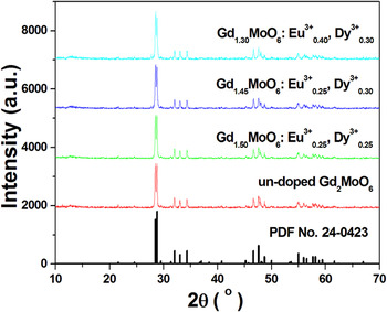
All the prepared phosphors were measured and analyzed by XRD. Figure 1 shows the comparison of the XRD patterns of un-doped Gd2MoO6, Gd1.50MoO6:Eu0.25 3+, Dy0.25 3+, Gd1.45MoO6:Eu0.25 3+, Dy0.30 3+, and Gd1.30MoO6:Eu0.40 3+, Dy0.30 3+ with PDF no. 24–0423. The XRD pattern of un-doped Gd2MoO6 is consistent with PDF no. 24–0423. Thus it can be indexed to Gd2MoO6 with single phase. With the introduction of Eu3+ and Dy3+, the XRD patterns can be still indexed to Gd2MoO6 with single phase when the total doping concentration of Eu3+ and Dy3+ is no more than 0.50 (x + y ≤ 0.50). When the total doping concentration is more than 0.50 (x + y > 0.50), diffraction peaks corresponding to impurity phase appears (especially for the peak at about 27.74°).
XRD patterns of un-doped Gd2MoO6 and several typical Eu3+ and Dy3+ doped samples.

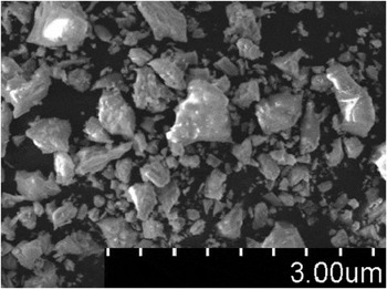
It is well known that the particle size can significantly affect emission intensity of phosphors. The general rule is that sample with larger particle size presents higher emission intensity. To explore the effect of particle size on the luminescent intensity, prepared samples were observed with a SEM. The SEM image of a typical Eu3+ and Dy3+ co-doped sample is shown in Fig. 2. From Fig. 2, it is observed that the particle size is inhomogeneous within ranges 0.5–4.0 um.
SEM image of Gd1.60MoO6:Eu0.25 3+,Dy0.15 3+ (with the optimal doping content).

B. Luminescence properties
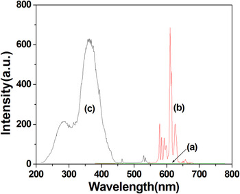
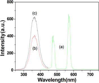
Figure 3(a) shows the emission spectrum of un-doped Gd2MoO6 under the excitation with 365 nm UV light. From Fig. 3(a), it is observed that un-doped Gd2MoO6 does not emit any visible light. However, when Eu3+ is introduced into host lattice (Gd1.75MoO6:Eu0.25 3+), some emission lines appear in the spectral range from 570 to 640 nm [Fig. 3(b)]. The stronger lines from 570 to 640 nm can be attributed to the transitions from excited state 5D0 to ground states 7F0 (579, 585 nm), 7F1 (592, 598 nm), 7F2 (611, 615, 626 nm), and 7F3 (658 nm), respectively. Reference Lei, Yan and Chen29–Reference Li, Wang, Quan, Liu, Yu, Wang and Lin31 In this emission spectrum, just the characteristic transition lines from the lowest excited 5D0 level of Eu3+ are observed. However, those from higher energy levels (5D1, 5D2, 5D3) of Eu3+ can’t be detected by our fluorescence spectrometer. The predominance of the emission lines from 5D0 to 7F J (J = 0, 1, 2, 3) might be attributed to the coupling of the vibration energy of the host lattice and the gaps between the higher energy levels (5D1 or 5D2) and the 5D0 level of Eu3+. Reference Lei, Yan and Chen29–Reference Li, Wang, Quan, Liu, Yu, Wang and Lin31 They are quenched by the multi-phonon relaxation.
Emission spectrum under 365 nm excitation (a) for Gd2MoO6 and (b) Gd1.75MoO6:Eu0.25 3+, excitation spectra monitoring the emission at 611 nm (c) for Gd1.75MoO6:Eu0.25 3+.

As is well-known, Eu3+ 5D0–7F2 emission belongs to hypersensitive transition with ΔJ = 2, which is strongly influenced by outside surroundings and can be served as a very efficient and sensitive structural probe. Reference Lei, Yan and Chen29–Reference Li, Wang, Quan, Liu, Yu, Wang and Lin31 If Eu3+ occupies a site in the crystal lattice with inversion symmetry, the magnetic dipole transition 5D0 → 7F1 of Eu3+ dominates; if there is no inversion symmetry at the site of Eu3+, the main emission is the electric dipole transition 5D0 → 7F2. The crystalline Gd2MoO6 belongs to the monoclinic phase with space group I2/a (15) whose structure is similar to that of the sheelite-type compound Bi2MoO6. Additionally, all the cations in this structure are eight-coordinated, and the anions are four-coordinated. Since the Gd coordination environment is less regular due to the wide variations in the O–Gd–O bond angles and length in Gd2MoO6, Reference Pan, Zhang, Zhao and Jiang32,Reference Chen, Wang, Liu, Kuang and Su33 it is expected that the Gd3+ ions are located at sites without inversion symmetry. The radii of the involved ions are as follows: R(Gd8 3+) = 1.053 nm, R(Mo8 6+) = 0.73 nm, R(Eu8 3+) = 1.066 nm, R(Dy8 3+) = 1.027 nm, Reference Shannon34 the subscript here denotes the coordination number (CN) of the cation. According to the difference of radii among these ions and the ratio of raw materials, it is assumed that most of Eu3+ and Dy3+ replace Gd3+. From Fig. 3, the red emission from transition 5D0 → 7F2 is dominant and there is nearly five times of orange emission from transition 5D0 → 7F1. This indicates that Eu3+ replaces Gd3+ and occupies a site in the crystal lattice without inversion symmetry in Gd2MoO6.
The excitation spectrum monitoring the emission at 611 nm (5D0 → 7F2) [Fig. 3(c)] clearly indicates strong and broad bands from 220 to 450 nm with a maximum at 365 nm and several excitation peaks located at 463, 530, and 535 nm. Sharp lines at 463 nm (7F0 → 5D2), 530 nm (7F0 → 5D1), and 535 nm (7F1 → 5D1) are due to the f–f transitions of Eu3+. The attribution of the broad excitation bands from 220 to 450 nm will be discussed later comparing with the counterpart of Dy3+ doped samples.
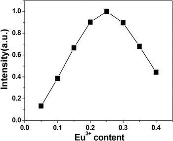
Figure 4 shows the relation between the integral emission intensity of 5D0 → 7F2 transition and Eu3+ content. As shown in Fig. 4, when the Eu3+ content increases, the emission intensity increases, and it reaches to maximum at x = 0.25, which was taken as the optimum concentration. When x > 0.25, emission intensity presents a clear tendency to saturation which suggests that at higher Eu3+ contents some quenching of the Eu3+ luminescence occurs.
Relation between the integral intensity of 5D0 → 7F2 transition and Eu3+ content.

Figure 5 shows the emission (a) and excitation spectra (b) and (c) of Dy3+ singly doped sample Gd1.85MoO6:Dy0.15. Two groups of emission peaks near 477 and 569 nm with 365 nm excitation are observed in Fig. 5(a), which can be attributed to the transitions from excited state 4F9/2 to ground states 6H15/2 and 6H13/2, respectively. Similar observation in Ca9Bi(PO4)7 Reference Zhang, Song, Yue, Zhong, Zhang, Ma and Liu35 and YVO4 Reference Foka, Dejene and Swart36 has also been reported. Just like Eu3+, the 4F9/2 → 6H15/2 transition is mainly magnetically allowed and hardly varies with the crystal field strength around the Dy3+ ion. The 4F9/2 → 6H13/2 transition belongs to a hypersensitive transition with ΔJ = 2, which is a forced electric dipole transition being allowed only at low symmetry with no inversion center. Reference Zhang, Song, Yue, Zhong, Zhang, Ma and Liu35,Reference Foka, Dejene and Swart36 When Dy3+ is located at low-symmetry local site (without an inversion center), this emission is often prominent in the emission spectrum. From the emission spectrum of Dy3+, it is observed that the yellow emission is obviously stronger that blue one. This proves that Dy3+ replace Gd3+ and occupy the site without inversion symmetry.
(a) Emission spectrum excited with 365 nm and excitation spectrum monitoring 477 (b) and 569 nm (c) emission of Dy3+ singly doped sample Gd1.85MoO6:Dy0.15.

Figure 5(b) and 5(c) shows the excitation spectra monitoring the emission at 477 and 569 nm, respectively. Generally, there exist some weak excitation lines from 290 to 400 nm (which can be attributed to the transitions among 4f energy levels of Dy3+ in the excitation spectra of Dy3+ singly doped phosphors without host absorption). However, in the excitation spectra of Gd1.85MoO6:Dy0.15 there is only a strong broad band from 300 to 450 nm peaking at about 365 nm.
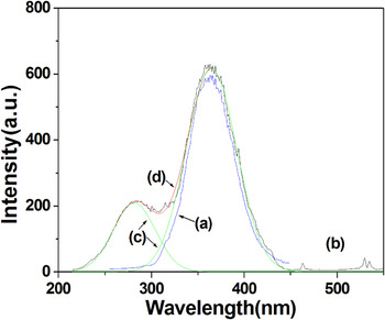
Figure 6 shows the excitation spectra of Gd1.85MoO6:Dy0.15 (monitoring the emission at 569 nm) and Gd1.75MoO6:Eu0.25 3+ (monitoring the emission at 611 nm). The excitation spectrum of Gd1.85MoO6:Dy0.15 is symmetric peaking at 365 nm. The excitation spectrum of Gd1.75MoO6:Eu0.25 3+ can be fitted well with two Gaussian bands peaking at 268 nm and 365 nm, respectively. It is worth noting that the intrinsic excitation of Eu3+ and Dy3+ might be annihilated by the strong broad band for their weak excitation intensity, thus they cannot be observed clearly in Figs. 3, 5, and 6. Generally speaking, there exists a charge transfer band of Eu3+ in the excitation spectrum of Eu3+ singly doped phosphors which peaks at from 250 to 300 nm. Reference Hao, Zhang, Zhang and Wang37–Reference Wang and Wang39 Thus, the former fitted broad band can be attributed to the charge transfer band of Eu3+. The latter fitted broad band peaking at 365 nm is very similar to the excitation broad band in Dy3+ singly doped sample. Obviously, both can be attributed to the host absorption of Gd2MoO6. The presentence of the strong Gd2MoO6 host absorption band in the excitation spectra of Eu3+ or Dy3+ doped samples indicates that there exists effective energy transfer from Gd2MoO6 host to the doped Eu3+ or Dy3+. From Fig. 3(a), un-doped Gd2MoO6 does not emit any visible light under the excitation of 365 nm UV light. This indicates that the host Gd2MoO6 can absorb near ultraviolet light around 365 nm and transfer absorbed energy to doped Eu3+ and Dy3+ effectively. This transferring introduces a broad excitation band in NUV spectral region. It is very advantageous to the combination of prepared phosphors with NUV LEDs.
Excitation spectra of Gd1.85MoO6:Dy0.15 monitoring the emission at 569 nm [(a), expressed in blue] and Gd1.75MoO6:Eu0.25 3+ monitoring the emission at 611 nm [(b), expressed in black], green lines (c) show two fitted Gaussian broad bands of the excitation of Gd1.75MoO6:Eu0.25 3+ and red line (d) shows its fitting curve.

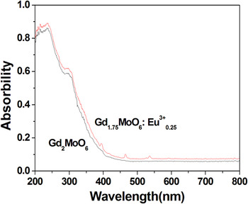
To prove that there exist host absorption in Gd2MoO6, the absorption spectra of Gd2MoO6 and Gd1.75MoO6:Eu0.25 3+ were measured and listed in Fig. 7. It is found that both Gd2MoO6 and Gd1.75MoO6:Eu0.25 3+ show a similar strong absorption band between 200 and 400 nm. This indicates that these absorption bands are not caused by Eu3+ but are mainly due to the host absorption of Gd2MoO6. Reference Pang, Liu and Lin40,Reference Neeraj, Kijima and Cheetham41 However, under UV excitation, the un-doped Gd2MoO6 does not show luminescence. This can be explained as follows. The excitation energy readily migrates through MoO6 6− groups to a quenching site (e.g., an impurity randomly located in the crystal lattice) and dissipates nonradiatively. Owing to the strong coupling between the electrons and the vibrations of the MoO6 6− center, and the relatively small Stokes shift in the MoO6 6− emission, the thermally activated energy migration is so efficient that the host Gd2MoO6 essentially does not show any visible emission at room temperature. When Eu3+ or Dy3+ is incorporated into Gd2MoO6, they function as activators and introduce a characteristic emission through an efficient energy transfer from the MoO6 6− groups to Eu3+ or Dy3+.
Absorption spectra of Gd2MoO6 and Gd1.75MoO6:Eu0.25 3+.

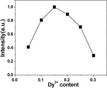
Figure 8 shows the relation between the integral emission intensity of Dy3+ and Dy3+ content. As shown in Fig. 8, while the Dy3+ content increases, the emission intensity increases, and it reaches to maximum at y = 0.15. When y > 0.15, there is a clear tendency to saturation for the emission intensity of Dy3+, which suggests that at higher Dy3+ contents some quenching of the Dy3+ luminescence occurs.
Dependence of integral emission intensity of Dy3+ on its doping content.

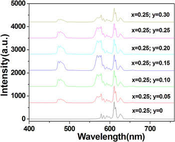
From Figs. 3 and 5, it can be observed that there is a large overlap between the excitation spectra of Eu3+ and Dy3+ in Gd2MoO6. In fact, overlapping parts are derived from the energy transfer from Gd2MoO6 to doped rare earth ions. Since the host Gd2MoO6 can transfer its absorbed energy to both Eu3+ and Dy3+, it is natural to co-doping Eu3+ and Dy3+ into Gd2MoO6. Figure 9 shows the excitation and emission spectra of Eu3+ and Dy3+ co-doped samples (x = 0.25; y = 0, 0.05, 0.10, 0.15, 0.25, and 0.30) under 365 nm excitation. From Fig. 9, except for the Eu3+ singly doped sample, all the emission spectrum includes three groups of emission lines. The former two groups of emission lines are located from 460 to 490 nm and from 560 to 590 nm with the strongest peaks at 477 and 569 nm, respectively. The latter group of emission lines is located from 570 to 640 nm with the strongest peaks at 611 nm. Apparently, the former two groups of emission lines come from doped Dy3+ and the latter from doped Eu3+. From Fig. 9, it can also be observed that with the introduction of Dy3+, the emission intensity of Eu3+ presents a slight decrement.
Emission spectrum excited with 365 nm of Eu3+ and Dy3+ co-doped samples Gd2−x−y MoO6:Eu x 3+,Dy y 3+ (x = 0.25; y = 0, 0.05, 0.10, 0.15, 0.25 and 0.30).

Eu3+ and Dy3+ co-doped samples can be excited by 365 nm UV light and emission blue, yellow and red light spontaneously. In general, blue and yellow light from Dy3+ emission can form white emission. However, this white emission presents a high color temperature for the short of red emission. However, in Eu3+ and Dy3+ co-doped samples, this shortcoming is improved by the co-doping of Eu3+. Thus, it is hoped that white emission from Eu3+ and Dy3+ co-doped samples presents better luminescence performance.
C. CIE analysis
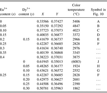
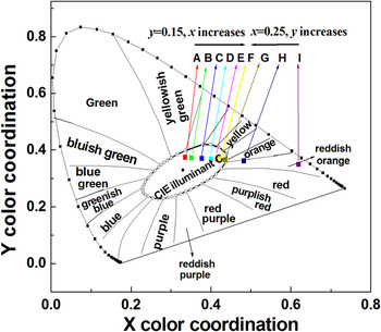
Emission spectra of co-doped samples with different Eu3+ and Dy3+ contents indicate that the emission ratio of blue, yellow, and red can be tuned conveniently by the doping contents of Dy3+ and Eu3+. The CIE 1931 chromaticity coordinates of the phosphors and color temperatures are calculated and listed in Table I. To show the luminescence performance more clearly, corresponding data dots are labeled in Fig. 10.
CIE chromaticity coordinates, color temperature and symbol in Fig. 10 of phosphors.

CIE 1931 chromaticity coordinates of the prepared phosphors.

From Table I and Fig. 10, when Dy3+ content is kept at y = 0.15, all the data points are located in white light region. However, color temperature of white light emission decreases remarkably with the increment of red emission content. It decreases from 5406 to 2878 °C (in this step, the red emission content increases), then increases from 2878 to 3875 °C (in this step, the red emission content decreases). This indicates that even if there is no Eu3+ in phosphors, Dy3+ can emit white light. However, this white light presents a high color temperature (cold white). The introduction of Eu3+ emission can remarkably decrease the color temperature of the white light emission (warm white).
When Eu3+ content is kept at 0.25 with a lower Dy3+ content (y = 0, 0.05), data points are located in red light region. Data point come into white light region with the increment of Dy3+ content (y = 0.10, 0.15). Thus, white light emission is mainly attributed to doped Dy3+. The function of Eu3+ doping is to decrease the color temperature of this white emission.
D. Thermal stability
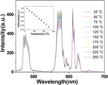
The thermal stability is one of the important technological parameters for phosphors used in white LEDs. The temperature-dependent luminescent properties of the phosphors Gd1.60MoO6:Eu3+ 0.25,Dy3+ 0.15 were measured at temperature in the range of 25–250 °C, as illustrated in Fig. 11. The emission intensity of the phosphors decreases as the temperature rises. This can be explained by the thermal quenching at configurational coordinate diagram. Reference Kim, Park, Kim, Choi and Park42 With increasing temperature, the nonradiative transition probability by thermal activation and release of the luminescent center through the crossing point between the excited state and the ground state increases. The nonradiative transition quenches the luminescence. However, the positions of emission peaks remain unchanged. At 150 °C, the emission intensity of the sample remains 83% of that measured at 25 °C, indicating that the phosphor has very low thermal quenching.
Temperature dependence of emission spectra of sample Gd1.6MoO6:Eu0.25 3+,Dy0.15 3+. The inset shows the temperature dependence of integral intensity of sample Gd1.6MoO6:Eu0.25 3+,Dy0.15 3+.

IV. CONCLUSIONS
A series of Eu3+ and Dy3+ doped Gd2MoO6 were synthesized with solid state reactions. The luminescence properties of Eu3+ and Dy3+ and the energy transfer from the host Gd2MoO6 to Eu3+ and Dy3+ were investigated. Eu3+ in Gd2MoO6 emit one group of emission peaks at about 611 nm under the excitation of ultraviolet light (365 nm) which is attributed to the transition from excited states 5D0 to ground state 7F2. Dy3+ in Gd2MoO6 emit two groups of emission lines near 477 and 569 nm respectively which are attributed to the transitions from excited state 4F9/2 to ground states 6H15/2 and 6H13/2. The co-doping of Eu3+ and Dy3+ into Gd2MoO6 yields the tri-color light at blue, yellow, and red region spontaneously. The remarkable enhancement of Eu3+ and Dy3+ emission proves the occurrence of effective energy transfer from the host to Eu3+ and Dy3+. Dy3+ singly doped phosphors can emit white light with a higher color temperature. The introduction of Eu3+ can remarkably decrease the color temperature of white light emission from cold white to warm light. At 150 °C, the emission intensity of the sample Gd1.60MoO6:Eu0.25 3+,Dy0.15 3+ remains 83% of that measured at 25 °C, indicating that the phosphor has very low thermal quenching.
AUTHORS’ NOTE
This work was done from the second half of 2015 to the first half of 2016. The manuscript was initially submitted to Journal of Materials Research on June 8th, 2015. Before we submit the revised manuscript, a paper about this material has been recently published in Journal of Materials Research. Reference Dutta and Sharma43 Here, we declare that this paper is an independent work. Some results involved in both papers are basically same. Two papers also have their own independent content. There is no essential conflict between the contents of the two papers.
ACKNOWLEDGMENTS
This work was financially supported by the Natural Science Foundation of Guangdong Province (Nos. S2013040013904).