Introduction
AD pathogenesis is multifaceted and difficult to pinpoint. Current genetic and cell biology research have led to the amyloid hypothesis, which posits that the beta-amyloid peptide (Aβ) plays a pivotal role in AD (Hardy and Selkoe, Reference Hardy and Selkoe2002). Aβ derives from the concerted action of BACE1 (β-secretase) and the γ-secretase complex on the cleavage of amyloid precursor protein (APP). In healthy subjects, the predominant route of APP processing via non-amyloidogenic pathway consists of successive cleavages by α and γ-secretases in the middle of Aβ region, thus releasing sAPPα – a structure with neurotrophic and neuroprotective functions (Bandyopadhyay et al., Reference Bandyopadhyay, Goldstein, Lahiri and Rogers2007). Several enzymes in the “a disintegrin and metalloproteinase” (ADAM) family, including ADAM9, ADAM10, and ADAM17, have α-secretase activity in vitro, although recent studies have demonstrated that ADAM10 is the major α-secretase that catalyses APP ectodomain shedding in the brain (Bernstein et al., Reference Bernstein, Steiner, Bogerts, Stricker and Reiser2014). Several studies have demonstrating the effects of serotonin receptors (5-HTRs) on APP processing and Aβ levels, showing that their activation increases non-amyloidogenic processing of APP in vitro (Arjona et al., Reference Arjona, Pooler, Lee and Wurtman2002; Shen et al., Reference Shen2011) and that chronic administration of SSRIs may reduce brain Aβ levels in vivo (Nelson et al., Reference Nelson2007; Cirrito et al., Reference Cirrito2011).
Our research group has been dedicated to the study of the platelet ADAM10 expression as a blood-based biomarker for AD patients. In the previous studies we (Manzine et al., Reference Manzine2013b) and other authors (Colciaghi et al., Reference Colciaghi2002; Reference Colciaghi2004) have reported a marked reduction in platelet ADAM10 protein levels in AD patients compared to cognitively healthy subjects. We have also demonstrated a positive correlation between ADAM10 levels, Mini Mental State Examination (MMSE) and clock drawing test and that ADAM10 levels decrease as the advance of the disease (Manzine et al., Reference Manzine, Barham, Vale Fde, Selistre-de-Araujo, Iost Pavarini and Cominetti2013a; Reference Manzine, Barham, Vale, Selistre-de-Araujo, Pavarini and Cominetti2014). Since ADAM10 is the most important α-secretase involved in cleavage of APP and antidepressant medications are frequently utilized by AD patients, the aim of this research was to investigate whether platelet ADAM10 protein levels would be related with the use of antidepressant drugs in older adults diagnosed with AD.
Methods
Study design and participants
Patients were form a convenience sample, which was recruited in reference (Public Centre of Specialties) and counter-reference (Family Health Units) health services in Brazil. Eligible participants were diagnosed with probable AD according to National Institute of Neurological Disorders and Stroke-Alzheimer Disease and Related Disorders Association (NINCS-ADRDA) criteria (McKhann et al., Reference McKhann, Drachman, Folstein, Katzman, Price and Stadlan1984). This study was carried out with 26 older adults diagnosed with probable AD, divided in two groups. The control group was composed of 13 AD patients, who were not taking any antidepressant drug and the SSRI group, which was formed of 13 AD patients already taking SSRIs medication, in a regular-basis, under medical prescription. Exclusion criteria for participants were: head trauma, metabolic dysfunctions, haematological diseases, alcohol abuse, drug abuse, delirium, mood disorders, and treatment with medications affecting platelet functions, i.e. anticoagulants, antiplatelet drugs, serotoninergic agonists-antagonists (for control group), and corticosteroids. All included participants were taking proper AD medication and had a standardized clinical workup based on neurological examinations, laboratory blood, and urine analysis, a neuroimaging study (Head Computed Tomography and/or Magnetic Resonance Imaging), and a neuropsychological assessment, including a MMSE according to scholarity levels of Brazilian population (Brucki et al., Reference Brucki, Nitrini, Caramelli, Bertolucci and Okamoto2003) and a Clinical Dementia Rating (CDR) (Montano and Ramos, Reference Montano and Ramos2005). Before enrolment, subjects or their legal caregivers filled out an informed consent, after the nature and possible consequences of the study were explained. The research project was approved by Brazil Platform Ethics Committee (CAAE: 02760312.0.0000.5504/ No: 112.543).
ADAM10 quantification
ADAM10 quantification was carried out according to our previous studies (Manzine et al., Reference Manzine2013b). Briefly, blood (8.5 mL) was collected in sodium citrate tubes (3.8% containing 136 mM glucose). The interval between the collection and the processing was 20 to a maximum of 30 min. The platelet-rich plasma (PRP) was obtained by centrifugation at 1,200 rpm for 10 min. From the PRP, platelets were collected by centrifugation at 2,400 rpm for 10 min at room temperature, then washed twice in phosphate buffered saline solution (PBS) and finally suspended in lysis buffer (200 mM NaCl, 10 mM EDTA, 10 mM Na2HPO4, 0.5% NP40, 0.1% SDS, and protease inhibitors). Bradford kit (1:4) (BioRad, Hercules, CA, USA) was used for protein measurement.
The necessary volume to obtain a total of 100 μg of protein for each sample was applied into a 10% SDS-PAGE gel (Laemmli, Reference Laemmli1970). Page-Ruler Prestained (Fermentas, Burlington, CA, USA) was used as a molecular marker in all gels. After the gel running, the proteins were transferred into nitrocellulose membranes (Sigma, St. Louis, MO, USA) for the period of 1 hour using the Mini Trans-Blot Cell transfer system (Bio-Rad, Hercules, CA, USA). The membranes were incubated with anti-ADAM10 mouse monoclonal primary antibody (Santa Cruz Biotechnology, Santa Cruz, CA, USA), followed by incubation with goat anti-mouse IgG-HRP secondary antibody (Santa Cruz Biotechnology). After these procedures, the membranes were developed using Clarity™ Western CCL Substrate (Bio-Rad). The antibody anti-β-actin conjugated to HRP (Santa Cruz Biotechnology) was used to detect β-actin protein, which was the endogenous control. The immunoreaction bands were quantified with ChemiDoc™ MP Imaging System (Bio-Rad).
Statistical analysis
A blind analysis of the participants was used for the ADAM10 sample quantification. In order to detect the sample normality level this study employed the Kolmogorov–Smirnov test (KS). Student's t test was used between groups and non-parametric Mann–Whitney U test was employed among subgroups. The figures were made using GraphPad Prism 5 (GraphPad Software Inc, La Jolla, CA, USA) software. A 5% significance level was chosen as standard.
Results
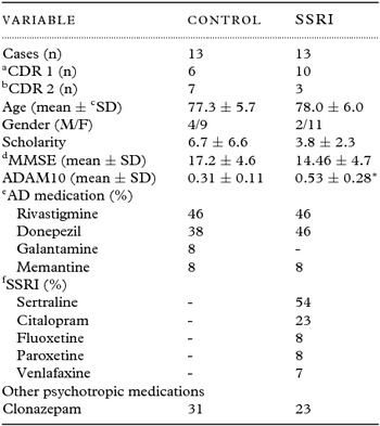
A total of 26 AD patients took part in this study. Socio demographic, clinical and cognitive data for all participants of this study are presented on Table 1. Patients enrolled in this study were all taking different types and dosages of acetylcholinesterase inhibitors (AChEIs). In both groups (control and SSRI) the most used AChEI (46%) was rivastigmine with a dosage ranging from 6–12 mg/day. The second most used AChEI (38% and 46%, for control and SSRI groups, respectively) was donepezil with dosages ranging from 5–10 mg/day. Memantine, an N-methyl-D-aspartate (NMDA)-receptor antagonist, was used (8% in both groups) at 10–20 mg/day and galantamine (8%, only in control group) at 16–24 mg/day. Among the SSRIs used in this study we found that 54% were sertraline (50 mg-100 mg/day); 23% citalopram (20–40 mg/day); 8% fluoxetine (20–40 mg/day); 8% paroxetine (20–40 mg/day), and 7% venlafaxine (75 mg/day) (Table 1).
Patients’ demographic and clinical variables

aMild AD (CDR1), bModerate AD (CDR2), cStandard Deviation, dMini-Mental State Examination, eAlzheimer's Disease medication, fSelective Serotonin Reuptake Inhibitors. *p < 0.001 compared to Control, multiple t test.
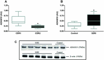
Blood was collected and analyzed for platelet ADAM10 expression in control and SSRI groups. Patients in more advanced stages of the disease (according to the CDR scores) had their ADAM10 platelet expression significantly reduced, confirming our previous results (Figure 1A). ADAM10 platelet levels were significant higher in patients taking SSRI medication, compared to controls (Table 1, Figure 1B). The ADAM10 expression measured by western blotting analysis and normalized by the endogenous control β-actin is presented on Figure 1C.
SSRI antidepressants positively influences ADAM10 expression. Platelet ADAM10 expression in AD patients according to CDR (A), control AD patients (B) or patients taking SSRI medication (B). Representative western blotting membranes showing platelet ADAM10 expression in five patients from SSRI, and in two patients from the control group (C). Platelets were collected, lysed and the protein content was applied to a 10% polyacrylamide gel and then transferred to a nitrocellulose membrane followed by the incubation with anti-ADAM10 or anti-β-actin antibodies. The bands were developed with the Clarity™ Western CCL Substrate (Bio-Rad) and then scanned for the measure of the reactive bands. Graph represents quantification of the ADAM10 normalized to β-actin bands; *p < 0.001, Mann–Whitney test.

Discussion
Alpha-secretase-mediated cleavage of APP releases the neuroprotective sAPPα fragment and prevents βA formation (Postina, Reference Postina2012). ADAM10 and ADAM17 are considered the main α-secretases involved in non-amyloidogenic pathway in AD (Fahrenholz, Reference Fahrenholz2010). Our previous studies had demonstrated that platelet ADAM10 levels were reduced in AD patients compared with cognitively healthy subjects (Manzine et al., Reference Manzine2013b) and that this reduction had a significant positive correlation with MMSE (Manzine et al., Reference Manzine, Barham, Vale Fde, Selistre-de-Araujo, Iost Pavarini and Cominetti2013a) and clock drawing (Manzine et al., Reference Manzine, Barham, Vale, Selistre-de-Araujo, Pavarini and Cominetti2014) tests. However, we also recently showed that this reduction was not resulting from differences in ADAM10 mRNA levels, suggesting that post-transcriptional or trafficking mechanisms could play a role in the regulation of ADAM10 expression (Manzine et al., Reference Manzine2015). Here we observed the same results, showing that with the advance of the disease, according to CDR, platelet ADAM10 expression is significantly decreased in AD patients. However, the platelet ADAM10 levels were increased in AD patients using SSRIs as antidepressant medication, compared with the control group.
Recent evidence suggests a strong relationship between depression and AD. A lifetime history of depression has been considered a risk factor for later development of AD and depressive symptoms can positively affect the conversion of mild cognitive impairment (MCI) into AD. In addition, neuritic plaques and neurofibrillary tangles are more pronounced in the brains of depressed AD patients, compared to non-depressed ones (Caraci et al., Reference Caraci, Copani, Nicoletti and Drago2010; Chi et al., Reference Chi, Wang, Jiang, Zhu, Yu and Tan2015).
Cochet and co-authors (Cochet et al., Reference Cochet2013) have demonstrated that 5-HTRs constitutively induce APP cleavage by ADAM10 with concomitant release of neuroprotective sAPPα both in HEK-293 cells and cortical neurons. Moreover, Cirrito and co-workers (Cirrito et al., Reference Cirrito2011) demonstrated that brain Aβ levels were significantly decreased following the administration of several SSRIs antidepressant drugs or the direct infusion of 5-HT in the brain of PS1APP transgenic mice, with the involvement of extracellular regulated kinase (ERK) signaling pathway, as the treatment with ERK inhibitors reversed the Aβ clearance. These authors also demonstrated an increased ADAM10 α-secretase enzymatic activity in antidepressant-treated mice.
The link between AD and depression could be related to 5-HT neurotransmitter and its receptor. One of the molecular mechanisms of depression is known to be the increased clearance of 5-HT from the synaptic cleft. SSRIs act inhibiting 5-HT reuptake, increasing its synaptic concentration and availability. It was demonstrated that G protein-coupled receptors (GPCRs) can affect Aβ peptide production by either modulating the cellular trafficking of APP or by influencing the activity and trafficking of α-, β-, and γ-secretases. Among GPCRs, 5-HTRs are transmembrane receptors that interact with ADAM10 and enhance sAPPα production by stimulating α-secretase activity (Thathiah and De Strooper, Reference Thathiah and Strooper2011). Moreover, activation of 5-HTRs stimulates acetylcholine release in prefrontal cortex and hippocampus and improves learning and memory in various preclinical paradigms of memory acquisition and retention (Bockaert et al., Reference Bockaert, Claeysen, Compan and Dumuis2011). These findings suggest that 5-HTR agonists like SSRIs might be used to improve cholinergic function and cognition, which may both be compromised in AD. In fact, it was already demonstrated that in AD patients treated with acetylcholinesterase inhibitors (AChEIs), SSRIs may exert some protection against the negative effects of depression on cognition (Rozzini et al., Reference Rozzini2010). On the other hand, there are reports showing that AChEIs activate specific subtypes of muscarinic acetylcholine receptors (mAChRs) that inhibit sAPPα release and aggravate amyloid-β generation (Thathiah and De Strooper, Reference Thathiah and Strooper2011). Our results emphasize the former hypothesis, showing that there is one plausible mechanism that could be related to the influence of these molecules on ADAM10 expression. It is important to mention that all AD patients in our study were using AChEIs. The exact molecular mechanisms underlying this positive effect of SSRIs on ADAM10 expression remain to be elucidated.
In this study the prevalence of patient's CDR was different in each group (control and SSRI), therefore, we analyzed the ADAM10 levels in both groups, classified for CDR scores. We observed that, for CDR1 subjects, ADAM10 levels were higher in SSRI, compared to control subjects. This was not true for CDR2 subjects, where no differences were found for ADAM10 levels between control and SSRI patients (data not shown). This raises an important question: are the SSRI effects on ADAM10 protein levels only effective for patients in mild stages (CDR1) of the disease? One of the limitations that avoid an immediate answer for this is the small number of the participants in this study, which does not allow subject matching for aging, sex, scholarity, or CDR levels. The lack of a measure for ADAM10 activity is also a limitation, since western blotting analysis only shows the protein quantity and not its activity, although usually these variables are positively related. Moreover, the effects of other antidepressant medications, such as MAO inhibitors or tricyclic drugs on ADAM10 expression were not analyzed in this study and could also be considered as a study limitation.
Conclusion
The results from this study provide relevant clinical implications by showing that serotonergic antidepressants positively influence the α-secretase of ADAM10 platelet expression in patients with AD. Studies on the molecular mechanisms of how platelet ADAM10 expression is positively influenced by SSRIs and whether this expression is reflected in the central nervous system should be carried out using a larger number of patients, and may represent an important step for the study of AD and for its more effective pharmacological treatment.
Conflicts of interest
None.
Description of authors’ roles
O.A.F.M. Bianco had contributed to data collection, P.R. Manzine and C.M.C. Nascimento had substantially contributed to data collection, analysis and interpretation of data, S.C.I. Pavarini and F.A.C. Vale substantially contributed to critically revising the paper with important intellectual content; and M.R. Cominetti contributed to drafting the paper and revising it critically for important intellectual content. All authors have given the final approval of the version to be published.
Acknowledgments
The authors thank all the subjects and their families. The authors are grateful for the financial support of Sao Paulo Research Foundation (FAPESP) grants 2010/09497-7 and 2013/06879-4. O.A.F.M. Bianco, P.R. Manzine, and C.M.C Nascimento have scholarships sponsored by FAPESP (grants 2012/08654-7, 2012/01936-7, and 2014/21066-2, respectively).