




No CrossRef data available.
Article contents
Can Copper-Coated Surfaces Prevent Healthcare-Associated Infections?
Published online by Cambridge University Press: 19 June 2017
Abstract
An abstract is not available for this content. As you have access to this content, full HTML content is provided on this page. A PDF of this content is also available in through the ‘Save PDF’ action button.
Information
- Type
- Commentary
- Information
- Copyright
- © 2017 by The Society for Healthcare Epidemiology of America. All rights reserved
References
REFERENCES
1.
Magill, SS, Edwards, JR, Bamberg, W, et al. Multistate point-prevalence survey of health care-associated infections. N Engl J Med
2014;370:1198–1220.Google Scholar
2.
Weinstein, RA. Epidemiology and control of nosocomial infections in adult intensive care units. Am J Med
1991;91:S179–S184.CrossRefGoogle ScholarPubMed
3.
Boyce, JM. Environmental contamination makes an important contribution to hospital infection. J Hosp Infect
2007;65(Suppl 2):50–54.Google Scholar
4.
Weber, DJ, Rutala, WA, Miller, MB, Huslage, K, Sickbert-Bennett, E. Role of hospital surfaces in the transmission of emerging health care-associated pathogens: norovirus, Clostridium difficile, and Acinetobacter species. Am J Infect Control
2010;38(5 Suppl 1):S25–S33.CrossRefGoogle ScholarPubMed
5.
Weber, DJ, Anderson, D, Rutala, WA. The role of the surface environment in healthcare-associated infections. Curr Opin Infect Dis
2013;26:338–344.CrossRefGoogle ScholarPubMed
6.
Otter, JA, Yezli, S, Salkeld, JA, French, GL. Evidence that contaminated surfaces contribute to the transmission of hospital pathogens and an overview of strategies to address contaminated surfaces in hospital settings. Am J Infect Control
2013;41(5 Suppl):S6–S11.Google Scholar
7.
Carling, PC. Optimizing health care environmental hygiene. Infect Dis Clin North Am
2016;30:639–660.CrossRefGoogle ScholarPubMed
8.
Donskey, CJ. Does improving surface cleaning and disinfection reduce health care-associated infections?
Am J Infect Control
2013;41(5 Suppl):S12–S19.CrossRefGoogle ScholarPubMed
9.
Otter, JA, Yezli, S, Perl, TM, Barbut, F, French, GL. The role of ‘no-touch’ automated room disinfection systems in infection prevention and control. J Hosp Infect
2013;83:1–13.CrossRefGoogle ScholarPubMed
10.
Doll, M, Morgan, DJ, Anderson, D, Bearman, G. Touchless technologies for decontamination in the hospital: a review of hydrogen peroxide and UV devices. Curr Infect Dis Rep
2015;17:498.Google Scholar
11.
Rutala, WA, Weber, DJ. Disinfection and sterilization in health care facilities: an overview and current Issues. Infect Dis Clin North Am
2016;30:609–637.Google Scholar
12.
Weber, DJ, Kanamori, H, Rutala, WA. ‘No touch’ technologies for environmental decontamination: focus on ultraviolet devices and hydrogen peroxide systems. Curr Opin Infect Dis
2016;29:424–433.Google Scholar
13.
Weber, DJ, Rutala, WA, Anderson, DJ, Chen, LF, Sickbert-Bennett, EE, Boyce, JM. Effectiveness of ultraviolet devices and hydrogen peroxide systems for terminal room decontamination: focus on clinical trials. Am J Infect Control
2016;44(5 Suppl):e77–e84.CrossRefGoogle ScholarPubMed
14.
Boyce, JM. Modern technologies for improving cleaning and disinfection of environmental surfaces in hospitals. Antimicrob Resist Infect Control
2016;5:10.Google Scholar
15.
Boyce, JM, Havill, NL, Otter, JA, et al. Impact of hydrogen peroxide vapor room decontamination on Clostridium difficile environmental contamination and transmission in a healthcare setting. Infect Control Hosp Epidemiol
2008;29(8):723–729.Google Scholar
16.
Vianna, PG, Dale, CR Jr, Simmons, S, Stibich, M, Licitra, CM. Impact of pulsed xenon ultraviolet light on hospital-acquired infection rates in a community hospital. Am J Infect Control
2016;44:299–303.Google Scholar
17.
Pegues, DA, Han, J, Gilmar, C, McDonnell, B, Gaynes, S. Impact of ultraviolet germicidal irradiation for no-touch terminal room disinfection on Clostridium difficile infection incidence among hematology-oncology -patients. Infect Control Hosp Epidemiol
2017;38:39–44.CrossRefGoogle ScholarPubMed
18.
Anderson, DJ, Chen, LF, Weber, DJ, et al. Enhanced terminal room disinfection and acquisition and infection caused by multidrug-resistant organisms and Clostridium difficile (the Benefits of Enhanced Terminal Room Disinfection study): a cluster-randomised, multicentre, crossover study. Lancet
2017;389:805–881.CrossRefGoogle ScholarPubMed
19.
Kundrapu, S, Sunkesula, V, Jury, LA, Sitzlar, BM, Donskey, CJ. Daily disinfection of high-touch surfaces in isolation rooms to reduce contamination of healthcare workers’ hands. Infect Control Hosp Epidemiol
2012;33:1039–1042.Google Scholar
20.
Weber, DJ, Rutala, WA. Self-disinfecting surfaces: review of current methodologies and future prospects. Am J Infect Control
2013;41(5 Suppl):S31–S35.Google Scholar
21.
Humphreys, H. Self-disinfecting and microbiocide-impregnated surfaces and fabrics: What potential in interrupting the spread of healthcare-associated infection?
Clin Infect Dis
2014;58:848–853.CrossRefGoogle ScholarPubMed
22.
Muller, MP, MacDougall, C, Lim, M. Antimicrobial surfaces to prevent healthcare-associated infections: a systematic review. J Hosp Infect
2016;92:7–13.Google Scholar
23.
Grass, G, Rensing, C, Solioz, M. Metallic copper as an antimicrobial surface. Appl Environ Microbiol
2011;77:1541–1547.CrossRefGoogle ScholarPubMed
24.
Weaver, L, Michels, HT, Keevil, CW. Survival of Clostridium difficile on copper and steel: futuristic options for hospital hygiene. J Hosp Infect
2008;68:145–151.CrossRefGoogle ScholarPubMed
25.
Souli, M, Antoniadou, A, Katsarolis, I, et al. Reduction of environmental contamination with multidrug-resistant bacteria by copper-alloy coating of surfaces in a highly endemic setting. Infect Control Hosp Epidemiol 2017;38:765–771.Google Scholar
26.
O’Gorman, J, Humphreys, H. Application of copper to prevent and control infection. Where are we now?
J Hosp Infect
2012;81:217–223.Google Scholar
27.
Hinsa-Leasure, SM, Nartey, Q, Vaverka, J, Schmidt, MG. Copper alloy surfaces sustain terminal cleaning levels in a rural hospital. Am J Infect Control
2016;44:e195–e20.CrossRefGoogle Scholar
28.
Schmidt, MG, von Dessauer, B, Benavente, C, et al. Copper surfaces are associated with significantly lower concentrations of bacteria on selected surfaces within a pediatric intensive care unit. Am J Infect Control
2016;44:203–209.Google Scholar
29.
von Dessauer, B, Navarrete, MS, Benadof, D, Benavente, C, Schmidt, MG. Potential effectiveness of copper surfaces in reducing health care-associated infection rates in a pediatric intensive and intermediate care unit: a nonrandomized controlled trial. Am J Infect Control
2016;44:e133–e139.Google Scholar
30.
Sifri, CD, Burke, GH, Enfield, KB. Reduced health care-associated infections in an acute care community hospital using a combination of self-disinfecting copper-impregnated composite hard surfaces and linens. Am J Infect Control
2016;44:1565–1571.Google Scholar
31.
Salgado, CD, Sepkowitz, KA, John, JF, et al. Copper surfaces reduce the rate of healthcare-acquired infections in the intensive care unit. Infect Control Hosp Epidemiol
2013;34:479–486.CrossRefGoogle ScholarPubMed
32.
Rivero, P, Brenner, P, Nercelles, P. Impact of copper in the reduction of hospital-acquired infections, mortality and antimicrobial costs in the Adult Intensive Care Unit. [In Spanish]. Rev Chilena Infectol
2014;31:274–279.Google Scholar
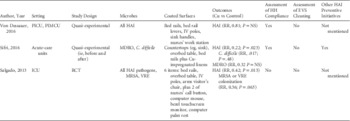
TABLE 1 Results of Selected Clinical Trials of Copper-Coated Versus Control Surfaces to Reduce Healthcare-Associated Infections
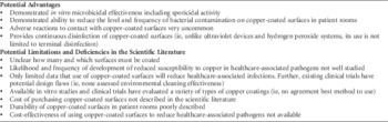
TABLE 2 Advantages and Limitations in the Use of Copper as a Self-Disinfecting Surface in Hospital Rooms
 You have
Access
- 8
- Cited by
(See the article by Souli et al, on pages 765–771.)
Despite dramatic decreases in the incidence of healthcare-associated infections (HAIs) in recent years, a large prevalence study of US acute-care hospitals estimated that ~722,000 HAIs occurred in 2011, resulting in ~75,000 deaths.Reference Magill, Edwards and Bamberg 1 Several decades ago, WeinsteinReference Weinstein 2 theorized that pathogens causing HAIs in the intensive care unit (ICU) had several sources: the patients’ endogenous flora (40%–60%), cross-infection via the hands of healthcare personnel (HCP; 20%–40%), antibiotic-driven changes in flora (20%–25%), and other causes (including contamination of the environment; 20%).Reference Weinstein 2 More recently, accumulating scientific evidence has indicated that contamination of environmental surfaces in hospital rooms plays an important role in the transmission of several key healthcare-associated pathogens, including methicillin-resistant Staphylococcus aureus (MRSA), vancomycin-resistant Enterococcus spp (VRE), Clostridium difficile, Acinetobacter spp, and norovirus.Reference Boyce 3 – Reference Otter, Yezli, Salkeld and French 6 In general, all of these pathogens share the following characteristics: ability to survive for prolonged periods of time on environmental surfaces, ability to remain virulent after environmental exposure, frequent contamination of the hospital environment, ability to colonize patients, ability to contaminate or transiently colonize the hands of HCP, and transmission via the contaminated hands of HCP.Reference Boyce 3 – Reference Otter, Yezli, Salkeld and French 6 Evidence supporting the role of contaminated surfaces in the transmission of key healthcare-associated pathogens includes the following observations. (1) The surfaces in rooms of colonized or infected patients are frequently contaminated with the pathogen.Reference Boyce 3 – Reference Otter, Yezli, Salkeld and French 6 (2) Contact with hospital-room surfaces or medical equipment by HCP frequently leads to contamination of hands and/or gloves.Reference Otter, Yezli, Salkeld and French 6 (3) The patient admitted to a room previously occupied by a patient colonized or infected with a pathogen (eg, MRSA, VRE, C. difficile, Acinetobacter) has a substantially increased likelihood (ie, 39%–353%) of developing colonization or infection with that pathogen.Reference Otter, Yezli, Salkeld and French 6 (4) Improved terminal cleaning of rooms decreases environmental pathogensReference Carling 7 and the rate of individual patient colonization and/or infections; it also leads to a decreased facility-wide rate of colonization and/or infection.Reference Donskey 8
Unfortunately, inadequate terminal room disinfection is common.Reference Carling 7 In fact, studies have demonstrated that <50% of room surfaces are cleaned.Reference Carling 7 Therefore, and not surprisingly, many room surfaces remain contaminated with a multidrug-resistant organisms (MDROs) following terminal room disinfection.Reference Weber, Anderson and Rutala 5 , Reference Otter, Yezli, Salkeld and French 6 Although enhanced education of environmental service staff with feedback regarding cleaning effectiveness (eg, fluorescent dye) improves cleaning effectiveness, overall cleaning remains suboptimal.Reference Carling 7 For this reason, “no-touch” methods of terminal room disinfection have been developed.Reference Otter, Yezli, Perl, Barbut and French 9 – Reference Boyce 14 No-touch methods that use ultraviolet (UV) light devices or hydrogen peroxide systems for terminal room disinfection when the occupant has been colonized or infected with a MDRO have been demonstrated to reduce the risk of the subsequent room occupant developing colonization and/or an HAI.Reference Rutala and Weber 11 , Reference Weber, Rutala, Anderson, Chen, Sickbert-Bennett and Boyce 13 Importantly, some studies have demonstrated a facility-wide reduction of HAIsReference Boyce, Havill and Otter 15 , Reference Vianna, Dale, Simmons, Stibich and Licitra 16 ; furthermore, these reductions were not related to improved hand hygiene or to environmental cleaning compliance.Reference Pegues, Han, Gilmar, McDonnell and Gaynes 17 , Reference Anderson, Chen and Weber 18
One of the limitations of currently available UV devices or hydrogen peroxide systems is that they can only be used for terminal disinfection because patients and HCP must evacuate the room prior to use. Because daily room disinfection has been shown to reduce contamination of HCP hands and gloves,Reference Kundrapu, Sunkesula, Jury, Sitzlar and Donskey 19 there has been interest in developing “no-touch” methods for decreasing MDRO surface contamination on a continuous basis.Reference Otter, Yezli, Perl, Barbut and French 9 Such methods have been reviewed and include self-disinfecting surfaces using heavy metals, light-activated photosensitizers, high-intensity narrow-spectrum light, low-dose continuous hydrogen peroxide, photocatalytic disinfection, bacteriophage-modified surfaces, and altered surface topography.Reference Weber, Kanamori and Rutala 12 , Reference Boyce 14 , Reference Weber and Rutala 20 – Reference Muller, MacDougall and Lim 22 Of these potential methods, the best studied is the use of heavy metals (eg, silver, copper) with copper-coated or impregnated surfaces; it is the only method that has been assessed in clinical trials.Reference Weber and Rutala 20 – Reference Muller, MacDougall and Lim 22
The studies assessing the effectiveness of no-touch methods including self-disinfecting surfaces for disinfection of room surfaces in hospitals can be divided into 3 general approaches. (1) the effectiveness of the self-disinfecting surface to kill or inhibit microbes artificially placed on the self-disinfecting surface, (2) the level of contamination on self-disinfecting surfaces in actual patient rooms compared to a “control” surface and (3) most importantly, clinical trials of a self-disinfecting surface to reduce HAIs.
Copper is an essential trace element in most living organisms, and >30 types of copper-containing proteins have been described.Reference Weber and Rutala 20 The use of copper by humans dates back to the fifth and sixth millennia bc.Reference Grass, Rensing and Solioz 23 Ancient civilizations used copper or copper compounds to prevent the growth of barnacles on the hulls of ships and as a medicinal agent.Reference Weber and Rutala 20 In the 19th and 20th centuries, inorganic copper preparations were used to treat chronic adenitis, eczema, impetigo, tuberculous infections, and syphilis.Reference Grass, Rensing and Solioz 23 The cause of bacterial cell death to exposure to copper may relate to its ability to accept and donate single electrons leading to the generation of reactive oxygen species, resulting is cell lysis.Reference Boyce 14 , Reference Weber and Rutala 20 , Reference Humphreys 21 , Reference Grass, Rensing and Solioz 23 In healthcare facilities, the most well-studied use of copper has been as a “self-disinfecting” surface, but many other uses have been studied, including copper-containing paints, fabrics, hand rubs, microfiber cleaning cloths, pens, and fins within air-conditioning units.Reference Weber and Rutala 20
Multiple studies have assessed the contact killing of microbes by copper surfaces.Reference Grass, Rensing and Solioz 23 Contact with copper has been demonstrated to kill a variety of healthcare-associated pathogens including Staphylococcus aureus (including methicillin-resistant S. aureus), Enterococcus spp, Escherichia coli, Klebsiella pneumoniae, Acinetobacter baumannii, Pseudomonas aeruginosa, and Mycobacterium tuberculosis. Reference Weber and Rutala 20 In general, contact killing of vegetative bacteria and fungi occurs rapidly with >6-log10 inactivation per hour. Spores (eg, C. difficile) are inactivated more slowly, but >3-log10 are inactivated within 24 hours.Reference Grass, Rensing and Solioz 23 , Reference Weaver, Michels and Keevil 24
In this issue of Infection Control and Hospital Epidemiology, Souli et alReference Souli, Antoniadou and Katsarolis 25 report on the frequency and level of contamination of multiple copper-coated surfaces (ie, patient beds, side table, IV pole, side cart handles, and manual antiseptic dispenser) compared to control surfaces in a patient room in an intensive care unit.Reference Souli, Antoniadou and Katsarolis 25 The study was performed in a Greek ICU setting in which evidence showed that the environment was considerably more contaminated than other settings and where highly resistant organisms predominated. For example, 62% of K. pneumoniae isolated from the environment were resistant to carbapenems and 100% were multidrug resistant; similarly, 98% of A. baumannii were resistant to carbapenems and 99% were multidrug resistant. Their study was conducted in 2 phases: In phase 1, enhanced environmental sampling was performed to identify the most microbiologically contaminated items to ‘copperize’; this targeted use of copper items is an important strength of the study. Phase 2 was the intervention phase; in phase 2a, copper-coated objects were placed next to uncoated objects (controls); and in phase 2b, cooper-coated objects were all placed in a single ICU compartment and non–copper-coated objects were placed in a different ICU compartment (controls). Combining the data for phases 2a and 2b revealed that copper-coated surfaces were less likely to be microbially contaminated (ie, 55.6% vs 72.5% for controls, P<.0001). Copper-coated surfaces were significantly less likely to be contaminated with gram-negative bacilli or Enterococcus spp, but no statistical reduction for A. baumannii, K. pneumoniae, or S. aureus was observed. Overall, the level of contamination was reduced by ~0.7-log10 (P=.049). Importantly, no significant reduction was observed in phase 2a, in which copper items were placed next to uncoated objects, suggesting a possible interaction between the number of copper items and the level of reduction, which requires further investigation. The statistical approach used did not take into account the full range of variables; most importantly, indicators of hand-hygiene compliance and patient-level variables could have been included in a model with copper items as a separate variable. Souli et al reported no difference in hand-hygiene compliance during the study, but the effectiveness of environmental clean/disinfection was not assessed. Other limitations include the use of alcohol spray for daily disinfection of surfaces, which is an unusual approach and may limit the generalizability of the study; lack of randomization of patients to beds with copper items; and the lack of clinical and cost-effectiveness outcomes.
Multiple studies of copper-coated surfaces or devices have been conducted in the healthcare setting comparing the level and frequency of surface contamination to control surfaces (for a review of studies, see Weber and Rutala,Reference Weber and Rutala 20 Humphreys,Reference Humphreys 21 Muller et al,Reference Muller, MacDougall and Lim 22 Weaver et al,Reference Weaver, Michels and Keevil 24 and O’Gorman and HumphreysReference O’Gorman and Humphreys 26 ). Studies have either used concurrent non–copper-coated control surfaces or a crossover design. Muller et alReference Muller, MacDougall and Lim 22 reported that 5 of 7 copper studies of copper-coated surfaces or fabrics reviewed demonstrated a significant reduction in quantitative bacterial contamination of <1-log10 (range, <1-log10 to 2-log10). Limitations of the studies noted by Muller et alReference Muller, MacDougall and Lim 22 included lack of blinding, lack of randomization, and in general, failure to measure potential confounding factors (eg, hand hygiene compliance, effectiveness of cleaning by environmental services) or reported any patient-level data. Similar to previous studies, 2 more recent studies also demonstrated significant reductions of microbial contamination on copper-coated surfaces compared to control surfaces of 1- to 2-log10.Reference Hinsa-Leasure, Nartey, Vaverka and Schmidt 27 , Reference Schmidt, von Dessauer and Benavente 28
In addition, 3 clinical trials have been published in the English literature that assessed the effectiveness of cooper-coated surfaces to reduce HAIs (Table 1).Reference Grass, Rensing and Solioz 23 – Reference Salgado, Sepkowitz and John 31 Of these 3 trials, 2 reported that the intervention arm (ie, copper) had a decreased incidence of HAIs,Reference Sifri, Burke and Enfield 30 , Reference Salgado, Sepkowitz and John 31 and 1 trial reported no significant reduction in HAIs.Reference von Dessauer, Navarrete, Benadof, Benavente and Schmidt 29 Importantly, none of the trials assessed the effectiveness of environmental cleaning and disinfection using fluorescent dye or ATPase measurements, and 1 did not assess compliance with hand hygiene.Reference Salgado, Sepkowitz and John 31 Although Salgado et alReference Salgado, Sepkowitz and John 31 described their trial as a randomized clinical trial, Muller et alReference Muller, MacDougall and Lim 22 described this study as “low-quality evidence due to inappropriate randomization and lack of blinding.” An additional clinical trial has been published in the Spanish literature; it reported no differences in the frequency of nosocomial infections as well as no difference in the rates of ventilator-associated pneumonia, catheter-associated urinary tract infections, or central line-associated bloodstream infections.Reference Rivero, Brenner and Nercelles 32 However, this study was limited by its failure to achieve its intended sample size.
Results of Selected Clinical Trials of Copper-Coated Versus Control Surfaces to Reduce Healthcare-Associated Infections
NOTE. Cu, copper; HH, hand hygiene; EVS, environmental service; HAI, healthcare-associated; RR, relative risk; PICU, pediatric intensive care unit; PIMCU, pediatric intermediate care unit; IV, intravenous; NS, not significant; MRSA, methicillin-resistant Staphylococcus aureus; VRE, vancomycin-resistant Enterococcus spp; RCT, randomized clinical trial.
The use of copper-coated surfaces to decrease contamination of hospital-room surfaces to decrease HAIs has several potential advantages over current technologies, but it also has several limitations, and several scientific questions remain unanswered (Table 2). Copper-coated surfaces have demonstrated the ability to inactivate many key pathogens associated with HAIs. Multiple studies have demonstrated that copper-coated surfaces can reduce the level of contamination on room surfaces by 1- to 2-log10. Importantly, unlike the “no-touch” methods that use UV devices or hydrogen peroxide systems (which currently can only be used for terminal disinfection), copper-coated surface provide continuous disinfection. To date, clinical trials have reported only rare adverse reactions related to contact with a copper-coated surface.
Advantages and Limitations in the Use of Copper as a Self-Disinfecting Surface in Hospital Rooms
However, currently available studies still leave many questions regarding the routine use of copper-coated surfaces in hospital rooms. First, it is unclear how many and which surfaces must be coated to achieve a reduction in HAIs. Second, the types of copper-coating that have been studied vary, and widely accepted standard for such surfaces do not exist. Third, only a limited number of clinical trials have been published that have assessed whether copper-coated surfaces reduce the incidence of HAIs, and among published studies, copper-coated surfaces have not uniformly been shown to reduce HAIs. Furthermore, the published studies have design flaws including failure to use a randomized design and failure to account for all possible confounding variables (eg, hand-hygiene compliance and cleaning effectiveness). Fourth, current studies investigating microbial reductions associated with copper-coated surfaces or clinical outcomes (ie, reduction of HAIs) have rarely assessed whether residual microbes had intrinsic or acquired resistance to copper. Survival on copper surfaces has been described and appears to be the consequence of endospore formation, survival on patches of dirt, or a special ability to endure a dry metallic copper surface.Reference Grass, Rensing and Solioz 23 The frequency and importance of reduced susceptibility to copper among healthcare-associated pathogens remains poorly defined but likely will not be clinically significant. Fifth, the cost of purchasing, installing, and maintaining copper-coated surfaces has not been delineated. Sixth, the durability of copper-coated surfaces has been poorly described. Finally, no cost-effective analysis of the use of copper-coated surfaces has been published.
In conclusion, the use of copper-coated surfaces to provide continuous room disinfection is promising, but additional carefully designed and appropriately powered studies are required before this technology should be routinely adopted to prevent HAIs.
ACKNOWLEDGMENTS
Potential conflicts of interest: J.A.O. is a consultant to Gama Healthcare. D.J.W. and W.A.R. are consultants for PDI Healthcare.
Financial support: J.A.O. would like to acknowledge support from the National Institute for Health Research Health Protection Research Unit (NIHR HPRU) in Healthcare-Associated Infection and Antimicrobial Resistance at Imperial College London in partnership with Public Health England (PHE), and the Imperial College Healthcare Trust NIHR Biomedical Research Centre (BRC).