Isolation of a subclavian artery is an uncommon congenital anomaly of the aortic arch in which one subclavian artery loses its connection with the aorta and originates from the homolateral pulmonary artery by way of a ductus arteriosus. Reference Stewart, Kincaid and Edwards1
Isolation of the subclavian artery, when present, is usually observed on the contralateral side of aortic arch sidedness. The involvement of the left subclavian artery is reported to be four times more frequently than the right, and is associated with intracardiac or aortic arch anomalies. Reference Stewart, Kincaid and Titus2 More than half of the reported cases of isolated left subclavian artery are associated with significant cardiac malformation. Reference Kutsche and Van Mierop3
The origin of the subclavian artery from the pulmonary artery is a rare anomaly of the aortic arch. It is defined as a loss of continuity between the subclavian artery and the aorta, with a persistent connection to the homolateral pulmonary artery through a patent ductus arteriosus. It is mostly associated with intracardiac or aortic arch anomalies. Reference Penkoske, Castaneda, Fyler and Van Praagh4,Reference Johnson, Watson, Strauss and Spray5
According to Edwards’ hypothetical double aortic arch model, isolated right subclavian artery with left aortic arch would be explained by interruption of the right aortic arch at two locations; between the right common carotid and the right subclavian arteries and between the right ductus arteriosus and the right dorsal aortic root. Reference Edwards6
We report the case of a child with an isolated right subclavian artery in a left aortic arch with a ventricular septal defect.
Case report
An asymptomatic 2-year-old-girl, who was referred to our hospital because of the presence of cardiac a murmur. On examination, the patient was active with no dysmorphic features. Vital signs are: blood pressure of 112/59, oxygen saturation of 97% on room air, heart rate of 98 beats per minute. The chest examination was clear; the cardiovascular exam showed normal first and second heart sounds with a pansystolic murmur, grade 3/6 with maximum intensity at the left lower sternal border. Abdomen was soft and lax with no hepatosplenomegaly. The right brachial pulse was faint and weaker than the left. A chest X-ray showed normal cardiac size with no congestion.
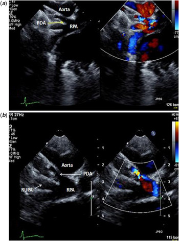
Echocardiography revealed an isolated right subclavian artery in a left aortic arch with a moderate-sized posterior muscular ventricular septal defect. The diagnosis was suspected when on a left aortic arch first branch did not bifurcate. The right subclavian artery was not seen originating from the aorta; isolated right subclavian artery was suspected when a right-sided ductus arteriosus was seen draining to the right pulmonary artery. Neither a left-sided ductus arteriosus nor an aortic coarctation was present (Fig 1).
( a and b ) A 2-D transthoracic echocardiography showed a right-sided patent ductus arteriosus (PDA) draining to the right pulmonary artery (RPA). RUPA = Right upper pulmonary artery.

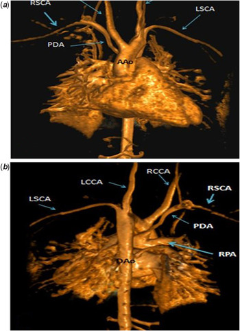
Cardiac MRI showed a left-sided aortic arch with an early take off of the right innominate artery. The vertebral left common carotid and left subclavian arteries were all seen originating normally. The right subclavian artery was anomalously originating from the main pulmonary artery connected via a right patent ductus arteriosus, completely isolated from the aortic arch. Distal aortopulmonary collateral was seen (Fig 2a and b). The patient was booked for surgery to ligate patent ductus arteriosus and reroute the right subclavian artery.
( a and b ) Cardiac Magnetic resonance imaging (CMRI) showed an isolated right subclavian artery (RSCA) connected to the right pulmonary artery branch via a right-sided patent ductus arteriosus (PDA). LSCA = Left subclavian artery; LCCA = Left common carotid artery; RCCA = Right common carotid artery.

Discussion
Isolation of the right subclavian artery is far less common than isolation of the left subclavian artery. The isolated artery is usually supplied from the vertebral artery; this reverse flow is responsible for stealing blood from cerebral circulation with the exercise of the arm and has been linked to dizziness and even syncope in adults. However, these symptoms are uncommon in children. When the ductus arteriosus is patent, steal to the pulmonary circulation is dependent on pulmonary arterial pressure and arteriolar resistance. If pulmonary arterial pressures are high due to an associated left-to-right shunting lesion (a ventricular septal defect in our patient); steal from the cerebral circulation would be unlikely; moreover, the arm will be perfused predominantly from the pulmonary circulation resulting in desaturation in the arm. Reference Mittal, Agarwal and Ghosh7,Reference Duncan, Freedom, Olley and Rowe8
The diagnosis of anomalous origin of right subclavian artery from the right pulmonary artery, which is connected via patent ductus arteriosus can be achieved by careful echocardiography. Other imaging modalities such as cine angiography, or magnetic resonance angiography have also been utilised. Reference Katayama, Shimizu, Tanaka, Narabayashi and Tamai9
Few cases were reported in the literature with different associations of intracardiac defects. We report a rare case of isolation of the right subclavian artery with an association of ventricular septal defect where the diagnosis was made by echocardiography and confirmed by MRI.
Acknowledgements
I would like to thank King Faisal Specialist Hospital and Research Center Staff, especially, Dr Ziad R. Bulbul (Senior Pediatric Cardiologist) for his bright editing of the manuscript.
Financial support
This research received no specific grant from any funding agency, commercial, or not-for-profit sectors.
Conflicts of interest
The author declares that there is no conflict of interest.