Introduction
It is well known that proper expression of the developmental genes is a key feature to determine embryo quality during the in vitro preimplantation period. During embryonic development, the embryo undergoes several morphogenetic events such as oocyte maturation, fertilization, the transition from the maternal to the embryonic genome, i.e. embryonic genome activation (EGA), embryo compaction, and blastocyst formation (Wrenzycki et al., Reference Wrenzycki, Herrmann and Niemann2007). Embryonic development is characterized by a minor and a major form of EGA, during which several developmentally important candidate genes are expressed in a stage-specific manner (Niemann and Wrenzycki, Reference Niemann and Wrenzycki2000). Therefore, the proper regulation of gene expression during such morphogenetic events is an important aspect for obtaining good quality blastocysts and for the maintenance of successful pregnancy (Watson et al., Reference Watson, Natale and Barcroft2004).
There are ∼7400 genes that are involved in the onset of embryonic transcription in bovine (Graf et al., Reference Graf, Krebs, Heininen-Brown, Zakhartchenko, Blum and Wolf2014). One of the important developmental genes is the glucose transporter-1 (GLUT1), which is associated with glucose uptake, transportation and metabolism in the blastocyst. Similarly, Connexin 43 (CX43) is the most abundant gap junctional protein, which is expressed in various tissues of mammalian species. Thermotolerance and resistance to protein denaturation is induced by heat shock proteins (HSPs). HSPs are the first proteins produced during embryonic development. HSP70.1 protein provides thermoprotection by refolding damaged proteins and protecting rRNA (Nover and Scharf, Reference Nover and Scharf1997). Another developmental gene is poly(A) polymerase, which is responsible for mRNA synthesis in developing embryos. The poly(A) tail is found at the 3′-ends of nearly all eukaryotic mRNAs, and is responsible for the initiation of translation and regulation of mRNA breakdown. Synthesis of the poly(A) tail is carried out by poly(A) polymerase (Wähle, Reference Wähle1991; Brevini-Gandolfi et al., Reference Brevini-Gandolfi, Favetta, Mauri, Luciano, Cillo and Gandolfi1999).
The other developmental important candidate genes are BMP15 (bone morphogenetic protein 15) and GDF9 (growth and differentiation factor 9), which are members of the transforming growth factor-beta (TGFβ) superfamily. The BMP15 and GDF9 transcripts are expressed in mammalian ovaries and oocytes, and therefore regulate folliculogenesis, ovulation and corpus luteum function (Shimasaki et al., Reference Shimasaki, Moore, Otsuka and Erickson2004; Bahire et al., Reference Bahire, Rajput, Kumar, Kumar, Kataria and Kumar2019; Kumar et al., Reference Kumar, Rajput, Bahire, Jyotsana, Kumar and Kumar2020). Interestingly, Survivin is an anti-apoptotic protein, which is expressed in all preimplantation stages of embryos and provides a role in the protection of embryos from apoptosis by inhibiting the apoptotic pathway through caspase activity (Kawamura et al., Reference Kawamura, Sato, Fukuda, Kodama, Kumagai, Tanikawa, Nakamura, Honda, Sato and Tanaka2003).
The expression of the developmentally important candidate genes has been examined in the oocytes and embryos of cattle, sheep and goats as species elsewhere. However, very limited studies have been carried out on the expression of certain pluripotency-associated genes in oocytes and embryos of buffalo (Kumar and Chauhan, Reference Kumar and Chauhan2021). Furthermore, scientific literature on the expression of the major developmental important candidate genes in different grades/stages of oocytes and embryos are missing in buffalo. Therefore, the present study was undertaken to analyze the relative abundance (RA) of the major developmental important candidate gene transcripts in different grades of immature oocytes and various stages of in vitro fertilized (IVF) produced buffalo embryos.
Materials and methods
Collection, aspiration and classification of buffalo oocytes
Buffalo ovaries were collected from the Delhi abattoir and these ovaries were washed three or four times with antibiotic-fortified (400 IU/ml penicillin and 500 μg/ml streptomycin) isotonic normal saline (32–37°C). The washed ovaries were transported to the laboratory within 6 h of slaughter. Immature oocytes were aspirated from surface follicles (2–8 mm diameter) of the buffalo ovary (Madan et al., Reference Madan, Das and Palta1996; Shahid et al., Reference Shahid, Jalalib, Khanc and Shamid2014) in aspiration medium (TCM-199 + 0.3% BSA + 0.0001 g/ml l-glutamine + 50 μg/ml gentamicin sulfate) with the help of an 18–19-gauge needle. The oocytes were searched and washed with washing medium [TCM-199 + 10% fetal bovine serum (FBS) + 0.81 mM sodium pyruvate + 0.0001 g/ml l-glutamine + 50 μg/ml gentamicin sulfate]. Finally, the aspirated immature oocytes were graded according to the following criteria. A-grade: compact cumulus–oocyte complexes (COCs) with an unexpanded cumulus mass having ≥5 layers of cumulus cells with homogenous evenly granular ooplasm. B-grade: oocytes with 2–4 layers of cumulus cells with homogeneous evenly granular ooplasm. C-grade: oocytes partially or wholly denuded or with expanded or scattered cumulus cells or with an irregular and dark ooplasm. D-grade: oocytes with deformed or irregular ooplasm without the cumulus cells. These different grades of immature oocytes were used to analyze expression of the developmentally important candidate genes.
In vitro maturation (IVM) of oocytes, in vitro fertilization (IVF), and in vitro culture (IVC) of embryos
The immature oocytes of buffalo were washed five or six times with washing medium and twice with IVM medium (TCM-199 + 10% FBS + 5 μg/ml porcine FSH + 1 µg/ml estradiol-17β + 0.81 mM sodium pyruvate + 0.0001 g/ml l-glutamine + 5% buffalo follicular fluid + 50 μg/ml gentamicin sulfate). Next ∼20 A-grade or B-grade immature oocytes were cultured in 100-μl droplets of IVM medium overlaid with sterile mineral oil in 35-mm Petri dishes and cultured for 24 h in a humidified CO2 incubator (5% CO2 in air) at 38.5°C (after removal of the mature oocytes from the IVM droplets, these droplets were kept at 38.5°C in a CO2 incubator to grow the shaded cumulus or granulosa cells on the surface of the droplets). These matured oocytes were assessed for cumulus cell expansion and then in vitro fertilized with proven frozen bull semen. Spermatozoan preparation was conducted in Brackett and Oliphant (BO) medium as described by earlier workers (Chauhan et al., Reference Chauhan, Singla, Palta, Manik and Madan1998). IVF was conducted in 50-µl droplets of BO medium for which 50 µl of processed semen was added in each droplet (∼1,50,000/- sperm per droplet) containing the mature oocytes. These droplets were overlaid with mineral oil and placed at 38.5°C in a CO2 incubator (5% CO2 in air) for 18 h.
At the end of sperm–oocyte incubation, the whole content was transferred to an Eppendorf tube and the cumulus cells attached to the mature oocytes were removed by gentle pipetting/vortexing. The presumptive fertilized oocytes/embryos were collected in 35-mm Petri dishes and washed in IVC medium [modified Charles Rosenkrans medium with amino acids (mCR2aa) + 0.6% BSA fraction V]. Embryos in the above IVM droplets were then cultured in vitro in IVC medium for 48 h post-fertilization (IVM droplets that had a bed of cumulus or granulosa cells which were chosen for COC maturation). After 48 h, 100 µl of IVC medium (mCR2aa + 0.6% BSA fraction V + 10% FBS) were replaced in the IVM droplets of the presumptive embryo culture. These embryos were cultured in the same droplets until day 7 post IVF in a humidified CO2 incubator at 38.5°C. The medium was replaced with 50% fresh IVC medium (mCR2aa + 0.6% BSA fraction V + 10% FBS) at 48 h intervals until day 7. Several trials were conducted to obtain the various stages of embryos, i.e. 2-cell, 4-cell, 8–16-cell, morula and blastocyst to analyze the expression of the developmental genes. Two-cell-stage embryos were collected at day 1 post IVF, 4-cell embryos on day 2, 8–16-cell embryos on day 3, morula on day 4 and blastocyst on day 8.
RNA isolation and cDNA preparation
Total RNA was isolated from different grades of immature oocytes and various stages of IVF-produced embryos using TRIzol reagent (Invitrogen). In total 10 immature oocytes of each grade (A-grade, B-grade, C-grade and D-grade) were taken for RNA isolation. The 2-cell (n = 90), 4-cell (n = 340), 8–16-cell (n = 205), morula (n = 135) and blastocyst (n = 129) stages of embryos were collected. The various stages of embryos were collected in different trials and pooled separately for isolation of the RNA. Here, ∼200 µl TRIzol reagent was used for the isolation of RNA for oocytes, while 500 µl TRIzol was used for each grade of embryos following the manufacturer’s instructions. The dried RNA pellet was dissolved in 10 µl (for immature oocytes) and 20 µl (for embryos) DEPC water. RNA was treated with 1 µl DNase I (Fermentas) at 37°C for 30 min to remove DNA contamination. DNase I was inactivated using DNase inactivation reagent (Ambion). The concentration of RNA samples was determined using a NanoQuant reader (Tecan). The concentration of total RNA of each sample (i.e. each grade of oocytes and each stage of embryos measured separately) was equalized for preparation of cDNA. Finally, 5 µl of total RNA (50 ng) from each sample was used to prepare cDNA, then was run on 0.8% agarose gels to check RNA integrity. cDNA was prepared using a first-strand cDNA synthesis kit (RevertAID, Fermentas). Finally, 5 µl (10 ng/µl) of total RNA was used for cDNA preparation. In the above sample, 1 µl oligo(dT) (supplied with the kit) and 6 µl DEPC water were mixed. The samples were incubated at 65°C for 5 min followed by 4 µl of 5× RT buffer, 1 µl of Ribolock (RNase inhibitor, 20 U/µl), 2 µl of 10 mM dNTPs mix, and 1 µl of MMLV (reverse transcriptase, 200 U/µl) added in each tube. Negative RT (all components except reverse transcriptase) was taken to assess DNA contamination in cDNA synthesis. cDNA quality was checked by readings for the optical density (OD) 260/280 wavelength ratio and the quantity of cDNA was calculated using a NanoQuant reader (Tecan).
Standardization of PCR for developmental important candidate genes
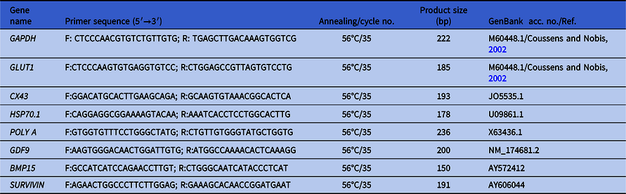
Primers were designed based on published sequences of mouse, human, sheep, goat, pig, cattle and buffalo using Primer3 software. Expression of the developmental genes [GLUT1, CX43, HSP70.1, poly(A) polymerase (POLY A), GDF9, BMP15, and Survivin] was analyzed in immature oocytes and the embryos using reverse transcription PCR (RT-PCR). Negative RT and negative PCR control were taken to confirm the authenticity of PCR amplification. PCR primer sequences, annealing temperature and their product sizes are given in Table 1.
Primer sequences for the developmental important candidate genes

Relative abundance of developmentally important candidate genes in buffalo oocytes and embryos
Reverse transcription qPCR (RT-qPCR) was performed on the CFX 96 system (Bio-Rad) using SYBR green, i.e. Light Cycler 480 SYBR Green I Master Mix (2×) (Roche Diagnostics) and Maxima SYBR green/fluorescein qPCR Master Mix (2×) (Fermentas). Both types of SYBR green mix were added in equal volume and used in whole RT-qPCR experiments. Here, 2.0 µl of cDNA (200 ng/µl), 5.0 µl of 2× SYBR green mix, and 0.2 µl of each forward and reverse primer were added and completed to a volume of 10 µl with nuclease-free water. The reaction was prepared in triplicate with two no-template controls (NTC) for each gene. The amplification conditions for all the genes were as follows: initial denaturation at 95°C for 5 min, followed by 45 cycles of denaturation at 95°C for 10 s, annealing X°C (X for each gene is given in Table 1) for 25 s and extension at 72°C for 25 s. A melting curve was plotted from 65 to 95°C with 0.5°C increments for 0.05 min. Fold difference in mRNA transcript for the target genes was calculated using the equation 2−ΔΔCt, by assuming the PCR amplification efficiency was two (Livak and Schmittgen, Reference Livak and Schmittgen2001).
Statistical analysis
Data were analyzed using SYSTAT 7.0 software (SPSS Inc. Chicago, IL, USA). Data were grouped and presented as mean ± SE and subjected to analysis of variance. Pairwise comparison of means was carried out using the least significance difference test in SYSTAT 7.0. Differences were considered significant at P-values < 0.05.
Results
In the present study, the RA of the developmental important candidate genes (GLUT1, CX43, HSP70.1, poly(A) polymerase, GDF9, BMP15 and Survivin) was studied in immature oocytes and embryos using RT-qPCR.
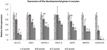
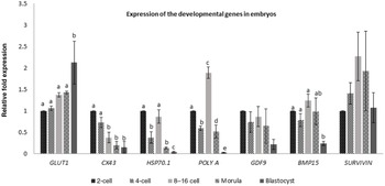
The RA of the GLUT1 transcript was significantly higher (P < 0.05) in A-grade than in C-grade and D-grade oocytes, but the value did not differ significantly from the B-grade oocytes (Fig. 1). Expression of GLUT1 was significantly higher (P < 0.05) in the blastocyst than in the other stages of embryos (Fig. 2). Similarly, RA of the CX43 transcript was higher in the A-grade oocytes than in the other grades of oocytes (Fig. 1). Expression of the CX43 transcript was almost similar in A- and B-grades of oocytes, but this was significantly different (P < 0.05) from the C-grade and D-grade oocytes. The expression of CX43 was detected in all stages of embryos but its expression was poorly detected in the blastocyst (Fig. 2). The RA of the HSP70.1 transcript was significantly higher (P < 0.05) in A-grade than the C-grade and D-grade oocytes (Fig. 1), but did not differ significantly from the B-grade of oocytes. It was found that HSP70.1 expression was higher in the 2-cell stage, but was significantly (P < 0.05) reduced in the 4-cell, morula and blastocyst stages, except for the 8–16-cell stage of embryos. The RA of HSP70.1 was significantly reduced (P < 0.05) in the blastocyst compared with other stages of embryos (Fig. 2). The RA of the poly(A) polymerase transcript was higher in the A-grade than the other grades of oocytes (Fig. 1), but no significant difference was detected between A-grade and D-grade oocytes. It was found that the RA of the poly(A) polymerase transcript was significantly higher (P < 0.05) in the 8–16-cell stage than in the other stages of embryos (Fig. 2). The RA of the GDF9 transcript was significantly higher (P < 0.05) in the A-grade than the in the C-grade and D-grade oocytes, but its expression was not significantly different from B-grade oocytes (Fig. 1), although its expression was higher in the 2-cell stage than the other stages of embryos (Fig. 2). Similarly, the RA of the BMP15 transcript was significantly higher (P < 0.05) in A-grade than the other grades of the oocyte (Fig. 1). The RA of BMP15 was significantly higher (P < 0.05) in the 8–16-cell stage than in the blastocyst stage, but it was similar in other stages of embryos (Fig. 2). Expression of BMP15 was slightly reduced at the 4-cell stage, but further increased in the 8–16-cell stage, and then reduced in the blastocyst stage (Fig. 2). The RA of the Survivin transcript was significantly higher (P < 0.05) in A-grade than the other grades of oocytes (Fig. 1). It was found that the C-grade and D-grade oocytes had very low Survivin expression. Survivin expression was higher in the 8–16-cell to morula stages, but was reduced in the later stages of development (Fig. 2).
Expression of developmentally important candidate genes [GLUT1, CX43, HSP70.1, poly(A) polymerase (POLY A), GDF9, BMP15, and Survivin] in immature oocytes of buffalo. a,b,cValues with different superscripts on a bar differ significantly (P < 0.05); no significant difference (P > 0.05).

Expression of developmentally important candidate genes [GLUT1, CX43, HSP70.1, poly(A) polymerase (POLY A), GDF9, BMP15, and Survivin] in various stages of buffalo embryos. a,b,cValues with different superscripts on a bar differ significantly (P < 0.05); no significant difference (P > 0.05).

Discussion
In the present study, the RA of developmentally important candidate genes was analyzed in different grades of immature oocytes and various stages of IVF-produced embryos using RT-qPCR. The expression of the GLUT1 transcript was found in all grades of buffalo immature oocytes and embryos. The present results are in line with the findings of other workers who showed that the GLUT1 transcript was expressed in the oocytes and preimplantation embryos to the blastocysts (Rajhans et al., Reference Rajhans, Kumar, Dubey and Sharma2010). The results are in support with the findings of Wrenzycki et al. (Reference Wrenzycki, Herrmann, Carnwath and Niemann1998) in which the hatched blastocysts expressed a higher amount of GLUT1 mRNA transcript compared with immature and mature oocytes, zygotes, 2–4-cell stage, and 8–16-cell stage embryos. Similarly, GLUT1 expression was higher in hatched blastocysts (Sadeesh et al., Reference Sadeesh, Selokar, Balhara and Yadav2016a), but it was reduced in mature oocytes and 2-cell stage embryos during the hot period (Sadeesh et al., Reference Sadeesh, Sikka, Balhara and Balhara2016b). It was indicated that expression of the GLUT1 transcript predominantly occurs in good quality immature oocytes and gradually increases from the 2-cell stage to the blastocyst stage in buffalo (Fig. 2).
Several connexins, which form the gap junctions, have been found in ovarian follicles (Read et al., Reference Read, Willhelm and Dyce2018). In the present study, the RA of the CX43 transcript was higher in A-grade than other grade of oocytes (Fig. 1), whereas expression of the CX43 transcript was very low in the buffalo blastocyst. This indicated that A-grade and B-grade oocytes may have well developed gap junctional proteins, required for the transport of ions and metabolites for several physiological processes. In other words, C-grade and D-grade oocytes may have less numbers of cumulus cell layers. Poor quality oocytes could have less established gap junctional proteins, therefore the developmental competence of C-grade and D-grade oocytes is likely to be small compared with A-grade and B-grade oocytes. The CX43 transcript is expressed in in vivo-developed morula and blastocyst in cattle (Wrenzycki et al., Reference Wrenzycki, Herrmann, Carnwath and Niemann1996). Similarly, it is also expressed in cumulus–oocyte complexes (COCs) to morulae, but not in blastocysts of cattle (Wrenzycki et al., Reference Wrenzycki, Herrmann, Carnwath and Niemann1998). In another study, the CX43 transcript was significantly expressed in cumulus cells of bovine COCs that had a higher developmental competence compared with cumulus cells in COCs that had low developmental competence (Read et al., Reference Read, Willhelm and Dyce2018). Therefore, it was indicated that expression of the CX43 mRNA transcript was predominantly found in good quality oocytes.
In the current study, the HSP70.1 transcript was expressed in all grades of immature oocytes and embryos, similar to findings from the study of Wrenzycki et al. (Reference Wrenzycki, Herrmann, Carnwath and Niemann1998) who reported HSP70.1 expression throughout preimplantation development to the blastocyst stage in cattle. In other studies, HSP70.1 expression was significantly higher in immature oocytes than mature oocytes and zygotes of cattle (Wrenzycki et al., Reference Wrenzycki, Herrmann, Carnwath and Niemann1999), while HSP70.1 expression was upregulated in in vitro-produced blastocysts compared with in vivo-produced embryos (Wrenzycki et al., Reference Wrenzycki, Herrmann, Korsawe, Hadeler and Niemann2000). Similarly, Sharma et al. (Reference Sharma, Nath, Prasad, Singhal, Singh, Gade, Dubey and Saikumar2012) showed a higher expression of HSP70.1 in in vitro-produced buffalo embryos compared with in vivo-derived embryos. In the present study, the RA of HSP70.1 was slightly increased at the 8–16-cell stage compared with other embryo stages (Fig. 2), which suggested that the several developmentally important candidate genes may be upregulated or downregulated during EGA (8–16-cell embryo) (Graf et al., Reference Graf, Krebs, Heininen-Brown, Zakhartchenko, Blum and Wolf2014). Therefore, the 8–16-cell stage of embryos in buffalo might express more HSP70.1 transcript to adjust the internal cellular environment. Expression of the HSP70.1 transcript may change, however, in different culture systems and environmental conditions.
In our study, the poly(A) polymerase transcript was expressed in all grades of immature oocytes and embryos. A similar finding was also documented by Wrenzycki et al. (Reference Wrenzycki, Herrmann, Carnwath and Niemann1998) who found that poly(A) polymerase was expressed in oocytes and preimplantation embryos to the hatched blastocyst in bovine. In the current study, the poly(A) polymerase transcript was higher in A-grade than in the other grades of oocytes, but no significant difference was recorded among other grades of oocyte. These results are similar to the study of Biase et al. (Reference Biase, Fonseca Merighe, Santos Biase, Martelli and Meirelles2008) who found higher expression of poly(A) polymerase in A-grade than B-grade oocytes in bovine. In the present study, the RA of poly(A) polymerase was significantly higher (P < 0.05) in the 8–16-cell stage embryos, while its expression was reduced in morulae and blastocysts. The reason for low poly(A) polymerase abundance in the morula and blastocyst is not clearly understood.
Poly(A) tail shortening of certain transcripts has been detected in the germinal vesicle stage of oocytes and the metaphase II stages of oocytes in bovine (Brevini-Gandolfi et al., Reference Brevini-Gandolfi, Favetta, Mauri, Luciano, Cillo and Gandolfi1999). This group found that the poly(A) tails of some physiological and developmentally associated genes [Connexin-43, glucose transporter type 1, heat shock protein 70, poly(A) polymerase, Oct4 and plakophilin genes] were shortened during oocyte maturation, however expression of the poly(A) tail of β-actin and pyruvate dehydrogenase phosphatase (PDP) did not differ in both types of oocytes. Furthermore, Brevini et al. (Reference Brevini, Lonergan, Cillo, Francisci, Favetta, Fair and Gandolfi2002) analyzed the poly(A) tail of several transcripts in the resumption of the meiosis stage of oocytes and the first cleavage stage of the oocytes in bovine. They found no change in the expression of β-actin and PDP, while gradual reduction and elongation of the other transcripts was observed between the oocytes and cleaved embryos. Poly(A) tail shortening may occur in matured oocytes or gene expression may be variable if we compare the gene expression of mature oocytes with the germinal vesicle stage of oocytes or cleaved embryos. In the present study, we analyzed the mRNA transcripts of several developmentally related genes in immature oocytes and IVF-produced embryos separately. Therefore the issue of poly(A) tail shortening of developmentally related genes may not arise in immature oocytes and would be randomly distributed in developing IVF embryos, if any. It is suggested that poly(A) tail shortening of the development-associated genes may not be the same in buffalo and the cattle; this aspect needs to be investigated in detail.
It is well known that the GDF9 is expressed in the oocytes and granulosa cells (Kumar et al., Reference Kumar, Rajput, Bahire, Jyotsana, Kumar and Kumar2020). In the present study, the GDF9 transcript was expressed in all grades of immature oocytes and embryos. It has been proposed that GDF9 transcripts are differentially expressed in mature oocytes (Kathirvel et al., Reference Kathirvel, Soundian and Kumanan2013), in immature oocytes and in in vitro-produced 5–8-cell embryos, but not at the later stages of development (Pennetier et al., Reference Pennetier, Uzbekova, Perreau, Papillier, Mermillod and Dalbies-Tran2004). The study by Lee et al. (Reference Lee, Kim, Hwang and Hyun2008) showed continuous expression of the GDF9 transcript from preimplantation embryos to the fetuses. In our study, expression of the GDF9 transcript was slightly higher at the 2-cell stage than in other embryo stages (Fig. 2). It is inferred from previous reports that the GDF9 transcript is exclusively expressed in the oocytes. Therefore, expression of the GDF9 transcript gradually decreased from the 2-cell stage to the blastocyst stage in buffalo. Differences in the transcript levels may be due to the developmental competence of the oocytes and the cell culture conditions used to culture oocytes and embryos.
In the present study, expression of the BMP15 transcript was found in all immature oocytes and embryos. These results are in agreement with a previous report that the BMP15 transcript was expressed in the oocyte and preimplantation embryos of cattle (Pennetier et al., Reference Pennetier, Uzbekova, Perreau, Papillier, Mermillod and Dalbies-Tran2004) and the COCs of buffaloes (Mahesh et al., Reference Mahesh, Gibence, Shivaji and Rao2017). The present results are in agreement with another study in which expression of the BMP15 transcript was found in oocytes of cattle (Hosoe et al., Reference Hosoe, Kaneyama, Ushizawa, Hayashi and Takahashi2011). This indicated that BMP15 transcript expression was confined more to oocytes, but its expression was irregular in buffalo embryos. This may be due to the effects of cell culture conditions, environmental surroundings and the developmental competence of the oocytes.
The Survivin transcript was expressed in all immature oocytes and embryos of buffalo. These results were in line with the study by Park et al. (Reference Park, Kim, Jeon, Cui, Lee, Kim, Park and Lim2007) who found the expression of Survivin mRNA and protein in the germinal vesicle, MII stage of oocytes, 1-cell, 2-cell, 4-cell, 8-cell stages, morula and blastocysts of bovine. Similarly, Jeon et al. (Reference Jeon, Kim, Tae, Lee, Lee, Kim, Jeong, Cho, Kim, Lee, Riu, Cho and Park2008) showed expression of Survivin in oocytes and all preimplantation stages of bovine embryos. It has been proposed that the Survivin protein is strongly detected in good quality COCs compared with poor quality COCs, which suggests that the Survivin may be a good marker for embryo quality in bovine (Jeon et al., Reference Jeon, Kim, Tae, Lee, Lee, Kim, Jeong, Cho, Kim, Lee, Riu, Cho and Park2008). In the present study, Survivin transcript expression was significantly higher (P < 0.05) in A-grade than in B-grade, C-grade and D-grade oocytes. Therefore, it was suggested that Survivin may be used as a marker for developmental competence of poor or good quality buffalo oocytes.
The expression patterns of developmentally associated genes are not usually directly associated with the developmental competence of oocytes. It is well known that proper regulation of developmental gene expression is a key feature when determining oocyte quality and its competency. It is assumed that A-grade oocytes contain more numbers of cumulus cell layers, which may have increased expression of the Cx43 transcript, and may offer more cell-to-cell communication to transport the ions and other metabolites required by cells. Therefore, A-grade oocytes would be more developmentally competent than other grades. It is presumed that if a particular developmentally related gene is expressed at normal levels in oocytes, then the oocyte would be adequately competent. But sometimes the temporal pattern of expression of a specific gene may also occur in a cell or tissue stage-specific manner. Therefore, the expression of developmental genes may also be influenced by environmental factors such as cell culture conditions, growth factors and the serum used in the culture medium.
It is concluded that most developmentally important genes are abundantly expressed in A-grade oocytes compared with B-grade, C-grade and D-grade oocytes. Expression of GLUT1 was higher in blastocysts, while the expression levels of CX43, GDF9, and HSP70.1 transcripts were greater in 2-cell stage embryos. Expression levels of poly(A) polymerase, BMP15 and Survivin transcripts were higher at the 8–16-cell stage compared with other stages of buffalo embryos.
Acknowledgements
The authors are highly grateful to the Director, ICAR-NDRI, Karnal, India for providing the necessary facilities for carrying out this study.
Author contributions
Satish Kumar: contributed to all experimental design, embryo culture and molecular work, and manuscript preparation. M.S. Chauhan: overall supervision and management of research funding.
Funding
This work was supported by the Department of Biotechnology, Govt. of India and National Agricultural Innovation Project (NAIP, ICAR) projects granted to M.S. Chauhan (project codes C-2067 and 075).
Conflict of interest
No potential conflict of interest was reported by the authors.
Ethical standards
Not applicable as ovaries were obtained from the abattoir.