Introduction
Anticipation is a basic human cognitive function involving the preparation for future events (Gilbert & Wilson, Reference Gilbert and Wilson2007). As we do not know what the future holds, we prepare for expected events in order to deal with the associated pleasant or unpleasant outcome. To be able to cope, it makes sense to consider the worst-case scenario. This appears to be valid also from an evolutionary perspective, as our antecedents had a better chance to survive when they were prepared to cope, for instance, with predators or a hard winter. Thus, pessimism, meaning to expect the disadvantageous outcome when facing events of unknown emotional impact, seems to have positive facets (Nesse, Reference Nesse2000), such as diminishing of risk behaviour (Gibson & Sanbonmatsu, Reference Gibson and Sanbonmatsu2004) and avoidance of disappointment by setting low expectations (Norem & Cantor, Reference Norem and Cantor1986; Shepperd & McNulty, Reference Shepperd and McNulty2002). However, the overly pronounced expectation that the future will be unpleasant also represents a key cognitive feature in major depression (Pyszczynski et al. Reference Pyszczynski, Holt and Greenberg1987; Lavender & Watkins, Reference Lavender and Watkins2004). This is expressed in the concept of the cognitive triad which comprises a negative attitude towards oneself, the environment and the future (Beck, Reference Beck1967). Recent reports have shown an altered emotion processing in depression compared with the healthy state concerning the perception and also the anticipation of emotional events (e.g. for reviews, see Drevets, Reference Drevets2001; Davidson et al. Reference Davidson, Pizzagalli, Nitschke and Putnam2002; Phillips et al. Reference Phillips, Drevets, Rauch and Lane2003; Leppanen, Reference Leppanen2006; for recent reports, see Keedwell et al. Reference Keedwell, Andrew, Williams, Brammer and Phillips2005; Abler et al. Reference Abler, Erk, Herwig and Walter2006; Johnstone et al. Reference Johnstone, van Reekum, Urry, Kalin and Davidson2007; Langenecker et al. Reference Langenecker, Kennedy, Guidotti, Briceno, Own, Hooven, Young, Akil, Noll and Zubieta2007; Lee et al. Reference Lee, Seong Whi, Hyung Soo, Lee, Choi, Lyoo and Ham2007; Dannlowski et al. Reference Dannlowski, Ohrmann, Bauer, Deckert, Hohoff, Kugel, Arolt, Heindel, Kersting, Baune and Suslow2008; Fales et al. Reference Fales, Barch, Rundle, Mintun, Snyder, Cohen, Mathews and Sheline2008; Grimm et al. Reference Grimm, Beck, Schuepbach, Hell, Boesiger, Bermpohl, Niehaus, Boeker and Northoff2008; Knutson et al. Reference Knutson, Bhanji, Cooney, Atlas and Gotlib2008; Mitterschiffthaler et al. Reference Mitterschiffthaler, Williams, Walsh, Cleare, Donaldson, Scott and Fu2008). However, the direct comparison of anticipating events of known positive and negative valence with an unknown valence as a model of ‘pessimistic’ expectation has not yet been performed in depressed patients. In a previous report in healthy subjects, we demonstrated that in the case of expecting an event of unknown emotional valence, emotion processing brain areas are activated in a way that is comparable with the expectation of an event known to be unpleasant but not with that of an event known to be positive (Herwig et al. Reference Herwig, Kaffenberger, Baumgartner and Jancke2007b). Further, distinct activations in that study correlated with individual depressiveness: the more depressed, the higher the activity. This finding of a ‘pessimistic’ bias towards expected unknown emotional events may be interpreted as a neural correlate of our propensity to prepare for a negative outcome. Given this context, we hypothesized that patients with major depression would show a ‘pessimistic’ bias in their brain activation as measured with functional magnetic resonance imaging (fMRI), meaning a higher activation in key brain regions during the expectation of events with negative and unknown valence than when expecting neutral or positive events. Those activations also were assumed to be more prominent in depressed patients compared with healthy subjects. Apart from the regions found in the healthy subjects to be related to ‘pessimism’, regions of key interest were also those known to be affected in depression and involved in planning and emotion processing, particularly the amygdala, dorsolateral prefrontal cortex (DLPFC), medial prefrontal cortex (MPFC) and anterior cingulate cortex (ACC) (e.g. Baxter et al. Reference Baxter, Schwartz, Phelps, Mazziotta, Guze, Selin, Gerner and Sumida1989; Fuster, Reference Fuster2000; Phillips et al. Reference Phillips, Drevets, Rauch and Lane2003; Paulus et al. Reference Paulus, Feinstein, Castillo, Simmons and Stein2005; Vogt, Reference Vogt2005; Abler et al. Reference Abler, Erk, Herwig and Walter2006; Siegle et al. Reference Siegle, Thompson, Carter, Steinhauer and Thase2007; Knutson et al. Reference Knutson, Bhanji, Cooney, Atlas and Gotlib2008).
Methods
Subjects
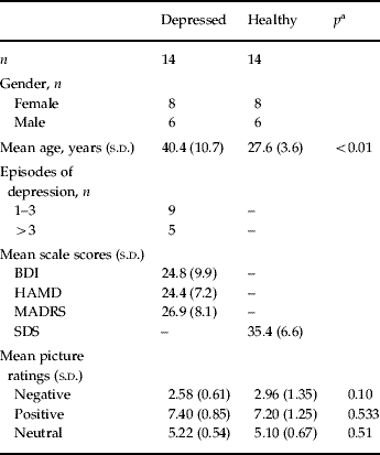
A total of 16 in-patients (14-included in analysis) with a current diagnosis of a major depressive episode were recruited at the Psychiatric University Hospital of Zurich, Switzerland (for demographic and psychometric data, see Table 1). A diagnosis of mildly to moderately severe depressive episode was made by trained psychiatrists according to ICD-10 and DSM-IV criteria. Psychiatric Axis I and other co-morbidities such as neurological disorders and MRI contraindications were excluded in a semi-structured interview prior to scanning. After receiving complete description of the study, all participants gave written informed consent. The study was approved by the local ethics committee. The patients were on stable antidepressant medication. They had been off benzodiazepines and neuroleptics for at least four half-lives, and they were not taking any mood-stabilizing medication. The patient group was analysed on its own and it was additionally compared with a group of 14 healthy subjects from a previous report (Table 1; Herwig et al. Reference Herwig, Kaffenberger, Baumgartner and Jancke2007b).
Demographic and psychometric data of included subjects

s.d., Standard deviation; BDI, Beck Depression Inventory; HAMD, Hamilton Depression Scale; MADRS, Montgomery–Åsberg Depression Rating Scale; SDS, Self-rating Depression Scale (score<50: not depressed).
a Difference by t test.
Experimental design
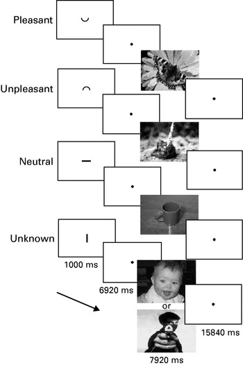
During fMRI scanning, the patients performed a cueing task (programmed with Presentation™, Neurobehavioral Systems, USA) consisting of 56 trials with expectation and presentation of emotional pictures (Fig. 1). The trials comprised of two main conditions: ‘known’ or ‘unknown’. The ‘known’ condition consisted of three subconditions (negative, positive, neutral). For each trial of the ‘known’ subconditions a small cue was presented that depicted either a smiling ‘⌣‘ (‘positive’ or ‘pleasant’, ps), a non-smiling ‘⌢‘ (‘negative’ or ‘unpleasant’, ng) or a neutral symbol ‘−‘ (nt) that indicated the emotional valence of the pictures presented after an anticipation period. In the ‘unknown condition’, ‘|‘ (uk), either pleasant or unpleasant pictures appeared randomly. The cues were of 1/40 of screen height and the pictures filled the screen. The cues were presented for 1000 ms followed by an anticipation period of a further 6920 ms [cue and anticipation: four times of MR volume repetitions; repetition time (TR) 1980 ms], during which a blank screen with a small fixation point was shown. Subsequently, emotional pictures (International Affective Picture System; Lang, Reference Lang1995) were presented for 7920 ms (4 TRs). During the following baseline period of 15 840 ms (8 TR) the blood oxygen level-dependent signal could wear off before the next trial. Altogether, 56 pre-cued pictures were shown, 14 for each ‘known’ trial as positive, negative and neutral, and 14 for ‘unknown’ (seven ps, seven ng). The different trials appeared in a randomized order. All participants were instructed to expect the emotional stimuli after the cue and to be aware of the emotional valence indicated, and to subsequently look at the following picture. The stimuli were matched for complexity, content of faces, scenery, food and nature, and concerning intensity of positive and negative valence with the same difference in valence ratings from neutral. Arousal was matched as far as possible for pleasant and unpleasant stimuli (Herwig et al. Reference Herwig, Baumgartner, Kaffenberger, Bruhl, Kottlow, Schreiter-Gasser, Abler, Jancke and Rufer2007a). All participants rated the presented pictures after the scanning according to the subjective valence on a visual analogue scale (1 most negative, 9 most positive).
Experimental task. Cues, presented for 1000 ms, indicated the valence of the picture which appeared after a delay of a further 6920 ms: ‘⌣‘, prior to a ‘pleasant’ picture; ‘⌢‘, prior to an ‘unpleasant’ picture; ‘−‘, prior to a ‘neutral’ picture; and ‘|’, prior to a picture of ‘unknown’ valence, either pleasant or unpleasant. The cues have been enlarged here for presentation purposes.

fMRI acquisition and data analysis
Imaging was performed with a 1.5 T Siemens Sonata whole-body scanner (Erlangen, Germany) equipped with a head coil. T1* weighted anatomical volumes (1×1×1 mm) were acquired for co-registration with the fMRI. T2* weighted functional magnetic resonance images were obtained using echoplanar imaging in an axial orientation (image size 64×64 pixels, 22 slices covering the whole brain, field of view of 220 mm, flip angle 90°, slice thickness 4 mm with 1 mm gap, voxel size 3.4×3.4×5 mm, repetition time/echo time (TR/TE) 1980/40 ms, 908 volumes). The subjects watched the stimuli in a mirror attached to the head coil and directed to a screen onto which the stimuli were projected by a video beamer.
fMRI data were analysed using BrainVoyager™ QX 1.8.6 (Brain Innovation, The Netherlands). Preprocessing included motion correction, slice scan time correction, high-frequency temporal filtering and removal of linear trends. Functional images were superimposed on the two-dimensional anatomical images and incorporated into three-dimensional (3D) datasets. The individual 3D datasets were transformed into Talairach space (Talairach & Tournoux, Reference Talairach and Tournoux1988) resulting in a voxel size of 3×3×3 mm and then spatially smoothed with an 8 mm Gaussian kernel for subsequent group analysis. Eight predictors representing the expectation (exp) and presentation (pres) of each valence (ng, uk, ps, nt) were used to build the design matrix. Expectation period and picture presentation periods were modelled as epochs using the standard two-γ haemodynamic response function provided by BrainVoyager (Glover, Reference Glover1999).
The fMRI data analysis based on the general linear model (GLM) comprised of the following steps: Fixed-effects analyses were calculated separately for each subject for the six contrasts comparing the emotion expectation conditions ‘negative versus neutral’, ‘positive versus neutral’, ‘unknown versus neutral’, ‘unknown versus negative’, ‘unknown versus positive’ and ‘negative versus positive’, resulting in summary images. The summary images were subjected to second-level analyses, separately for both the groups of the depressed patients and of the healthy subjects. Addressing our main question to evaluate which of the ‘known’ emotion expectation conditions (ng, ps, nt) revealed activations similar to those in the ‘unknown’ expectation condition (uk), we applied conjunction analyses based on the single contrasts. These were done with fixed-effects conjunction analyses with separate subject predictors in order to address the minimum-t problem (Nichols et al. Reference Nichols, Brett, Andersson, Wager and Poline2005). We defined ‘unknown’ expectation-related activity in a certain brain region as being similar to one of the ‘known’ expectation-related activations, when in this region both activations were significantly different from the activations associated with both remaining ‘known’ emotion expectation conditions, but were not different in the corresponding single contrast. For example, addressing similarity of the ‘unknown’ and the ‘negative’ condition, the ‘unknown’ condition had to differ significantly from ‘positive’- (contrast exp uk>ps) and also ‘neutral’ (exp uk>nt)-related activity, and ‘negative’ also had to differ from ‘positive’ (exp ng>ps) and ‘neutral’ (exp ng>nt) expectation-related activity. These contrasts therefore also comprised the direct comparisons between the ‘known’ expectation conditions as ‘negative versus positive’. The revealed brain regions further should not differ in the direct comparison, for instance when using the contrast exp ng>uk.
Accordingly, the following conjunctions were analysed:
(1) The ‘pessimism-contrast’: expecting unknown events to be negative should be reflected by activity during exp uk resembling the activity during exp ng, but differ from ps and nt: exp uk>ps and uk>nt and ng>ps and ng>nt.
(2) The ‘optimism-contrast’: assuming the upcoming unknown event to be positive, the brain activity during the exp uk should be similar to the activity during exp ps: exp uk>ng and uk>nt and ps>ng and ps>nt.
(3) The ‘indifference-contrast’: if subjects anticipate ‘unknown’, thus either positive or negative events, like neutral events, brain activity might be similar to the ‘neutral’ expectation: exp uk>ng and uk>ps and nt>ng and nt>ps.
The statistical threshold for these conjunctions was set at a level of p<0.001 (uncorrected considering the conjunction approach with the application of four single contrasts together) with a cluster size of 135 voxels of 1×1×1 mm corresponding to 5 voxels of 3×3×3 mm. The results obtained from the healthy subjects have been reported previously (Herwig et al. Reference Herwig, Kaffenberger, Baumgartner and Jancke2007b). For the depressed patients, further explorative results with lower cluster size are reported when they complied with the results from the healthy subject group. An analysis of the presentation period and main-effect analyses of emotion and expectation were performed on an exploratory basis not reported here.
Questionnaires and correlation statistics
The patients completed a handedness questionnaire (Annett, Reference Annett1967) and a depression self-rating [Beck's Depression Inventory (BDI); Beck et al. Reference Beck, Ward, Mendelson, Mock and Erbaugh1961]. Further, depressiveness was assessed by the 21-item Hamilton Depression Scale (HAMD; Hamilton, Reference Hamilton1960) and the Montgomery–Åsberg Depression Rating Scales (MADRS; Montgomery & Åsberg, Reference Montgomery and Åsberg1979). Immediately after scanning, the patients rated the emotional valence of the pictures (presented again as printouts) on a visual analogue scale. Using Pearson's correlation, we correlated the rating score results of the depression scales with the individual β-weights (mean from all voxels of the respective activated cluster) of the emotion expectation conditions in those regions resulting from the conjunction analyses. Thus, within the regions of interest (ROIs) derived from the conjunction analyses, we correlated the different individual β-weights of the emotion conditions in each ROI with the individual psychometric data. In order to control for an influence of age, we also correlated the β-weights with the age in both groups, healthy and depressed participants, and performed an exploratory partial correlation analysis with age as the control variable.
Comparison between depressed patients and healthy subjects
Activations in depressed patients and healthy subjects were compared. Within the activated clusters in the depressed patients and in the healthy subjects random-effects analyses of the ‘emotion versus neutral’ contrasts were performed by using separate subject predictors and implementing the factor of being depressed or healthy in the GLM. Additionally, we explored group differences of the single contrasts in the bilateral insula based on ROIs derived from the contrast ps>nt in the depressed patients in order to assess whether failing to find hypothesized activations in the ‘pessimism’-contrast might be due to an activation during the positive expectation.
Results
Participants and behavioural data
From the 16 depressed patients, one had to be excluded because of movement artefacts (>3 mm in at least one direction) and one patient reported after scanning that she had not been able to identify the cues correctly. Thus, 14 depressed patients were entered into the analysis. All were right-handed. They were also compared with 14 healthy subjects (Herwig et al. Reference Herwig, Kaffenberger, Baumgartner and Jancke2007b), who were younger than the patients. The picture ratings were not different (Table 1). Medication in the depressed patients included mirtazapin (15–45 mg, n=5), venlafaxin (75–150 mg, n=2), citalopram/escitalopram (60/20 mg, n=2), citalopram/escitalopram (20–40 mg) plus mirtazapin (30–60 mg) (n=3), trazodone (100 mg, n=1) and no medication (n=1).
Conjointly activated areas during ‘unknown’ and ‘known’ emotion expectation
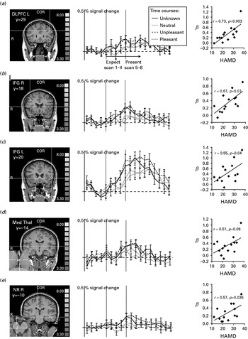
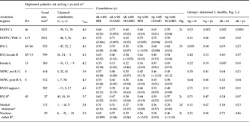
We performed conjunction analyses to discover which of the ‘known’ emotion expectation conditions revealed activations similar to those in the ‘unknown’ condition. Regarding the comparison ‘negative’ and ‘unknown’ in the depressed patients we found activity within the MPFC [Brodmann area (BA) 8], left DLPFC (BA 9/46), bilateral inferior frontal gyrus (IFG, BA 45) and insula (BA 13), in a region adjacent to the anterior thalamus and head of the caudate nucleus best fitting the area comprising the bed nucleus of the stria terminalis (BNST; being aware that this structure is small for the applied fMRI resolution), in the anterior and medial thalamus, and in the nucleus ruber (NR; Table 2, Fig. 2). These areas were not different in the direct contrast exp ng>uk in either direction. The equivalent analyses of the relation of ‘unknown’ and ‘positive’ expectation (‘optimism-contrast’) and of ‘unknown’ and ‘neutral’ expectation (‘indifference-contrast’) did not deliver any results even at a level of p<0.01 uncorrected. Other regions such as the amygdala were active in the single emotion expectation contrasts not reported here. Results from the healthy subjects have been reported earlier (Herwig et al. Reference Herwig, Kaffenberger, Baumgartner and Jancke2007b). In brief, activated areas in that group in the ‘pessimism’-contrast were within the right IFG, insular regions, medial thalamus, BNST region, NR and temporo-occipital cortex.
Comparison of ‘known’ and ‘unknown’ expectation. Conjunction analysis of the conditions ‘unknown’ and ‘unpleasant’ expectation versus ‘pleasant’ and versus ‘neutral’ (exp uk>ps and uk>nt and ng>nt and ng>ps). The activated voxels were co-registered using structural magnetic resonance imaging and colour coded (consider cross-hair, see coloured version of this figure in Supplementary online material) according to their significance (t-value). Presented are coronal (COR) slices of (a) the left dorsolateral prefrontal cortex (DLPFC L), (b) the right inferior frontal gyrus (IFG R), (c) the left IFG (IFG L), (d) the medial thalamus (Med Thal) and (e) the right nucleus ruber (NR R) and cut-outs of the axial slices of the thalamus and NR R in the lower left of the respective pictures. Further, on the right, the time courses of the activities within the marked regions of interest are shown in percentage BOLD signal change, indicating the conjoint activity of the ‘unpleasant’ expectation with the ‘unknown’ expectation. The vertical grey bars represent the beginning of the expectation (Expect) and presentation (Present) periods, comprising each four volumes. When interpreting the time courses, the delayed haemodynamic response function has to be considered. Further, the correlations of the β-weights of the activity during the ‘unknown’ expectation condition with the Hamilton depression scores (HAMD) are presented.

Conjunction analyses of emotion expectation contrasts, correlations of β-weights with rating scores, and comparison of depressed and healthy groups

uk, Unknown expectation; ng, negative expectation; ps, positive expectation; nt, neutral expectation; BA, Brodmann area; BDI, Beck depression inventory; HAMD, Hamilton depression scale; MADRS, Montgomery–Åsberg depression rating scale; DLPFC, dorsolateral prefrontal cortex; L, left; PMC, premotor cortex; IFG, inferior frontal gyrus; R, right; MPFC, medial prefrontal cortex; ant, anterior; post, posterior; BNST, bed nucleus striae terminals.
On the right, results from the random-effects group comparison of activations within the regions of interest from the ‘pessimism’-contrast.
a Activated regions according to the conjunction analysis of expectation ‘unknown’ and ‘negative’ stimuli, both versus expecting ‘positive’ and versus ‘neutral’ stimuli. Indicated are: the amount of voxels (mm3); Talairach coordinates (x, y, z) of the centre of mass of the activation; and maximal t-values of the voxels within the regions.
b Results from explorative analysis.
Correlation of brain activation with depression ratings
The assumption was made that subjects with a higher level of depressiveness may exert stronger activity in areas associated with ‘unknown’ and ‘negative’ expectation. We tested this by correlating psychometric data obtained from the BDI, HAMD and MADRS with the mean β-weights obtained from the significantly activated clusters of the conjunction analyses (one BDI missing). This analysis revealed significant positive correlations in the MPFC, left DLPFC, IFG/insula regions and NR during ‘unknown’ and ‘negative’ expectation (Fig. 2, Table 2). We did not find any correlation of the activations with age, neither in the depressed, nor in the healthy group, and the exploratory correlation analysis controlled for age provided essentially equivalent results as without this control variable.
Comparison between depressed patients and healthy subjects
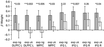
We compared the activity in the revealed clusters from the conjunction analysis in the depressed patients with the activations in healthy subjects (Fig. 3, Table 2). Compared with the healthy subjects, depressed patients showed increased activations during both the negative and unknown conditions in the left DLPFC and anterior MPFC, and for the unknown conditions also in the bilateral IFG/insular regions and left BNST. Concerning regions activated in healthy subjects but not in the patients, the right BNST (p=0.04) and left insula (p=0.04) were more strongly activated in the respective contrasts in the healthy subjects. The exploratory analysis of group differences in the single contrasts within the ROIs based on the ps>nt contrast found a stronger insular activation in the depressed patients (see Supplementary Fig. 1, available online).
Comparison of activity in regions of interest (β-weights) in depressed patients ( ) compared with healthy subjects (□). Values are means, with standard errors represented by vertical bars. For the different regions and conditions, p values are given for the difference between the depressed and healthy subjects (* p<0.05, ** p<0.01). DLPFC L, left dorsolateral prefrontal cortex; exp uk, expectation unknown; exp ng, expectation negative; MPFC, medial prefrontal cortex; IFG L, left inferior frontal gyrus; IFG R, right inferior frontal gyrus.

Discussion
Assuming a ‘pessimistic’ attitude in depression when expecting upcoming events, our hypothesis was that patients with major depression would show brain activation while expecting events of unknown emotional valence as if these events are known to be unpleasant. Further, we assumed that distinct emotion-processing brain areas known to be affected in depression would be activated differently compared with healthy subjects. Our main findings were: (i) while anticipating pictures of unknown emotional valence, the brain activity resembled that observed when expecting pictures known to be negative but not when expecting positive or neutral pictures; (ii) the activity within a prominent part of the corresponding brain regions, particularly within the DLPFC, MPFC and insula/IFG correlated with the grade of depression; and (iii) the activation in these regions differed significantly from that in healthy subjects. This supports the assumption of considering a negative outcome for future scenarios to form a key cognitive feature of depression with distinct neural representations.
Brain regions involved in ‘pessimistic’ expectation
Activations within the DLPFC and MPFC were associated with ‘pessimistic’ expectation in depressed patients, which was not the case in the healthy subjects. Further, the activity in these areas correlated with the depression scores: the more depressed, the higher the activation. Notably, the activation of the DLPFC differed between the groups in all, and in the anterior MPFC in most of the contrasts. This provides evidence that the ‘pessimistic’ expectation in depression may be associated with the functional domains of the DLPFC and MPFC as a final evaluation of situations, response planning, and cognitive and executive control (Fuster, Reference Fuster2000; Gross & John, Reference Gross and John2003). The DLPFC has been associated with hypometabolism in depression (Soares & Mann, Reference Soares and Mann1997), or with hypofunctionality in cognitive tasks (Siegle et al. Reference Siegle, Thompson, Carter, Steinhauer and Thase2007), possibly leading also to compensatory activation (Harvey et al. Reference Harvey, Fossati, Pochon, Levy, Lebastard, Lehericy, Allilaire and Dubois2005). One may assume that DLPFC resources in depression might be absorbed due to a bias towards depressive cognition by focusing on corresponding situations and stimuli (Grimm et al. Reference Grimm, Beck, Schuepbach, Hell, Boesiger, Bermpohl, Niehaus, Boeker and Northoff2008), for instance, due to a negative attitude towards future events according to the cognitive triad (Beck, Reference Beck1967). This may be represented within neural network concepts by information processing modules in the DLPFC acting as attractors (Yuste et al. Reference Yuste, MacLean, Smith and Lansner2005) for depressive cognition. We also found the MPFC to be active during unknown and negative expectation, with the ‘unknown’-related activity being more prominent in depressed than in healthy subjects. Whereas the DLPFC is involved in evaluating and preparing action in the external world (Fuster, Reference Fuster2000), the MPFC was reported to be involved in the cognitive control of internal emotional processes (Gross & John, Reference Gross and John2003; Herwig et al. Reference Herwig, Baumgartner, Kaffenberger, Bruhl, Kottlow, Schreiter-Gasser, Abler, Jancke and Rufer2007a). It might be recruited in depression in order to exert cognitive control onto subcortical emotion-associated areas, possibly with an ineffective compensatory over-activation. Basically, a complex disturbance of the information processing within and between functional systems for integrating emotional–cognitive information is assumed (Pessoa, Reference Pessoa2008). In this context, one may consider primary modulating regions on emotion processing such as the DLPFC or the amygdala working as ‘connector hubs’ for information processing (Sporns et al. Reference Sporns, Honey and Kotter2007).
In our previous study with healthy subjects, we found the IFG, insular regions and the medial thalamus to be associated with ‘pessimistic’ expectation (Herwig et al. Reference Herwig, Kaffenberger, Baumgartner and Jancke2007b). This was essentially also the case in the depressed patients, with the exception that certain insular regions revealed different activation patterns. The IFG, in the region of the ventrolateral prefrontal cortex, was assumed to represent multi-modal sensory cues with high emotional salience (Yamasaki et al. Reference Yamasaki, LaBar and McCarthy2002) and to be involved in emotional–cognitive integration (Mayberg, Reference Mayberg2003). It may provide a link between the evaluation of the internal state associated with an emotional situation and its cognitive processing, for instance, by propagating ‘internal’ information to upstream prefrontal areas. Medial thalamic regions receive input from viscero-sensitive and pain-mediating brainstem areas and are considered to form a relay within the visceroceptive pathway towards, for example, insular regions (Craig, Reference Craig2002; Vogt, Reference Vogt2005). The insula was found to be involved in the processing of multimodal sensory and emotional stimuli, supporting views of a general role of the insula in emotion processing (Damasio et al. Reference Damasio, Grabowski, Bechara, Damasio, Ponto, Parvizi and Hichwa2000; Calder et al. Reference Calder, Lawrence and Young2001; Craig, Reference Craig2002; Critchley et al. Reference Critchley, Wiens, Rotshtein, Ohman and Dolan2004; Paulus & Stein, Reference Paulus and Stein2006; Simmons et al. Reference Simmons, Matthews, Paulus and Stein2008). It was proposed that the physical sensation of emotional responses depends on sensations from the viscera represented in the insula (Critchley et al. Reference Critchley, Wiens, Rotshtein, Ohman and Dolan2004). In this context, the insula may be involved in the mediation of bodily viscero-sensitive signals for decision making and behavioural planning according to emotional valences. We found activations in the depressed patients during both the ‘unknown’ and the ‘negative’ expectation as hypothesized, but also increased activation during the ‘positive’ expectation compared with the healthy subjects. This may be interpreted as an insular ‘unpleasantness’-signal not only during ‘negative’ or ‘unknown’ expectation but also during ‘positive’ expectation, and may be associated with anhedonia (Keedwell et al. Reference Keedwell, Andrew, Williams, Brammer and Phillips2005). Being depressed is often associated with unpleasant somatic feelings and a negative self-perception. This may result from a dysregulation of interoceptive areas such as the insula due to a misinterpretation of external cues based on unpleasant previous experiences, inducing subjective unpleasantness (Davidson et al. Reference Davidson, Irwin, Anderle and Kalin2003). These considerations lead to further concepts of the insula to be involved in self-representation and ‘self-awareness’ (Churchland, Reference Churchland2002; Craig, Reference Craig2002) and to disturbed ‘self’ functions in depression (Northoff & Bermpohl, Reference Northoff and Bermpohl2004).
At the brainstem level, we found, in both the depressed and the healthy group, a region covering the NR to be associated with ‘negative’ and ‘unknown’ expectation. The NR has been assumed to be involved in psychomotor modulation and in behavioural attendance (Horn et al. Reference Horn, Pong, Batni, Levy and Gibson2002). During expectation, it may prime or facilitate the readiness for a later motor reaction.
Two main brain regions hypothesized as being associated with functional disturbance in depression were not found to be activated in the ‘pessimism’-contrast: the amygdala (e.g. Abler et al. Reference Abler, Erk, Herwig and Walter2006; Lee et al. Reference Lee, Seong Whi, Hyung Soo, Lee, Choi, Lyoo and Ham2007) and the ACC (Bermpohl et al. Reference Bermpohl, Pascual-Leone, Amedi, Merabet, Fregni, Gaab, Alsop, Schlaug and Northoff2006; Nitschke et al. Reference Nitschke, Sarinopoulos, Mackiewicz, Schaefer and Davidson2006). For the amygdala, this might be explained by the fact that it also showed activation during the positive expectation condition (Herwig et al. Reference Herwig, Kaffenberger, Baumgartner and Jancke2007b) and thus not fulfilling the criteria for the ‘pessimism’-contrast but being more generally associated with valence-independent emotional arousal (Anderson et al. Reference Anderson, Christoff, Stappen, Panitz, Ghahremani, Glover, Gabrieli and Sobel2003; McClure et al. Reference McClure, York and Montague2004). However, though being difficult to distinguish anatomically, a region covering and best fitting the bed nucleus of the stria terminalis was activated in the ‘pessimism’-contrast. This region was considered to form a functional unit with the ‘extended’ amygdala (Heimer & Van Hoesen, Reference Heimer and Van Hoesen2006) and may be a link to further emotion-processing circuits. The ACC may be activated more in the context of conflict monitoring between functional state and perceived new information with potential affective or motivational consequences (Carter et al. Reference Carter, Macdonald, Botvinick, Ross, Stenger, Noll and Cohen2000; Vogt, Reference Vogt2005) and not in the specific context of ‘pessimistic’ expectation.
Principally, one may assume that a circuit for biasing the organism towards potentially disadvantageous or even dangerous events makes evolutionary sense. Whenever we may also have an optimistic bias for certain personal long-term attitudes towards the future (Sharot et al. Reference Sharot, Riccardi, Raio and Phelps2007), the chances of survival for our early ancestors who for instance anticipated a predator when hearing a rustling sound in the vicinity, and prepared for it appropriately, were conceivably higher than for those who did not. Hence, one may speculate that a certain degree of depressiveness may be of evolutionary advantage (Nesse, Reference Nesse2000; Panksepp, Reference Panksepp2006), in that uncertain future events are more likely to be evaluated as unpleasant and that the person is better prepared for when negative events actually do occur.
Limitations
In reflecting on possible limitations, we addressed the issue of the differing age of both groups. Whenever the main analysis of this report was based on the depressed group, we also performed a group comparison with the healthy subjects. One may argue that the higher activation in the depressed patients might be accounted for by their higher age. This, however, would be in contrast with findings of attenuated emotion processing-related brain activity with increasing age (Erk et al. Reference Erk, Walter and Abler2008). Furthermore, age-dependence is unlikely in view of the fact that the activations correlated with depressiveness, but not with age, which would have been the case if age had an influence on the activation. The same holds true for the medication in the depressed patients. The fact of being medicated does not explain the correlations with psychopathology, such that depressiveness, not age or medication, can be accounted for by our results. Nevertheless, our findings should be interpreted as applying to medicated patients because any influence of medication on activations cannot be excluded entirely.
Emotional arousal (McClure et al. Reference McClure, York and Montague2004) or experience of uncertainty (Simmons et al. Reference Simmons, Matthews, Paulus and Stein2008) may also contribute to the revealed activation. However, again, the correlations with depressiveness point to an association of the activation with depression and not to these factors. Further, valence-independent arousal would also have occurred through positive expectation and thus would have led to non-fulfilling the criteria of ‘pessimism’. Nevertheless, uncertainty and heightened arousal towards unpleasant events might be inherent factors for ‘pessimistic’ expectation (Simmons et al. Reference Simmons, Matthews, Paulus and Stein2008).
The experimental task was designed to focus on the mental process during expectation. Therefore, the highly abstract and graphically comparable cues were intuitively understandable, so that no prominent working memory component was necessary to establish their meaning. Further, no motor reaction was required, the preparation and exertion of which might have interfered with processes of emotional anticipation. However, the task thus did not comprise an implemented behavioural control. We decided not to use such a control, for instance by motor reaction, in order to avoid interfering and noisy other brain activation. On the other hand, a behavioural measure implemented in our task demonstrating a ‘pessimistic’ bias also on the level of behavioural outcome could have further strengthened the assumption of ‘pessimistic’ expectation in depression. However, here we focused on the neural correlates considering that neural aspects, and also subjective emotional experience, must not necessarily be reflected by observable behaviour. We asked the subjects and patients afterwards whether if they had been able to attend to the task and we attempted to verify this by checking appropriate activation in the visual cortex. If the participants had not attended, one would have expected noisy data and no specific activations.
In conclusion, the similarity of ‘unknown’ and ‘unpleasant’ expectation-related brain activity may be attributed to a ‘pessimistic’ bias towards future events as expressed particularly in depressed patients. This supports cognitive models of a ‘pessimistic’ attitude in depression at the neurobiological level. Dorsolateral and medial prefrontal regions, known to be involved in executive and cognitive control, and viscero-sensitive areas such as the insula appear to play a key role in biasing the organism towards preparation for the worst-case scenario and to contribute to depressiveness.
Acknowledgements
The study was performed at the Psychiatric University Hospital Zürich in Switzerland. U.H. and L.J. were funded by the Swiss National Foundation (SNF no. 3200B0-112631). The SNF had no further role in study design, in collection, analysis or interpretation of the data, in writing the report and in the decision to submit the paper for publication. We are grateful to Dr D. Huber and C. Sauerwald, Hirslanden Clinic, Zürich, for providing the fMRI scanning.
Note
Supplementary material accompanies this paper on the Journal's website (http://journals.cambridge.org/psm).
Declaration of Interest
None.